The clinical translation of tissue-engineered biomaterials is currently hindered by the lack of standardized, high-throughput methods for characterizing complex, sub-micron, 3-dimensional pore architectures, critical for governing cellular infiltration and tissue integration, and ultimately, successful treatment of disease. This study validates an efficient, multidisciplinary workflow using micro-computed tomography (Micro-CT) enhanced by deep-learning super-resolution and segmentation to characterize heterogenous biomaterial scaffolds. Using the Zeiss DeepScout module, we upscaled low-resolution scans (1.38μm voxel size) by a factor of 2.55x, to a super-resolved resolution of 0.54μm. This approach achieved a 5.15x larger lateral field of view, and a 26.6x increase in total slice area compared to traditional high-resolution imaging. By stacking multiple experimental groups, the equivalent "brute-force" scan time was reduced by approximately 40.2x, decreasing acquisition time from 6.16 days to 220 minutes. To overcome beam-hardening and phase-propagation artifacts, an Attention U-Net deep-learning architecture was implemented, providing superior phase separation compared to traditional Otsu thresholding. Validation via Representative Elementary Volume (REV) analysis and pore metric distributions confirmed near-identical structural integrity between super-resolved and high-resolution scans. Deep-learning enhanced micro-CT characterisation of biomaterial scaffolds offers a scalable, high-throughput, high-resolution platform for precise, preclinical biomaterial assessment.
This study describes the derivatization of Rapamycin (Ra) with acryloyl chloride (AcCl) and iodoacetic acid (IAA), yielding hydrolysis-susceptible esters designed for controlled drug release at physiological pH. These esters were further conjugated to thiolated polyethylene glycols (PEGs), yielding compounds with enhanced water solubility, pendant thiol groups and with variation in the number of methylene groups between the ester and thioether moieties. Hydrogels were subsequently formed via conjugate addition reactions using multi-arm PEG macromers, specifically 8-arm PEG acrylates or vinyl sulphones, alongside thiolated PEG crosslinkers. The primary focus was to elucidate the impact of structural modifications surrounding the thioether ester linker on drug release kinetics. In vitro release studies demonstrated zero-order Ra elution over 7-19 days, modulated by gel architecture. Notably, Ra incorporated via α-thioether ester bonds exhibited significantly faster release than their β-thioether ester counterparts, with release rate increases of 11% and 31%, respectively, across the gel assemblies examined. This behavior was attributed to the electron-withdrawing effect of the adjacent thioether group, which enhanced ester hydrolysis. Additionally, creating a hydrogel more prone to swelling and degradation (by using the PEG acrylate multi-arm instead of the PEG vinyl sulphone equivalent) increased the overall drug release rate due to higher water uptake within the gel matrix. An alternative strategy involved Ra-based crosslinking, where Ra, di-functionalized with IAA, acted as a crosslinker for the PEG thiol multi-arm molecules. This assembly exhibited a biphasic release profile, initially mimicking the linear zero-order release of Ra mono-iodoacetic ester crosslinked with PEG acrylates, followed by an exponential burst phase. These findings provide critical insights into hydrogel design strategies for tailoring drug release kinetics, paving the way for advanced controlled drug delivery applications.
Background:Successful vascular tissue regeneration in vascular and soft-tissue biomaterials is governed not by bulk volumetric porosity, but by the existence of continuous, ingrowth-permissive pathways traversing the full scaffold thickness, termed angio-permissivity. Conventional structural metrics often fail to capture these functional conduits, leading to unpredictable in vivo outcomes and a disconnect between scaffold design and biological integration. Methods:We developed a transmural space characterization workflow integrating micro-computed tomography, deep-learning-assisted super-resolution reconstruction and segmentation, and pore-network modeling. Three architecturally distinct electrospun scaffold groups were thresholded for continuous pathways (>10 μm) and analyzed for vascular ingrowth permissivity. Findings were validated in a subcutaneous rat model (7 and 21 days) to correlate architectural parameters with extracellular matrix remodeling and neovascularization. Results:Quantitative modeling identified a "porosity paradox," where architectures with the highest total volumetric void space remained functionally isolated due to internal partitioning and sub-critical bottlenecks (<10 μm). Only scaffolds exhibiting a dense, continuous network of surface-to-surface growth tunnels supported robust transmural integration. In vivo, these continuous spatial configurations facilitated deep neovascularization and a rapid maturation "catch-up" to native glycosaminoglycan levels by day 21. In contrast, partitioned architectures restricted vessel recruitment and demonstrated impaired extracellular matrix preservation, regardless of high initial porosity. Conclusions:Transmural connectivity is a primary architectural determinant of tissue and vascular integration. This non-destructive, scalable framework enables the engineering-led design of tissue-engineered biomaterials by prioritizing the specific spatial and architectural requirements of the target physiological niche over stochastic volumetric metrics. Validation here uses a subcutaneous model that isolates architecture from cardiovascular hemodynamics - the next requirement for translation.
Background and Aims:Bioprosthetic heart valves (BHVs) are inherently susceptible to structural degeneration, driven by a combination of mechanical stress, lipid infiltration, glutaraldehyde-induced crosslinking instability, and progressive calcification. Recent evidence has implicated the αGal antigen (galactose-α-1,3-galactose) as an additional contributor to BHV deterioration through activation of innate immune pathways. The present study aims to: 1) perform a quantitative assessment of the residual presence of xenoantigens, specifically αGal, in a range of commercial BHV models; 2) evaluate the efficacy of an experimental polyphenol-based treatment in neutralizing these antigenic determinants; and 3) investigate the long-term stability of glutaraldehyde fixation concerning the potential re-exposure of αGal epitopes. Methods:Twelve distinct BHV models were subjected to in vitro analysis for αGal antigen quantification both before and following application of an experimental polyphenol treatment. Additionally, glutaraldehyde-fixed bovine pericardial tissues were incubated in a physiologically mimetic, blood-like environment for up to 9 years in real-time to simulate the long-term behavior of BHV materials and assess antigen unmasking associated with glutaraldehyde degradation. Results:The average count of the αGal epitope in original pericardial valve models was 4.18 ± 0.72 × 10¹¹/10 mg of tissue, whereas porcine valve-derived prostheses exhibited a higher mean value of 8.51 ± 2.17 × 10¹¹/10 mg. Treatment with the polyphenol formulation resulted in a marked reduction (approximately 99%) in detectable αGal epitopes. Furthermore, glutaraldehyde fixed pericardial tissues subjected to prolonged incubation demonstrated up to 60% re-exposure of previously masked αGal antigens after 9 years, consistent with a progressive compromise of glutaraldehyde crosslinking integrity. Conclusion:The data confirm that commercially available BHVs retain a substantial immunogenic burden attributable to αGal xenoantigens. Importantly, the overtime degradation of glutaraldehyde crosslinks facilitates the gradual re-exhibition of these epitopes, potentially undermining long-term valve performance. The pronounced efficacy of polyphenol-based treatment in inhibiting αGal antigens highlights its promise as a biocompatibility-enhancing pretreatment strategy for next-generation BHVs.
This study introduces a biodegradable silk-fibroin/DegraPol DP30 (SF-DP30) hybrid scaffold for heart-valve replacement and wider cardiovascular tissue engineering. By coupling SF’s tensile strength with DP30’s elasticity and controlled degradation, we targeted a construct that withstands cyclic loading while supporting cell integration. Mechanical testing of electrospun sheets and tri-leaflet prototypes showed tensile strength of 0.4–1.1 MPa, toughness of 0.1–0.6 MJ m −3 , and strain of 12%–90%, with 10 wt.% SF/90 wt.% DP30 blends offering the most balanced performance. Pulse-duplicator assays revealed orifice areas, pressure gradients, and closing dynamics equivalent to CE-approved polymeric and bioprosthetic valves, confirming hydrodynamic suitability. In vitro, cocultured human endothelial and fibroblast cells achieved confluent coverage, pore infiltration, and expression of vWF and CD31, indicating a hospitable microenvironment for endothelialization and remodeling. Key translational hurdles persist. Long-term risks of calcification, thrombogenicity, and inflammatory degradation must be quantified in large-animal models. The current reliance on the cytotoxic solvent hexafluoroisopropanol would complicate regulatory approval, necessitating greener processing routes such as benign-solvent or melt-electrospinning methods. Extended studies of degradation kinetics, immune modulation, and hemocompatibility are especially critical for pediatric implants that must accommodate somatic growth. Overall, SF-DP30 scaffolds combine mechanical resilience with demonstrated cytocompatibility, positioning them as promising—but not yet clinically validated—candidates for next-generation cardiovascular implants.
Cardiovascular tissue engineering has made significant strides since the 1970s, when initial efforts sought to reduce the thrombogenicity of total artificial hearts using cultured endothelial layers. Over the decades, however, the field has experienced repeated cycles of rediscovery and setbacks. Unlike other disruptive medical innovations, its greatest challenge remains the failure to achieve broad clinical translation. While research enthusiasm has spread globally and engaged diverse scientific communities, it is time for the clinician-scientist to reassert a central role - bridging mechanistic insights with therapeutic application. This is essential for two reasons. First, to restore an understanding of the distinct healing-biology of cardiovascular prostheses in humans. Animal models have often misrepresented human healing responses, leading to incorrect assumptions and divergent, non-translatable research paths. Second, clinical needs have changed. Whereas early efforts were driven by the need for coronary bypass grafts, priorities have shifted to transcatheter valve therapies, where durable soft-leaflet solutions are urgently needed. Despite past setbacks, a coherent framework has emerged that can guide global efforts. Central to this is renewed recognition of human-specific healing impediments: complete transanastomotic endothelialization does not occur in humans, and compacting fibrinogen-rich thrombus formation inhibits transmural tissue ingrowth. Against this backdrop stand key insights from decades of research: Autologous in vitro endothelialization markedly improves prosthetic performance in patients. Transmural tissue ingrowth is vital for in-situ endothelialization. Such sprouting must precede hostile fibrin accumulation that would otherwise prevent integration. Healing must conclude before scaffold degradation induces fibrosis that blocks further ingrowth. All core elements are now in place, backed by extensive clinical proof-of-principle. The path forward requires the reintegration of the clinician-scientist to lead this promising field toward successful therapeutic translation.
There is growing recognition in regenerative cardiovascular tissue engineering that transmural vessel ingrowth is the predominant—if not exclusive—mechanism for achieving in-situ endothelialization in prosthetic vascular grafts and heart valves in humans This process requires continuous ingrowth channels with dimensions sufficient to accommodate capillaries or even arterioles. While a variety of methods—such as electrospinning—exist to create porous scaffolds, current characterization techniques fail to determine whether the resulting structures offer well-defined and consistently continuous ingrowth spaces. Drawing on principles from geological porous media research, we applied a combination of nano-computer-tomography, deep-learning segmentation and super-resolution algorithms, and pore network modelling, to characterize the full thickness pore space morphology of electrospun scaffolds. Scaffolds were non-destructively reconstructed at high resolution (0.54 microns) and large fields of view, 57x faster than a brute-force approach, achieving total sample volumes greater than 1x10⁸ um3 in just a few hours. Electrospun scaffolds showed a median pore size and median pore volume of 5.51um (IQR: 5.15)/418.07um2 (IQR: 1153.74), n = 15 698, for the 16% polymer weight percentage group; 5.40um (IQR: 6.23)/412.24um2 (IQR: 1485.24), n = 13 437, for 18%; and 5.40um (IQR: 4.22)/356.34um2 (IQR: 826.53), n = 28 620, for 20%. On deeper analysis, continuous, interconnected pore networks of <10 microns in minimum diameter were extracted, with the 18% group showcasing the most extensive, surface-to-surface networking. This analysis highlights the limited ability of single-needle electrospinning to produce sufficient growth space for reliable transmural capillary endothelialisation. With the advent of cutting-edge additive and reductive manufacturing techniques, alternative methods for porous scaffold construction show promise.
IntroductionThe development of transcatheter aortic valve devices critically depends on preclinical testing in large animal models, yet key anatomical differences between these models and humans remain insufficiently defined. This study evaluated the translational relevance of ovine and porcine models by comparing aortic root anatomy with that of healthy individuals and patients with aortic valve disease.MethodsSilicone root casts and ECG-gated computed tomography (CT) imaging were used to assess annular, sinus of Valsalva (SOV), and sinotubular junction (STJ) dimensions, as well as coronary ostial height and eccentricity. ResultsPigs and sheep exhibited significantly lower and more laterally displaced left coronary ostia compared to humans—features that may predispose to coronary obstruction during valve implantation. Body weight correlated with key root dimensions, but wide individual variability precludes its use for selecting individual animals. However, it remains a useful filter for defining cohorts from which suitable subjects can be selected using CT. Sheep demonstrated flatter sinuses and lower STJ heights than pigs and humans, further reducing coronary inflow clearance. In contrast, coronary heights in humans were consistent across valve pathologies, with sinus dimensions being the most variable feature.ConclusionValidation of ex vivo silicone casting against in vivo CT confirmed its suitability for scalable anatomical assessment while aligning with animal welfare principles. These findings support refinement of animal selection strategies and provide an anatomically grounded framework for preclinical evaluation of transcatheter valve technologies.
Background. Heart transplantation in South Africa faces numerous challenges related to organ scarcity and unequal access to advanced heart therapy. There is an urgent need to analyse the current transplant referral pathway to optimise equitable access to transplantation. Objectives. To provide an audit of heart transplant referrals to Groote Schuur Hospital, Cape Town, over a 23-year period, focusing on patient demographics, indications for referral, waiting-list dynamics, and transplant referral outcomes. Methods. The study utilised a retrospective patient folder review for the period 1 January 1997 - 31 December 2019 and audited the trends in heart transplant referrals and associated outcomes of the referral at a tertiary academic hospital. Results. A total of 625 recipients were referred for heart transplantation, with the majority being male (n=412; 65.9%), while gender was undocumented for 69 cases (11.0%). The mean age was 38.1 (14.6) years, and 153 (24.5%) were listed for transplant, while 215 (34.4%) were deemed ineligible for listing. Contraindications for listing included social (n=106; 49.3%), medical (n=83; 38.6%) and psychological (n=26; 12.0%) factors, while 134 patients (21.4%) were considered too well. Poor social circumstances (n=38; 39.6%), poor insight (n=28; 29.2%) and poor compliance (n=21; 21.9%) were the most common non-medical reasons for not listing recipients, while obesity (n=30; 31.3%) and smoking (n=23; 24.0%) were notable medical contraindications. Forty-nine patients (7.8%) died during work-up, while 130 (85.0%) of the listed patients received a heart transplant. Of the 429 donor referrals, 139 (32.4%) were accepted for organ procurement. Reasons for declining donors included unsuitability for transplantation (30.3%), lack of capacity (1.8%), and recipient-donor mismatch (66.9%). Conclusion. Three-quarters of the referred patients were deemed unsuitable for heart transplantation for medical and/or social reasons. The ratio of referral to listing has decreased over time. However, once listed, the likelihood of receiving a transplant was high.
Informed by the almost unimaginable unmet need for cardiac surgery in the developing regions of the world, leading surgeons, cardiologists, editors in chief of the major cardiothoracic journals as well as representatives of medical industry and government convened in December 2017 to address this unacceptable disparity in access to care. The ensuing “Cape Town Declaration” constituted a clarion call to cardiac surgical societies to jointly advocate the strengthening of sustainable, local cardiac surgical capacity in the developing world. The Cardiac Surgery Intersociety Alliance (CSIA) was thus created, comprising The Society of Thoracic Surgeons (STS), the American Association for Thoracic Surgery (AATS), the Asian Society for Cardiovascular and Thoracic Surgery (ASCVTS), the European Association for Cardio-Thoracic Surgery (EACTS) and the World Heart Federation (WHF). The guiding principle was advocacy for sustainable cardiac surgical capacity in low-income countries. As a first step, a global needs assessment confirmed rheumatic heart disease as the overwhelming pathology requiring cardiac surgery in these regions. Subsequently, CSIA published a request for proposals to support fledgling programs that could demonstrate the backing by their governments and health care institution. Out of 11 applicants, and following an evaluation of the sites, including site visits to the 3 finalists, Mozambique and Rwanda were selected as the first Pilot Sites. Subsequently, a mentorship and training agreement was completed between Mozambique and the University of Cape Town, a middle-income country with a comparable burden of rheumatic heart disease. The agreement entails regular video calls between the heart teams, targeted training across all aspects of cardiac surgery, as well as on-site presence of mentoring teams for complex cases with the strict observance of “assisting only.” In Rwanda, Team Heart, a US and Rwanda-based non-governmental organization (NGO) that has been performing cardiac surgery in Rwanda and helping to train the cardiac surgery workforce since 2008, has agreed to continue providing mentorship for the local team and to assist in the establishment of independent cardiac surgery with all that entails. This involves intermittent virtual conferences between Rwandan and US cardiologists for surgical case selection. Five years after CSIA was founded, its “Seal of Approval” for the sustainability of endorsed programs in Mozambique and Rwanda has resulted in higher case numbers, a stronger government commitment, significant upgrades of infrastructure, the nurturing of generous consumable donations by industry and the commencement of negotiations with global donors for major grants. Extending the CSIA Seal to additional deserving programs could further align the international cardiac surgical community with the principle of local cardiac surgery capacity-building in developing countries.
The majority of patients requiring heart valve replacement in low- to middle-income countries (LMICs) need it for rheumatic heart disease (RHD). While the young age of such patients largely prescribes replacement with mechanical prostheses, reliable anticoagulation management is often unattainable under the prevailing socioeconomic circumstances. Cases of patients with clotted valves presenting for emergency surgery as a consequence of poor adherence to anticoagulation control are frequent. The operative mortality rates of reoperations for thrombosed mechanical valves are several times higher than those for tissue valves, and long-term results are also disappointing. Under-anticoagulation prevails in these regions that has clearly been linked to poor international normalised ratio (INR) monitoring. In industrialised countries, safe anticoagulation is defined as >60%–70% of the time in the therapeutic range (TTR). In LMICs, the TTR has been found to be in the range of twenty to forty percent. In this study, we analysed >20,000 INR test results of 552 consecutive patients receiving a mechanical valve for RHD. Only 27% of these test results were in the therapeutic range, with the vast majority (61%) being sub-therapeutic. Interestingly, the post-operative frequency of INR tests of one every 3–4 weeks in year 1 had dropped to less than 1 per year by year 7. LMICs need to use clinical judgement and assess the probability of insufficient INR monitoring prior to uncritically applying Western guidelines predominantly based on chronological age. The process of identification of high-risk subgroups in terms of non-adherence to anticoagulation control should take into account both the adherence history of >50% of patients with RHD who were in chronic atrial fibrillation prior to surgery as well as geographic and socioeconomic circumstances.
Coronary artery bypass grafting (CABG) on cardiopulmonary bypass (CPB) is associated with myocardial ischemia–reperfusion injury (IRI), which may limit the benefit of the surgery. Both experimental and clinical studies suggest that Intralipid, a lipid emulsion commonly used for parenteral nutrition, can limit myocardial IRI. We therefore aimed to investigate whether Intralipid administered at reperfusion can reduce myocardial IRI in patients undergoing CABG on CPB. We conducted a randomized, double-blind, pilot trial in which 29 adult patients scheduled for CABG were randomly assigned (on a 1:1 basis) to receive either 1.5 ml/kg Intralipid 20
Hydrogel-based localised drug delivery minimises systemic side effects and a linear release profile ensuring a sustained drug release over time, crucial for long-term therapy. The current paper describes the use of the Copper(I)-catalyzed Azide-Alkyne Cycloaddition (CuAAc) to append azidified Dexamethasone (Dex) onto dendrons of first- and second-generation PEGs. Crosslinking with thiolated PEGs using either thiol-acrylate or nucleophilic addition reactions yielded gels containing β-thio-ether ester groups that imparted enhanced hydrolytic susceptibility. In vitro gel degradation was followed gravimetrically and expressed as swelling ratios. Thiol-acrylate crosslinked hydrogels exhibited zero-order Dex release kinetics over 11, 27, and 16 days (G1, G1-star, and G2). Crosslinking the G1-gels by nucleophilic addition also resulted in linear release and the end point was reached in 5 days. Hydrolysis was accounted as the main release mechanism for covalently bound Dex, while physically incorporated Dex showed undefined rapid burst or first-order release, with most of the drug released in the initial 1–3 days. Eluates from covalently bound Dex maintained high activity, whereas Trap-Dex gels lost activity over time, as detected by the upregulation of luciferase expression from a transformed cell line. This novel chemistry combination offers precise drug release control applicable beyond Dex to drugs with suitable nucleophilic groups.
Socioeconomic factors such as poor health and poor nutrition in low- and middle-income countries (LMICs) may favour inflammatory reactions, thus contributing to the recurrence of rheumatic fever (RF) and thereby modifying trends in rheumatic heart disease (RHD). Apart from epidemiological studies, studies of HIV infections in RHD patients are limited. This systematic review synthesises data on the prevalence and impact of HIV infections or AIDS on RHD from PubMed, Scopus, Web of Science databases up to April 2021. The outcomes were managed using PRISMA guidelines. Of a total of 15 studies found, 10 were eligible for meta-analyses. Meta-analysis found that 17% (95 % CI 8–33, I2 = 91%) of adults in cardiovascular disease (CVD) cohorts in Southern Africa are HIV positive. The proportion of RHD diagnosed among people living with HIV was 4% (95% CI 2–8, I2 = 79%) for adults but lower [2% (95% CI 1–4, I2 = 87%)] among perinatally infected children. Despite limited reporting, HIV-infected patients with RHD are prone to other infections that may enhance cardiac complications due to poor immunological control. PROSPERO registration number: CRD42021237046.
OBJECTIVES:The purpose of this study was to evaluate the impact of a polyphenols-based treatment on the extrinsic mechanisms responsible for early bioprosthetic heart valve (BHV) degeneration. Structural degeneration can be driven by both extrinsic and intrinsic mechanisms. While intrinsic mechanisms have been associated with inherent biocompatibility characteristics of the BHV, the extrinsic ones have been reported to involve external causes, such as chemical, mechanical and hydrodynamic, responsible to facilitate graft damage.METHODS:The chemical interaction and the stability degree between polyphenols and pericardial tissue were carefully evaluated. The detoxification of glutaraldehyde in commercial BHVs models and the protective effect from in vivo calcification were taken into relevant consideration. Finally, the hydrodynamic and biomechanical features of the polyphenols-treated pericardial tissue were deeply investigated by pulse duplicator and stress-strain analysis.RESULTS:The study demonstrated the durability of the polyphenols-based treatment on pericardial tissue and the stability of the bound polyphenols. The treatment improves glutaraldehyde stabilization's current degree, demonstrating a surprising in vivo anti-calcific effect. It is able to make the pericardial tissue more pliable while maintaining the correct hydrodynamic characteristics.CONCLUSIONS:The polyphenols treatment has proved to be a promising approach capable of acting simultaneously on several factors related to the premature degeneration of cardiac valve substitutes by extrinsic mechanisms.
Introduction:Preformed antibodies against αGal in the human and the presence of αGal antigens on the tissue constituting the commercial bioprosthetic heart valves (BHVs, mainly bovine or porcine pericardium), lead to opsonization of the implanted BHV, leading to deterioration and calcification. Murine subcutaneous implantation of BHVs leaflets has been widely used for testing the efficacy of anti-calcification treatments. Unfortunately, commercial BHVs leaflets implanted into a murine model will not be able to elicit an αGal immune response because such antigen is expressed in the recipient and therefore immunologically tolerated.Methods:This study evaluates the calcium deposition on commercial BHV using a new humanized murine αGal knockout (KO) animal model. Furtherly, the anti-calcification efficacy of a polyphenol-based treatment was deeply investigated. By using CRISPR/Cas9 approach an αGal KO mouse was created and adopted for the evaluation of the calcific propensity of original and polyphenols treated BHV by subcutaneous implantation. The calcium quantification was carried out by plasma analysis; the immune response evaluation was performed by histology and immunological assays. Anti-αGal antibodies level in KO mice increases at least double after 2 months of implantation of original commercial BHV compared to WT mice, conversely, the polyphenols-based treatment seems to effectively mask the antigen to the KO mice's immune system.Results:Commercial leaflets explanted after 1 month from KO mice showed a four-time increased calcium deposition than what was observed on that explanted from WT. Polyphenol treatment prevents calcium deposition by over 99% in both KO and WT animals. The implantation of commercial BHV leaflets significantly stimulates the KO mouse immune system resulting in massive production of anti-Gal antibodies and the exacerbation of the αGal-related calcific effect if compared with the WT mouse.Discussion:The polyphenol-based treatment applied in this investigation showed an unexpected ability to inhibit the recognition of BHV xenoantigens by circulating antibodies almost completely preventing calcific depositions compared to the untreated counterpart.
 Deon Bezuidenhout合作论文数Cardiovascular Research Unit, University of Cape Town;Department of Surgery, University of Cape Town;Strait Access Technologies78
 Thomas Franz合作论文数Faculty of Health Sciences, University of Cape Town45
 Roland Fasol合作论文数44M Deutsch合作论文数32
 Paul Human合作论文数Christian Barnard Department of Cardiothoracic Surgery/Cardiovascular Research Unit, University of Cape Town Medical School31
 Neil Davies合作论文数Department of Earth Sciences, University of Cambridge31
 Scherman Jacques合作论文数23P Human合作论文数21
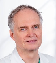 Theodor Fischlein合作论文数Klinik für Herzchirurgie, Klinikum Nürnberg;Cardiovascular Center, Paracelsus Medical University20
 Pennel Timothy合作论文数Faculty of Health Sciences, University of Cape Town19
 Johan Brink合作论文数Faculty of Health Sciences, University of Cape Town and Groote Schuur Hospital19Johann Meinhart合作论文数17
 Karen Sliwa合作论文数Cape Heart Institute, Faculty of Health Sciences, University of Cape Town;Division of Cardiology, Department of Medicine, Groote Schuur Hospital, University of Cape Town;Private Academic Hospital, University of Cape Town;Hatter Institute for Cardiovascular Research in Africa16D. Bezuidenhout合作论文数15Mona Bracher合作论文数15
 Ralph Bolman合作论文数14
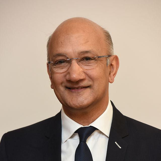 Dayanand Reddy合作论文数University of Cape Town13
 David F. Williams合作论文数Holdings Pty Ltd;University of Liverpool;Wake Forest Institute of Regenerative Medicine13M Kadletz合作论文数12Petra Preiss合作论文数12Gregor Wollenek合作论文数11
 Peter Groscurth合作论文数Institute of Anatomy, University of Zurich-Irchel;Fax: +41-1-2575702 CH;Institute of Anatomy, University of Zurich-Irchel10
 Gunther Laufer合作论文数Department of Cardiac Surgery, Medical University of Vienna10U Vonoppell合作论文数9Friedhelm Beyersdorf合作论文数9
 Percy Boateng合作论文数9Ernst Wolner合作论文数9Michael Wolf合作论文数9Uo Vonoppell合作论文数8
 Liesl Zühlke合作论文数Faculty of Health Sciences, University of Cape Town;Division of Paediatric Cardiology, Red Cross War Memorial Children’s Hospital;Children’s Heart Disease Research Unit, University of Cape Town8
 Robert Higgins合作论文数8Ulrich Von Oppell合作论文数8Jeroen Kortsmit合作论文数8B Reichart合作论文数7
 Psalms Ofoegbu合作论文数Department of Surgery, University of Ilorin Teaching Hospital7Melanie Black合作论文数7
 Christian A P Schmidt合作论文数7Anel Oosthuysen合作论文数7Manfred Deutsch合作论文数7C Weissenstein合作论文数6T Eberl合作论文数6Zachary Obinna Enumah合作论文数Department of Surgery, Johns Hopkins Hospital6
 Jithan Jacob Koshy合作论文数6
 Mark Yeoman合作论文数School of Pharmacy and Biomolecular Sciences, University of Brighton6J. Swanevelder合作论文数Department of Anaesthesia, Glenfield Hospital6Nasser Rafiee合作论文数6Maximilian Y. Emmert合作论文数Department of Cardiovascular Surgery, University Hospital Zurich6W Müller-Glauser合作论文数5E Minar合作论文数5




