Castleman’s disease presents as a peculiar type of lymph node hyperplasia. Traditionally, the disease has been classified on clinical grounds (solitary or multicentric) and by histologic appearance (hyaline vascular pattern, plasma cell predominance, or mixed lesions). It is now increasingly clear that there are different etiologies for each of these different subtypes. Reported associations include POEMS syndrome (polyneuropathy, organomegally, endocrinopathy, monoclonal gammopathy, and skin changes), paraneoplastic pemphigus, Hodgkin’s disease, and follicular dendritic cell sarcoma. We present a case of Castleman’s disease associated with myasthenia gravis, the third reported case in the literature. We discuss Castleman’s disease and review the literature.
Venous bullet embolism to the heart is a rare complication of penetrating gunshot trauma. There are little data regarding long-term follow-up of missiles retained in the right ventricle. We report a rare case of right ventricular bullet embolus following a left-sided thoracic gunshot wound. The patient presented with delayed onset of cardiac irritability symptoms 4 years after injury.
Pulmonary blastoma is a rare primary lung tumour associated with poor prognosis. Despite modern diagnostic imaging and biopsy techniques, the diagnosis is usually not considered initially. A case of a 22-year-old man with a large biphasic left-sided pulmonary blastoma, extending to the mediastinum, is reported. It was treated by pneumonectomy and pericardiectomy with radical tumour resection from the anterior mediastinum, under cardiopulmonary bypass. Adjuvant radiotherapy was used. 3 years postoperatively there is no sign of recurring disease.
Cardiopulmonary bypass is often followed by pulmonary dysfunction as assessed by measuring the alveolar-arterial oxygenation gradient, intrapulmonary shunt, degree of pulmonary edema, pulmonary compliance, and pulmonary vascular resistance. It is also regarded as a risk factor for development of acute respiratory distress syndrome. On the other hand, cardiopulmonary bypass is associated with a whole body inflammatory response, which involves activation of complement, leukocytes, and endothelial cells with secretion of cytokines, proteases, arachidonic acid metabolites, and oxygen free radicals. Leukocyte adhesion to microvascular endothelium, leukocyte extravasation, and tissue damage are the final steps. Although the inflammatory response to cardiopulmonary bypass often remains at subclinical levels, it can also lead to major organ dysfunction and multiple organ failure. This review article summarizes the recent literature on the molecular and cellular mechanisms involved in the phenomenon of pulmonary dysfunction after cardiopulmonary bypass. It also summarizes reports on the prevalence and mortality of acute respiratory distress syndrome after cardiac surgery.
Background Marked cerebral swelling visible on magnetic resonance images has been found immediately after hypothermic (28 degrees C) cardiopulmonary bypass. The mechanism is unknown, but indices of cerebral ischemia are seen during rewarming from hypothermic bypass that are not present with normothermic bypass (37 degrees C). Methods T1-weighted and fluid-attenuated inversion recovery magnetic resonance images were taken of seven patients undergoing routine coronary artery bypass surgery before, 1 h, and 7 days after the operation using normothermic bypass. Results Marked cerebral swelling was seen in fluid-attenuated inversion recovery images in five of seven patients 1 h after bypass. Scans in four patients taken 7 days after bypass showed that the cerebral swelling had returned to normal. There was no change in cerebral ventricular size, and all patients had uncomplicated postoperative courses. Conclusions Normothermic bypass is followed by acute postoperative cerebral swelling. However, the amount of swelling was similar to that found in a previous study after hypothermic bypass. The mechanism of swelling is still obscure, and its relation to neurologic outcome is unknown.
OBJECTIVE:Some form of organic and functional cerebral deficit may occur in up to one third of patients following cardiopulmonary bypass surgery. This study was designed to assess cerebral functional deficit in cardiac surgical patients. METHODS:Neuropsychological and quantitative electroencephalographic (EEG) changes were assessed in 62 first time coronary artery bypass graft surgery patients before surgery and within 1 week and 2 months after surgery. Patients underwent surgery with a standard Hammersmith Hospital anaesthesia and hypothermic cardiopulmonary bypass (28 degrees C), using either bubble (Harvey 1700, n = 28) or membrane (Cobe CML, n = 34) oxygenators with arterial line filters (Pall 40 microm). Neuropsychological performance was assessed using a well established battery of ten tests. Four EEG relative power frequency bands; delta (1-3.5 Hz), theta (4-7.5 Hz), alpha (8-11.5 Hz), and beta (12-23 Hz), were determined using Fast Fourier Transformation (FFT). RESULTS:Neuropsychological and EEG deficits were found in 48% of patients 1 week after surgery and in 34% 2 months after surgery. Post-operative deficits were not associated with duration of perfusion, type of oxygenator used in surgery or patient age. Neuropsychological and EEG deficits were associated 2 months after surgery, but not 1 week after surgery. Post-operative EEG deficit was associated with pre-operative deficit. CONCLUSIONS:Cerebral functional deficit was found following CABG surgery using quantitative EEG and neuropsychological assessments. Patients who had neuropsychological deficit were also more likely to show EEG deficit. EEG deficit before and after surgery suggests vulnerability of patients with already compromised cerebral function to the effects of CPB procedure.
OBJECTIVE:Quantitative electroencephalography was used during cardiopulmonary bypass surgery to determine the point in time of most neuronal functional change which may result in postoperative neuropsychological deficit. It was also used to determine any relationship between quantitative electroencephalography changes and type of oxygenator used in surgery.METHODS:We studied 61 coronary artery bypass graft patients. Anaesthesia included thiopental, fentanyl and N2O. Surgery was performed with hypothermic bypass (28 degrees C), arterial pressure of 50-70 mmHg, and alpha-stat, using bubble (Harvey 1700), or membrane (Cobe CML) oxygenators, both with arterial line filters (Pall 40 microns).RESULTS:The main finding was a significant increase in delta power at the end of perfusion (P < 0.01), which showed a positive association with delta power before the start of perfusion. Marked quantitative electroencephalography change at the end of perfusion was not related to systemic hypotension, temperature, type of oxygenator, bypass time, or patient age. Intraoperative quantitative electroencephalography changes found in most patients were transient and could not be related to postoperative cerebral function. However, 16 of the 18 patients who had neuropsychological deficit 2 months after surgery, also had a significant quantitative electroencephalography change at the end of perfusion.CONCLUSIONS:While no difference in anaesthetic technique was found between patients, the variation in quantitative electroencephalography power before perfusion may indicate a difference in individual response to anaesthetic. Usefulness of quantitative electroencephalography to predict postoperative cerebral functional deficit remains doubtful.
Imaging the native coronary vessels using contrast angiography can be difficult in the context of redo coronary operations when native disease is severe. When previous vein grafts undergo aneurysmal dilatation, imaging of the native vessel is restricted by the inability to infuse a sufficient volume of contrast medium through the graft and into the native vessel. We present a case of a patient whose redo coronary artery bypass graft operation was planned on the basis of magnetic resonance imaging of his native coronary arteries and vein graft after unsuccessful coronary angiography.
Lynn and associates [11] presented a review of 1,000 patients who had undergone coronary artery bypass grafting. They noted a postoperative serious stroke rate of approximately 2.7%. Has t he incidence of major stroke after a cardiac procedure decreased in the last 10 years? The answer is probably no. While obtaining a consent from patients for a cardiac operation, most surgeons would still, I believe, quote a stroke risk of approximately 2%. This has been unchanged for many years. This stroke risk however, is not an obligatory one that surgeons can do nothing about. Most cases of stroke after a cardiac surgical intervention with cardiopulmonary bypass are due to macroemboli, u sually particulate and usually atheromatous or calcific in nature. These emboli can be associated with the aorta or a calcified disorganized valve. Some strokes are due to macroemboli associated with clot that may be attached to the left ventricular surface after a myocardial infarction or associated in the left atrium with mitral valve disease. Other strokes are associated with air emboli, as in cases where the cardiac chambers are opened and vigorous deairing has not successfully removed them at the end of the procedure. Strokes from air emboli are often less severe and m ore transitory than those from particulate emboli.
Objectives: To detect co-expression of genes coding for components of the renin-angiotensin system and investigate the potential for variation in the level of angiotensin converting enzyme (ACE) gene expression in the right atrial appendage of patients undergoing heart surgery.Design: The right atrial appendage was collected at the time of surgery from 30 randomly chosen patients and was rapidly frozen in liquid nitrogen prior to extraction of messenger (m)RNA. Surgical samples of heart valve (n = 6) and papillary muscle (n = 3) were also examined.Methods: Aliquots of purified mRNA were reverse-transcribed for analysis of gene expression by a polymerase chain reaction amplification assay. Primers specific for angiotensinogen, renin, ACE, cardiac chymase, atrial natriuretic peptide, glyceraldehyde-3-phosphate dehydrogenase, adenosine deaminase and the transferrin receptor were used for a qualitative analysis of co-expression of these genes with in the same sample. In a subgroup of eight patients, a quantitative comparison of the relative levels of ACE gene expression was performed using a competitive polymerase chain reaction.Results: Angiotensinogen and ACE expression were detected in all atrial, valve and ventricular samples examined, at levels similar to those of 'housekeeping' genes such as the transferrin receptor. Atrial renin and chymase expression were more difficult to detect, being demonstrable in only 70 and 63% of the samples, respectively. Higher levels of chymase were detected in ventricular samples than in atrial tissues. A quantitative analysis of ACE expression in eight atrial samples provided evidence of interindividual variation in the relative level of atrial ACE expression.Conclusions: The essential components of the renin-angiotensin system are co-expressed at a low level in the right atrial appendage and are detectable in other regions of the human heart. Renin and chymase genes are expressed at a lower level than the angiotensinogen and ACE genes and exhibit regional differences in expression. Interindividual variation in the relative level of ACE expression can be detected by a competitive polymerase chain reaction.
Cerebral morbidity is a problem after cardiac surgery. Although neuropsychological tests and imaging techniques have been applied to cardiac patients, the relationship between them has not been considered. In the preliminary investigation, we studied 15 patients (11 male, mean age 59 years) having coronary artery bypass graft (CABG) surgery. Before surgery, patients had magnetic resonance (MR) imaging and neuropsychological assessment with a battery of 10 tests. During surgery, cardiopulmonary bypass was maintained at 28 degrees C with a flow rate of 2.4 L/m2/min-1 and at a mean arterial pressure of 50-70 mm Hg. Bubble or membrane oxygenators with in-line filters were used. Arterial blood gases were maintained using a pH-stat protocol. Fourteen of the 15 patients showed MR abnormalities before surgery. One week after surgery, four patients had additional MR changes. Six patients had significant postoperative neuropsychological deficit in memory (verbal and nonverbal) and attention. The four patients with new MR abnormalities all had significant neuropsychological deficit. In addition to corroborating previous observations that a high proportion of patients undergoing elective CABG have MR abnormalities before surgery, these preliminary data suggest a promising concordance between structural brain changes and cerebral function after CABG.
Gastric mucosal tonometry was used to determine the adequacy of gastrointestinal perfusion in 10 patients undergoing elective myocardial revascularization. Patients were prospectively randomized to receive either pulsatile or nonpulsatile flow during cardiopulmonary bypass. All patients showed a reduction in gastric mucosal perfusion during bypass, manifested by a reduction in the gastric mucosal pH, which occurred independently of variations in the arterial pH. In the group of patients receiving nonpulsatile flow, this reduction was significantly greater (p < 0.05). Cardiopulmonary bypass using nonpulsatile flow is associated with the development of a gastric mucosal acidosis, which may have implications for the development of postoperative complications.
Six patients undergoing routine coronary artery bypass surgery were examined by magnetic resonance imaging of the brain before surgery, immediately afterwards, and 6-18 days later. Brain swelling was visible in all six patients on the immediate postoperative scan. In five patients who had later scans the swelling had subsided. No major neurological deficits were seen, and the patients were extubated successfully within 3 h of the operation. The mechanism of the cerebral swelling is uncertain, but it may provide insight into the cause of neurophysiological deficits seen after coronary artery surgery.
PURPOSETo present a computer method that can be used to combine the images from a sequence of fluorescein angiograms of the retinal microcirculation so that a composite image can be generated and a color image illustrating circulation at all points in the vascular network can be computed. This should enable more accurate comparison of retinal vascular occlusions that occur during cardiopulmonary bypass surgery.METHODSPhotographic negatives of the macular region from two angiographic sequences, one taken before surgery and the other taken just before the end of bypass, were digitized, background shade corrected, and registered. Composite images were generated as minimum projection images and the filling images generated from parameters of a smooth curve fit to the filling data at every point.RESULTSThe composite images showed a filling pattern that more accurately reflected the maximum fluorescence at every point than any single image. The images generated from the filling data provide a new way to visualize and quantify changes in the retinal circulation.CONCLUSIONSThe technique demonstrates that problems arising from selection of a single frame from a filling sequence can be overcome by combining images. The technique used to generate the color-coded filling image should prove useful for any image sequence in which differential filling is of interest.
Fluorescein angiography has been used to image blood flow in the retinal microcirculation during cardio pulmonary bypass and has revealed areas of ischaemia consistent with microembolic events. Techniques to measure capillary dropout from a comparison of high resolution scans of the foveal region of retinal angiograms are presented.
Thirty-three intraabdominal complications occurred in 27 patients over a 16-year period in 4,629 patients who underwent cardiopulmonary bypass (0.58% incidence). The mortality was 14.8% for the intraabdominal complication group compared with 3.4% for the control group of patients (p less than 0.01). The most common complication was gastrointestinal hemorrhage (n = 20), of which esophagitis (n = 6) was the most common cause. However, patients with duodenal ulcer (n = 4) had the highest mortality; 2 patients who underwent truncal vagotomy and pyloroplasty subsequently died. Two further patients underwent operation for perforated anterior duodenal ulcers without further morbidity. Cholecystitis developed in 5 patients and acute pancreatitis in 4; all were managed nonoperatively with no mortality. Multisystem organ failure developed in 2 patients, of whom 1 died. There was a significant correlation between intraabdominal complications and prolonged bypass time. The mean bypass time was 96.7 +/- 28.6 minutes for the patients with gastrointestinal complications, compared with 81.7 +/- 48.4 minutes for the whole group (p less than 0.01). No correlation was demonstrated for type of operation undergone or the age of the patient. In the last 5 years, 2,145 patients underwent cardiopulmonary bypass, of whom 562 received pulsatile and 1,583 nonpulsatile flow. The incidence of intraabdominal complications was 0.18% (n = 1) in the pulsatile group compared with 0.63% (n = 10) for the nonpulsatile group (p = 0.14). Intraabdominal complications, although of low incidence, carry a significantly high mortality, and the clinician must be alert in the postoperative period to institute early therapy.
 Km Taylor合作论文数4I Toner合作论文数2C Blauth合作论文数2r jagoe合作论文数2Pj Ell合作论文数2Dnf Harris合作论文数2mjg harrison合作论文数2G Asimakopoulos合作论文数2
 R. J. Wootton合作论文数Institute of Biological, Environmental and Rural Sciences, Aberystwyth University2Tom Treasure合作论文数2CP RATNATUNGA合作论文数1J V Arnold合作论文数1Jrs Day合作论文数1K M Taylor合作论文数1c m oakley合作论文数1Andrea Schneidau合作论文数1SK Hamid合作论文数1Jm Polak合作论文数1P Joseph合作论文数1
 Kevin Morgan合作论文数Ctr Cardiovasc & Metab Res, Univ Hull1
 John Wharton合作论文数Imperial Coll London, London, England1Sp Newman合作论文数1Peden Carol J合作论文数1K. M. Taylor合作论文数1Lucy Wales合作论文数1
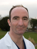 David Jenkins合作论文数Royal Papworth hospital1
 Bruce Keogh合作论文数National Institute for Clinical Outcomes Research;University College London;National Institute for Clinical Outcomes Research, University College London1
 Roberto Dina合作论文数Imperial College School of Medicine, Hammersmith Hospital1




