Introduction: Vestibular schwannomas (VS) are the most common tumor of the cerebellopontine angle. As VSs arise from the vestibular nerve, most displace CN VII anteriorly. An anteriorly displaced CN VII does not obscure access to the presenting pole of the VS via a suboccipital or translabyrinthine approach. Rarely, CN VII is found on the posterior, presenting, surface of the VS—adding difficulty to the surgical resection. We present surgical and clinical results in a series of patients undergoing microsurgery for VS in whom CN VII was displaced posteriorly.
Meningiomas, the most prevalent primary benign intracranial tumors, often exhibit complicated levels of adhesion to adjacent normal tissues, significantly influencing resection and causing postoperative complications. Surgery remains the primary therapeutic approach, and when combined with adjuvant radiotherapy, it effectively controls residual tumors and reduces tumor recurrence when complete removal may cause a neurologic deficit. Previous studies have indicated that slip interface imaging (SII) techniques based on MR elastography (MRE) have promise as a method for sensitively determining the presence of tumor-brain adhesion. In this study, we developed and tested an improved algorithm for assessing tumor-brain adhesion, based on recognition of patterns in MRE-derived normalized octahedral shear strain (NOSS) images. The primary goal was to quantify the tumor interfaces at higher risk for adhesion, offering a precise and objective method to assess meningioma adhesions in 52 meningioma patients. We also investigated the predictive value of MRE-assessed tumor adhesion in meningioma recurrence. Our findings highlight the effectiveness of the improved SII technique in distinguishing the adhesion degrees, particularly complete adhesion. Statistical analysis revealed significant differences in adhesion percentages between complete and partial adherent tumors (p = 0.005), and complete and non-adherent tumors (p<0.001). The improved technique demonstrated superior discriminatory ability in identifying tumor adhesion patterns compared to the previously described algorithm, with an AUC of 0.86 vs. 0.72 for distinguishing complete adhesion from others (p = 0.037), and an AUC of 0.72 vs. 0.67 for non-adherent and others. Aggressive tumors exhibiting atypical features showed significantly higher adhesion percentages in recurrence group compared to non-recurrence group (p = 0.042). This study validates the efficacy of the improved SII technique in quantifying meningioma adhesions and demonstrates its potential to affect clinical decision-making. The reliability of the technique, coupled with potential to help predict meningioma recurrence, particularly in aggressive tumor subsets, highlights its promise in guiding treatment strategies.
The lateral skull base is comprised of the squamous and petrous temporal bones. Fractures through the petrous pyramid account for approximately 20% of all traumatic skull fractures. They can be classified into two types, based upon the involvement of the otic capsule, otic capsule sparing, and otic capsule disrupting. The eloquent structures within and surrounding the petrous temporal bone lead to significant morbidity associated with these fractures. They can lead to cerebrospinal fluid (CSF) leak, facial nerve injury, hearing loss (both conductive and sensorineural), and cerebrovascular injury. Early recognition of a lateral skull base fracture, based upon clinical signs (e.g., Battle's sign) and imaging, is key to further management. Examination of the external ear for CSF leak or extruded brain parenchyma and examination of facial movement are the most important physical exam findings to determine need for early surgical intervention. Facial weakness is treated conservatively for the first 7 days after injury. Acute-onset, complete facial weakness, though, is a poor prognostic sign and can be an indication for early surgical intervention to decompress the nerve. Most CSF leaks will resolve spontaneously within the first week after injury and will not require surgical intervention. Hearing loss is managed conservatively with delayed audiogram. Sensorineural hearing loss has a poor prognosis for recovery. Persistent conductive hearing loss for more than 6 months can be treated with exploratory tympanotomy. Cerebrovascular injury is treated with antithrombotic therapy. In conclusion, the timing of onset of facial weakness is the most important determining factor in surgical intervention, while most sequelae of lateral skull base trauma can be treated conservatively.
Introduction: Tumor size and hearing status are the factors most heavily considered in surgical decision making for vestibular schwannomas (VS), but there is a paucity of literature identifying additional complicating factors in surgical outcomes for this pathology. Patient age has been explored with respect to outcomes and recurrence but to date has not been studied in the context of length of surgery and blood loss during microsurgical resection of VS. We have observed that younger patients with VS often have more vascular and fibrous tumors making surgical resection more challenging.
Objective Dizziness is one of the most prevalent and debilitating symptoms associated with vestibular schwannoma (VS), and there are little data on contributing or alleviating factors in the perioperative setting. In this study, we aimed to evaluate whether vestibular nerve sectioning or labyrinthectomy concomitant with surgical resection would improve dizziness in the postoperative period. Methods This is a retrospective study of a consecutive series of VS patients who underwent resection at a large tertiary care center between 2009 and 2023. Dizziness at 6 months was the primary endpoint, whereas facial nerve and hearing outcomes were secondary endpoints. Fisher's exact test was used to identify significant differences between categorical variables, and multivariate logistic regression analysis was performed to identify predictors of dizziness as well as facial nerve and hearing outcomes. Results A total of 333 patients underwent resection of VS at our institution. There was no significant difference in binary reported dizziness based on surgical approach (p = 0.14). However, patients reported significantly less dizziness at discharge (p < 0.01) as well as 6-month (p = 0.02) and 1-year (p < 0.01) follow-up in the trans-labyrinthine group. On the other hand, patients who underwent labyrinth-sparing approaches reported a significant increase in dizziness that remained up to 1 year from the time of surgery. Conclusions Our data suggest that labyrinthectomy may improve dizziness symptoms in patients with VS. Selective VN sectioning does not appear to affect dizziness or hearing outcomes. Finally, surgical approach does not affect facial nerve outcomes.
OBJECTIVE:To describe the experience and results from coordinated and closely scheduled radiosurgery and cochlear implantation (CI) in a vestibular schwannoma (VS) cohort. PATIENTS:Patients with VS who underwent radiosurgery followed by CI on the same or next day. INTERVENTIONS:Interventions included sequential radiosurgery and CI. MAIN OUTCOME MEASURES:Tumor control defined by tumor growth on posttreatment surveillance and audiometric outcomes including consonant-nucleus-consonant words and AzBio sentences in quiet. RESULTS:In total, six patients were identified that met the inclusion criteria, with an age range of 38 to 69 years and tumor sizes ranging from 2.0 to 16.3 mm. All patients successfully underwent radiosurgery and CI on the same or immediately successive day. Postoperatively, all patients obtained open-set speech recognition. Consonant-nucleus-consonant word scores ranged from 40 to 88% correct, and AzBio scores ranged from 44 to 94% correct. During posttreatment magnetic resonance imaging surveillance, which ranged from 12 to 68 months, all tumors were noted to be adequately visualized, and no tumor progression was noted. CONCLUSION:Coordinated radiosurgery and CI can be safely performed in patients with VS on the same or next day, serving to decrease burden on patients and increase access to this vital rehabilitative strategy.
Background: Neurofibromatosis Type 2 (NF2) is a debilitating neurocutaneous disorder with a prevalence of 1 in 80,000 worldwide with ~5,000 patients affected in the United States. Patients present with multiple schwannomas, meningiomas, and ependymomas affecting the entirety of the neuro-axis. The care of these patients, by necessity, is complex and requires multiple specialties to work together in a coordinated fashion. As a result, it can be challenging for patients to obtain comprehensive care in a single center, which can lead to delayed treatment and increased neurologic morbidity. There are 83 centers in 34 states recognized by the NF network.
Pituitary microadenomas may be challenging to identify on MRI, precluding focal resection in some cases and increasing the risk of surgery.(1-4) Corticotroph adenomas in particular present as MRI-negative in up to 40% of cases.(4,5) Seven-tesla (7-T) MRI offers a greater level of detail than 1.5-T and 3-T and may be used to identify smaller pituitary lesions.(6,7) Intraoperative ultrasound may offer real-time image guidance.(8,9) In this video, the authors demonstrate a case of Cushing disease that was considered MRI-negative on 3-T imaging where 7-T MRI and intraoperative ultrasound supported the diagnosis and surgical management. The patient was a 60-year-old woman who presented with signs and symptoms typical of Cushing syndrome including weight gain, fatigue, and headache as well as refractory hypertension, and prediabetes. Preoperative laboratory workup was suggestive of Cushing disease; however, the patient was identified as MR-negative based on 3-T imaging. 7-T MRI showed a small region of hypoenhancement in the anterior-inferior pituitary gland suggestive of pituitary microadenoma. The patient provided consent and underwent endoscopic endonasal resection of the pituitary lesion with intraoperative ultrasound guidance. The surgery was uncomplicated, and histopathology confirmed a corticotroph adenoma. At 3-month follow-up, the patient had significant improvement in her symptoms, and laboratory studies supported biochemical cure. In this case, the combination of 7-T MRI and intraoperative ultrasound guided the endoscopic endonasal resection of a pituitary micro adenoma, thereby minimizing the destruction of the healthy gland and ensuring the best outcome for the patient. Written informed consent was obtained from the patient for the publication of this work. (C) Congress of Neurological Surgeons 2023. All rights reserved.
OBJECTIVE:Preoperative differentiation of facial nerve schwannoma (FNS) from vestibular schwannoma (VS) can be challenging, and failure to differentiate between these two pathologies can result in potentially avoidable facial nerve injury. This study presents the combined experience of two high-volume centers in the management of intraoperatively diagnosed FNSs. The authors highlight clinical and imaging features that can distinguish FNS from VS and provide an algorithm to help manage intraoperatively diagnosed FNS.METHODS:Operative records of 1484 presumed sporadic VS resections between January 2012 and December 2021 were reviewed, and patients with intraoperatively diagnosed FNSs were identified. Clinical data and preoperative imaging were retrospectively reviewed for features suggestive of FNS, and factors associated with good postoperative facial nerve function (House-Brackmann [HB] grade ≤ 2) were identified. A preoperative imaging protocol for suspected VS and recommendations for surgical decision-making following an intraoperative FNS diagnosis were created.RESULTS:Nineteen patients (1.3%) with FNSs were identified. All patients had normal facial motor function preoperatively. In 12 patients (63%), preoperative imaging demonstrated no features suggestive of FNS, with the remainder showing subtle enhancement of the geniculate/labyrinthine facial segment, widening/erosion of the fallopian canal, or multiple tumor nodules in retrospect. Eleven (57.9%) of the 19 patients underwent a retrosigmoid craniotomy, and in the remaining patients, a translabyrinthine (n = 6) or transotic (n = 2) approach was used. Following FNS diagnosis, 6 (32%) of the tumors underwent gross-total resection (GTR) and cable nerve grafting, 6 (32%) underwent subtotal resection (STR) and bony decompression of the meatal facial nerve segment, and 7 (36%) underwent bony decompression only. All patients undergoing subtotal debulking or bony decompression exhibited normal postoperative facial function (HB grade I). At the last clinical follow-up, patients who underwent GTR with a facial nerve graft had HB grade III (3 of 6 patients) or IV facial function. Tumor recurrence/regrowth occurred in 3 patients (16%), all of whom had been treated with either bony decompression or STR.CONCLUSIONS:Intraoperative diagnosis of an FNS during a presumed VS resection is rare, but its incidence can be reduced further by maintaining a high index of suspicion and undertaking further imaging in patients with atypical clinical or imaging features. If an intraoperative diagnosis does occur, conservative surgical management with bony decompression of the facial nerve only is recommended, unless there is significant mass effect on surrounding structures.
Inverted papilloma is the most common benign sinonasal neoplasm. These tumors are locally invasive causing hyperostosis at the site of attachment. The standard treatment is complete resection due to the high rate of recurrence with incomplete resection.(1) The goal of the resection is complete removal of the tumor along with a rim of normal bone and mucosa. They most commonly occur in the lateral nasal wall or maxillary sinus and carry a 5% to 15% chance of malignant transformation.(2) Only 10% to 14% occurring in the sphenoid sinus.(3) We present a 2-dimensional video of endoscopic endonasal resection of a sphenoid sinus inverted papilloma with bony erosion overlying the paraclival internal carotid artery (ICA) bilaterally. A 66-year-old male patient presented with progressive headaches. A computed tomography head revealed a mass filling and expanding the sphenoid sinus with erosion of a portion of the sphenoid body leading to exposure of the bilateral paraclival ICAs. The MRI with contrast revealed a weakly enhancing mass with linear T1 hypointensities suggestive of an inverted papilloma. The patient elected to proceed with surgical resection of the mass. The patient consented to the procedure. The right sphenoid was cleared first to identify and protect the right ICA and obtain a Doppler signal form the left ICA. A gross total resection was obtained, and the surrounding mucosa and bone were removed from the site of attachment. Bilateral ICAs were preserved. Final pathology revealed an inverted papilloma without dysplasia. No further treatment is warranted at this time.
Background: Vestibular schwannomas (VS) between 1.5 and 2.5 cm (i.e., medium-sized) in cisternal diameter present a unique challenge. Recent evidence suggests with increasing cisternal diameter between 1.4 and 2.0 cm the facial nerve and hearing outcomes decline with microsurgery. In this size cohort the hearing preservation is ~10%; therefore, hearing is often not a significant factor in surgical decision making. Given this, the choice between retrosigmoid (RS) and translabyrinthine (TL) approach is contentious with each skull base team having their own preference. In this series, we combined the microsurgical data between two high-volume VS centers with opposing approach biases for medium-sized VS to assess the difference between the approaches in extent of resection (EOR) and long-term facial nerve outcomes.
Tumors situated in both the posterior fossa and the region of Meckel's cave have traditionally been approached with an anterior petrosectomy. For lesions mostly situated in the posterior fossa with extension into the middle fossa or Meckel's cave, a retrosigmoid craniotomy with suprameatal extension can be used. Despite several descriptions of a retrosigmoid craniotomy with suprameatal extension in the literature, videos detailing its operative steps and highlighting the critical anatomy involved in its execution are lacking. In this video, we present the case of a 27-year-old man with a trigeminal schwannoma extending from the posterior fossa into Meckel's cave who underwent a retrosigmoid approach with suprameatal extension. Gross total resection of the lesion was achieved. The patient gave informed consent for surgery and video recording.
OBJECTIVE:To compare cochlear implant (CI) and auditory brainstem implant (ABI) performance in patients with NF2-related schwannomatosis (NF2). STUDY DESIGN:Historical cohort. SETTING:Tertiary academic center. PATIENTS:A total of 58 devices among 48 patients were studied, including 27 ABIs implanted from 1997 to 2022 and 31 CIs implanted from 2003 to 2022. Three patients had bilateral ABIs, three had bilateral CIs, three had an ABI on one side and a CI on the other, one had a CI that was later replaced with an ipsilateral ABI, and one had an ABI and CI concurrently on the same side. INTERVENTIONS:CI or ABI ipsilateral to vestibular schwannoma. MAIN OUTCOME MEASURES:Open-set speech perception, consonant-nucleus-consonant word scores, and AzBio sentence in quiet scores. RESULTS:Among all patients, 27 (47%) achieved open-set speech perception, with 35 (61%) daily users at a median of 24 months (interquartile range [IQR], 12-87 mo) after implantation. Comparing outcomes, CIs significantly outperformed ABIs; 24 (77%) CIs achieved open-set speech perception compared with 3 (12%) ABIs, with median consonant-nucleus-consonant and AzBio scores of 31% (IQR, 0-52%) and 57% (IQR, 5-83%), respectively, for CIs, compared with 0% (IQR, 0-0%) and 0% (IQR, 0-0%), respectively, for ABIs. Patients with ABIs were younger at diagnosis and at implantation, had larger tumors, and were more likely to have postoperative facial paresis. CONCLUSION:Many patients with NF2-associated vestibular schwannoma achieved auditory benefit with either a CI or an ABI; however, outcomes were significantly better in those patients who were able to receive a CI. When disease and anatomy permit, hearing rehabilitation with a CI should be considered over an ABI in these patients. Tumor management strategies that increase the ability to successfully use CIs should be strongly considered given the high risk of losing bilateral functional acoustic hearing in this population.
Background: Preoperative differentiation of facial nerve schwannomas (FNS) from vestibular schwannoma (VS) can be challenging, particularly when the FNS is limited to cerebellopontine angle (CPA) and/or internal auditory canal. Failure to correctly identify a FNS preoperatively can lead to avoidable facial nerve injury.[1]
INTRODUCTION: Meningioma prognostication and treatment continues to evolve with an increasing understanding of tumor biology. METHODS: This is a retrospective study of a consecutive series of patients with WHO grade 1–3 meningioma resected at UT Southwestern from 1994 to 2015. Time to meningioma recurrence (RFS) was the primary endpoint measured. Kaplan-Meier curves were constructed and compared using log-rank tests. Cox univariate and multivariate analyses were performed to identify predictors of RFS. RESULTS: A total of 703 consecutive patients with meningioma underwent resection at UT Southwestern between the years 1994 and 2015. The median age of the cohort was 56 years (range 16-88 years) and was 68.7% (n = 483) female. Median follow-up was 45 months (range 0-289 months). There was not a significantly increased risk of recurrence in patients with evidence of brain invasion, in patients with otherwise WHO grade 1 meningioma (p = .82). Adjuvant radiation to subtotally resected WHO grade 1 meningiomas did not prolong the time to recurrence. Location (midline skull base, lateral skull base, and paravenous) was significantly associated with RFS (p < .01) on univariate analysis. In patients with high grade (WHO grade 2 or 3) meningiomas, location was predictive of RFS (p = .03) with paravenous meningiomas exhibiting the highest rates of recurrence. Location was not significant on multivariate analysis. CONCLUSIONS: Our data suggests that brain invasion does not increase the risk of recurrence in otherwise WHO grade 1 meningioma. Adjuvant radiation to subtotally resected WHO grade 1 meningiomas did not prolong the time to recurrence. Location categorized by distinct molecular signatures did not predict RFS in a multivariate model.
Background: Hemifacial spasm (HFS) is characterized by involuntary and irregular contractions of muscles innervated by the facial nerve and is commonly caused by an aberrant or ectatic vessel compressing the facial nerve at its root exit zone. The offending vessel is usually the anterior inferior cerebellar (AICA), posterior inferior cerebellar (PICA), or vertebral artery. Surgical intervention frequently consists of microvascular decompression and placement of Teflon pledgets between the facial nerve and compressing vessel. If the vertebral artery is large and dolichoectatic ([Fig. 1A]), adequate mobilization with Teflon alone may be inadequate. Herein, we present a novel technique for durable mobilization of the vertebral artery during microvascular decompression for HFS.
OBJECTIVE:Meningioma prognostication and treatment continues to evolve with an increasing understanding of tumor biology. In this study, the authors aimed to test conventional predictors of meningioma recurrence, histopathology variables for which there exists some controversy (brain invasion), as well as a novel molecular-based location paradigm.METHODS:This is a retrospective study of a consecutive series of patients with WHO grade I-III meningioma resected at The University of Texas Southwestern Medical Center between 1994 and 2015. Time to meningioma recurrence (i.e., recurrence-free survival [RFS]) was the primary endpoint measured. Kaplan-Meier curves were constructed and compared using log-rank tests. Cox univariate and multivariate analyses were performed to identify predictors of RFS.RESULTS:A total of 703 consecutive patients with meningioma underwent resection at The University of Texas Southwestern Medical Center between the years 1994 and 2015. A total of 158 patients were excluded for insufficient follow-up (< 3 months). The median age of the cohort was 55 years (range 16-88 years) and 69.5% (n = 379) were female. The median follow-up was 48 months (range 3-289 months). There was not a significantly increased risk of recurrence in patients with evidence of brain invasion, in patients with otherwise WHO grade I meningioma (Cox univariate HR 0.92, 95% CI 0.44-1.91, p = 0.82, power 4.4%). Adjuvant radiosurgery to subtotally resected WHO grade I meningiomas did not prolong the time to recurrence (n = 52, Cox univariate HR 0.21, 95% CI 0.03-1.61, p = 0.13, power 71.6%). Location (midline skull base, lateral skull base, and paravenous) was significantly associated with RFS (p < 0.01, log-rank test). In patients with high-grade meningiomas (WHO grade II or III), location was predictive of RFS (p = 0.03, log-rank test), with paravenous meningiomas exhibiting the highest rates of recurrence. Location was not significant on multivariate analysis.CONCLUSIONS:The data suggest that brain invasion does not increase the risk of recurrence in otherwise WHO grade I meningioma. Adjuvant radiosurgery to subtotally resected WHO grade I meningiomas did not prolong the time to recurrence. Location categorized by distinct molecular signatures did not predict RFS in a multivariate model. Larger studies are needed to confirm these findings.
Objective: The midline location and relatively large size of olfactory groove meningiomas at diagnosis present a unique surgical challenge, with transcranial subfrontal, both unilateral and bilateral, and endoscopic endonasal approaches employed to access these tumors. While endoscopic approaches have been well-explored in the recent literature, few studies directly explore the difference between bilateral and unilateral approaches in accessing and treating olfactory groove meningiomas. In this study, we aim to compare extent of resection and clinical outcomes in patients treated for OGMs with bilateral and unilateral subfrontal approaches.
Introduction Decompression of the optic nerve within the optic canal is indicated for compressive visual decline. The two most common approaches utilized for optic canal decompression are a medial approach with an endoscopic endonasal approach and a lateral approach with a craniotomy. Our study is a cadaveric anatomical study comparing the length and circumference of the orbit decompressed via an endoscopic endonasal approach versus a frontotemporal craniotomy. Methods Five cadaveric specimens were utilized. Predissection computed tomography (CT) scans were performed on each specimen. On each specimen, a standard frontotemporal craniotomy with anterior clinoidectomy and superolateral orbital decompression was performed on one side and an endoscopic endonasal approach with medial wall decompression was performed on the contralateral side. Post-dissection CT scans were performed. An independent radiologist provided measurements of the length (mm) and circumference (degrees) of optic canal decompression bilaterally. Results The mean length of optic canal decompression for open and endoscopic approach was 13mm (range 12-15mm) and 12.4mm (range 10-16mm), respectively. The mean circumference of decompression for open and endoscopic approaches was 252.8 degrees (range 205-280 degrees) and 124.6 degrees (range 100-163 degrees), respectively. Conclusion The endoscopic endonasal and the transcranial approaches provide a similar length of optic canal decompression, but the transcranial approach leads to greater circumferential decompression. The endoscopic endonasal approach has the benefit of being minimally invasive, though. Ultimately, the surgical approach decision should be based on the location of the pathology and the surgeon's comfort.
 Tarek Y El Ahmadieh合作论文数Department of Neurological Surgery, Northwestern University Feinberg School of Medicine23
 Salah Aoun合作论文数Department of Neurological Surgery, The University of Texas Southwestern Medical Center21
 Samuel L. Barnett合作论文数Neurological Institute, Cleveland Clinic Brain Tumor and Neuro-Oncology Center16
 Om J Neeley合作论文数10Patel Toral R合作论文数9
 Robert Timmerman合作论文数Department of Radiation Oncology, The University of Texas Southwestern Medical Center9
 Michael J. Link合作论文数Department of Neurologic Surgery, Mayo Clinic9
 Babu Guai Welch合作论文数9
 Jonathan A. White合作论文数Department of Neurological Surgery, The University of Texas Southwestern Medical Center9Ankur R. Patel合作论文数8Brian A. Neff合作论文数Department of Otolaryngology, Mayo Clinic8
 Bruce Mickey合作论文数Department of Neurological Surgery, The University of Texas Southwestern Medical Center;Baylor University Medical Center8
 Jamie J. Van Gompel合作论文数Mayo Clinic8Matthew L Carlson合作论文数7Benjamin Kafka合作论文数7
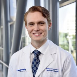 Jeffrey I Traylor合作论文数Department of Neurological Surgery, UT Southwestern Medical School7
 Henry Hunt Batjer合作论文数Department of Neurological Surgery, University of Texas Southwestern Medical Center7
 Zabi Wardak合作论文数Puneeth Iyengar, University of Texas Southwestern Medical Center;University of Colorado School of Medicine;University of Texas Southwestern Medical Center, University of Colorado School of Medicine6
 Colin L W Driscoll合作论文数Dept Otorhinolaryngol, Mayo Clin6Eva M Wu合作论文数5James P Caruso合作论文数5
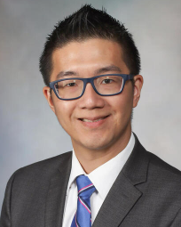 Vin Shen Ban合作论文数Mayo Clinic;Stanford University5McDougall Cameron M合作论文数5Garzon-Muvdi Tomas合作论文数Department of †Neurosurgery;‡Oncology Center-Chemical Therapeutics, The Johns Hopkins University School of Medicine;§Technion Faculty of Medicine, Technion-Israel Institute of Technology;Technion-Israel Institute of Technology, The Johns Hopkins University School of Medicine5Maria Peris Celda合作论文数4
 Kristopher A Lyon合作论文数4
 DaiWai Olson合作论文数Neurology Department, Medical School, The University of Texas Southwestern Medical Center;Neurological Surgery Department, Medical School, The University of Texas Southwestern Medical Center4Jack Raisanen合作论文数4Jameson T Mendel合作论文数3James R Dornhoffer合作论文数3
 Tu Dan合作论文数3
 El Tecle Najib E合作论文数Department of Neurological Surgery, Northwestern University Feinberg School of Medicine3Nedzi Lucien合作论文数3Halderman Ashleigh合作论文数3Beecher Jeffrey S合作论文数3
 Andrew Holmes合作论文数Laboratory of Behavioral and Genomic Neuroscience, National Institute on Alcohol Abuse and Alcoholism, National Institute of Health3Robert A. Ruchinskas合作论文数3
 Andrew T King合作论文数2Collin Driscoll合作论文数2
 Pooja Venkatesh合作论文数2Kim L Rickert合作论文数2
 Alexander S. Fabiano合作论文数2Scott Connors合作论文数2Gillian Potter合作论文数2Abhinav Thummala合作论文数2
 A. Yohan Alexander合作论文数Department of Neurologic Surgery, Mayo Clinic2
 Cathal John Hannan合作论文数Manchester Centre for Clinical Neurosciences2Shivani Bindal合作论文数2
 Hassan Fadel合作论文数2




