Endocrine neoplasms of the pancreas differ from the more common adenocarcinomas of the pancreas not only in histologic appearance, but also in clinical presentation and biologic behavior. Chromosomes were analyzed from nine fresh pancreatic endocrine neoplasms. Clonal chromosomal abnormalities were found in five; all were malignant neoplasms. One showed only a loss of the Y chromosome and another had a small triploid population of cells in addition to a normal mainline, with a karyotype of 61–66,XX,−X,−1,−2,−3,−4,+5,−6,+7,−11,−14,+17,+18,+20,+mar1,x2,+mar2,inc. Three neoplasms had near-haploid clones. One neoplasm had a composite karyotype of 31–36◁n▷,X,+1,+3,+5,+7,+9,+10, +17,+18,+mar. Two were from the same patient, who had the autosomal dominant syndrome MEN-1. The same clone, described as 29◁n▷,X,+add(1)(p12),+5,+7,+8,+18,+19, was found in both the primary pancreatic neoplasm and in the metastatic tumor. To our knowledge, this is the first report of a haploid clone in both a primary and metastatic solid tumor, and suggests that the near-haploid state is at least compatible with metastasis. These data, combined with the limited reports of cytogenetic data from endocrine pancreatic neoplasms, suggest that at least half of such neoplasms will have an abnormal karyotype.
Thirty-three meningeal neoplasms were karyotyped, and the results were compared with histologic features. Thirteen neoplasms had no discernible abnormality or sex chromosome loss only; nine had monosomy or structural abnormality involving only chromosome 22; and 11 had other chromosome abnormalities with or without chromosome 22 involvement. Histologic evidence of invasion was not associated with an abnormal karyotype in the three angioblastic tumors examined. All seven fibroblastic meningiomas had abnormal karyotypes, with monosomy 22 the most common change. Abnormal karyotypes were detected in 76% of syncytial and 55% of transitional meningiomas. When these results were combined with those from 259 meningeal tumors reported since 1987, abnormal karyotypes were detected in at least half of all histologic types. Chromosome changes secondary to those involving chromosome 22 may indicate additional areas of the genome that play a role in tumor progression. In the combined series, chromosome losses were most frequently observed in meningiomatous and transitional histologies; chromosomes 1, 6, 14, 18, and Y each were lost in 10 or more meningiomas, whereas only chromosome 20 was gained at the same frequency. Structural abnormalities most frequently involved chromosome 1. These changes are distinctly different from those observed in other common intracranial neoplasms, specifically astrocytic neoplasms.
The purpose of this study was to determine if fluorescence in situ hybridization for the Y-chromosome can be used to detect cells of recipient origin in allografted hearts following cardiac transplantation. Formalin-fixed, paraffin-embedded tissue sections of coronary arteries from two hearts surgically explanted from heart transplant recipients undergoing retransplantation because of accelerated arteriosclerosis were examined by fluorescence in situ hybridization for the presence of cells containing the Y-chromosome using a biotinylated Y-chromosome cocktail probe. In both cases, the recipients were male and the original donor hearts were obtained from female donors. Hybridization was detected in cells morphologically recognizable as infiltrating lymphocytes, macrophages, and mast cells, establishing that these cells in the donor hearts were of recipient origin. In contrast, hybridization was not detected in cardiac myocytes, in vascular smooth muscle cells, or in the majority (>95%) of endothelial cells, suggesting that these cells were of donor origin. Although hybridization was detected in rare flattened cells lining vascular lumina, these cells did not stain for factor VIII, suggesting that they were, in fact, flattened inflammatory cells and not endothelial cells. These results demonstrate that, when the recipient and donor are of the opposite sex, fluorescence in situ hybridization for the Y-chromosome can be used to detect graft chimerism in transplanted hearts.
The cytogenetic evaluation of prostatic adenocarcinoma has shown no consistent cytogenetic abnormalities. Despite manipulation of culture conditions, the majority of low-stage, untreated prostatic adenocarcinomas show a normal karyotype. We have performed cytogenetic analysis on eight primary prostate adenocarcinomas, using several control measures to increase the probability that any normal karyotype was derived from neoplastic cells rather than accompanying normal cells. Tumors were grown in media that encourages epithelial growth; DNA ploidy studies were performed before and after tissue culture; and immunohistochemical confirmation of the prostatic and epithelial nature of the cells was done following culture. Percentage of tumor on tissue sections adjacent to those submitted for culture was >75% in all cases. Seven of eight cases were evaluable, and six cases showed no clonal abnormalities and were diploid. One tumor showed a population of tetraploid cells, without structural abnormalities. Three additional tumors showed evidence of tetraploidy by DNA analysis. One case showed nonclonal marker chromosomes and was aneuploid. This patient was pathologic Stage D. We conclude that the majority of prostatic adenocarcinomas at their inception may not show routinely detectable cytogenetic abnormalities. However, tetraploidy may play a role in the evolution of prostatic adenocarcinoma.
Ependymomas, oligodendrogliomas, and low-grade astrocytomas are slow-growing central nervous system (CNS) tumors that occur in both adults and children, whereas craniopharyngiomas and choroid plexus papillomas occur predominantly in children. We examined karyotypes of 32 of these low-grade tumors, including ten oligodendrogliomas, six ependymomas, 11 low-grade astrocytomas, four craniopharyngiomas, and one choroid plexus papilloma. Only normal karyotypes were obtained from 6 oligodendrogliomas. The rest had normal stemlines; three tumors had 45,X,-Y sidelines and one tumor had a sideline of monosomy 22. The most frequent abnormalities in the ependymomas were +7 (three tumors), -21 (two tumors), -22 (two tumors), and del(9)(p22) (two tumors). Gains of chromosome 7 and deletions of 9p were found more often in high-grade gliomas. Seven low-grade astrocytomas had normal stemlines, two had chromosome 7 abnormalities, a pilocystic astrocytoma had +der(15), and one tumor had a -Y sideline. The four craniopharyngiomas and one choroid plexus tumor were all apparently normal. The cytogenetics of low-grade CNS tumors differ from higher grade gliomas in that most low-grade tumors show little deviation from the normal karyotype.
Chromosome analysis of short-term culture of melanoma cells from a chroidal melanoma showed a karyotype of 46,XY,−21,+t(6p21q). Trisomy 6p has been observed in cutaneous melanomas; this case suggests that chromosome abnormalities in ocular melanomas may be similar to those from cutaneous melanoma.
 Constance A. Griffin合作论文数11
 Ralph Hruban合作论文数Department of Oncology, Johns Hopkins University School of Medicine;The Sol Goldman Pancreatic Cancer Research Center, Johns Hopkins University7
 Elizabeth J. Perlman合作论文数Department of Pathology, Feinberg School of Medicine, Northwestern University;Ann and Robert H. Lurie Children's Hospital of Chicago;Northwestern Memorial Hospital5
 Henry Brem合作论文数Department of Neurosurgery, School of Medicine, The Johns Hopkins University5
 Charles J. Yeo合作论文数Department of Surgery, Sidney Kimmel Medical College, Thomas Jefferson University4Laura A. Morsberger合作论文数3T ELLINGHAM合作论文数3
 Stuart Grossman合作论文数Johns Hopkins Sidney Kimmel Comprehensive Cancer Center;The Johns Hopkins Hospita3B S Carson合作论文数2La Morsberger合作论文数2WA BAUMGARTNER合作论文数2
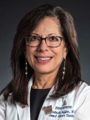 Elizabeth M. Jaffee合作论文数Department of Pathology, School of Medicine, Johns Hopkins University;Institute for Clinical & Translational Research, Johns Hopkins University2
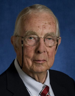 Grover M. Hutchins合作论文数School of Medicine, The Johns Hopkins University2KL Baughman合作论文数1KL BAUGHMAN合作论文数1Fadia Douna-Issa合作论文数1MH Haddadin合作论文数1GM HUTCHINS合作论文数1R Lo合作论文数1Al Hawkins合作论文数1
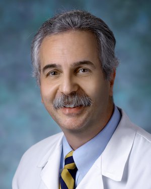 Rafael Tamargo合作论文数Department of Neurosurgery, School of Medicine, The Johns Hopkins University1Hauda K M合作论文数1
 Jeffrey Alan Cohen合作论文数Mellen Center, Cleveland Clinic ;Cleveland Clinic Lerner College of Medicine1
 Kathleen M Murphy合作论文数Department of Oncology, Johns Hopkins University School of Medicine1Dawn Depew合作论文数1Peggy Volz合作论文数1Stefan K. Bohlander合作论文数1NR MILLER合作论文数1Benjamin Carson合作论文数1




