Supplementary Text from Genetic Alterations in the Tyrosine Kinase Transcriptome of Human Cancer Cell Lines
AbstractProtein tyrosine kinases (PTKs) play a critical role in the manifestation of cancer cell properties, and respective signaling mechanisms have been studied extensively on immortalized tumor cells. To characterize and analyze commonly used cancer cell lines with regard to variations in the primary structure of all expressed PTKs, we conducted a cDNA-based sequence analysis of the entire tyrosine kinase transcriptome of 254 established tumor cell lines. The profiles of cell line intrinsic PTK transcript alterations and the evaluation of 155 identified polymorphisms and 234 somatic mutations are made available in a database designated “Tykiva” (tyrosine kinome variant). Tissue distribution analysis and/or the localization within defined protein domains indicate functional relevance of several genetic alterations. The cysteine replacement of the highly conserved Y367 residue in fibroblast growth factor receptor 4 or the Q26X nonsense mutation in the tumor-suppressor kinase CSK are examples, and may contribute to cell line–specific signaling characteristics and tumor progression. Moreover, known variants, such as epidermal growth factor receptor G719S, that were shown to mediate anticancer drug sensitivity could be detected in other than the previously reported tumor types. Our data therefore provide extensive system information for the design and interpretation of cell line–based cancer research, and may stimulate further investigations into broader clinical applications of current cancer therapeutics. [Cancer Res 2007;67(23):11368–76]
C73 Hepatocellular carcinoma (HCC) is a major malignancy inflicting most ethnicities with close to half a million fatalities annually. While the etiology of the disease is relatively well-established (mostly due to viral hepatitis and chronic liver injuries), the progression and the treatment of the disease is still poorly understood and under-explored. Hence, we investigated the contributory role of FGFR4 to HCC, a member of the fibroblast growth factor receptor family that has been recently associated with progression of melanoma, breast and head and neck carcinoma. FGFR4 also presents a unique interest to HCC because liver is the tissue type with the highest mRNA expression of FGFR4. We performed a comprehensive resequencing of the entire coding region of FGFR4 in 62 tumor-normal HCC tissue pairs, and also quantified their respective transcript expression by real-time PCR. We found 4 novel genetic alterations (D126N, T179A, G426D, D709G) as well as a dominant representation of two known polymorphisms among the patients (V10I and G338R). Increased FGFR4 mRNA expression was observed in the tumor tissues as compared to the adjacent normal tissue in 31.6% of the patients. Correlation studies with available clinical follow-ups revealed an increased α-fetoprotein (AFP) production in patients bearing the homozygous Arg388 allele, an observation suggestive of the role of FGFR4 in disease progression. Hence, in vitro studies were subsequently performed to confirm the role of FGFR4 in HCC progression. FGFR4 siRNA administration significantly reduced the production of AFP in HuH7, a high AFP-producing cell line. Stimulation of HepG2 cells (low AFP-producing) with FGF19, a specific FGFR4 ligand, also increased the production of AFP. These effects were independent from the impact of FGFR4 activities on cell proliferation. In summary, our data (both in vivo and in vitro) demonstrated that FGFR4 may represent a drug target against the progression of HCC. The specific role of FGFR4 in HCC and the signal transduction pathways implicated will require further investigation.
 S. Streit合作论文数3Stefan Hart合作论文数3Hooi Linn Loo合作论文数3
 S. Pok合作论文数Med Sch, Australian Natl Univ3
 Han Kiat Ho合作论文数Department of Pharmacy and Pharmaceutical Sciences, Faculty of Science, National University of Singapore3
 Axel Ullrich合作论文数Department of Molecular Biology, Max Planck Institute of Biochemistry;Centre for Molecular Medicine, Agency for Science, Technology and Research3Jens E. Ruhe合作论文数3Ming Luo合作论文数2Shu Jing Lim合作论文数2Liang Seah Tay合作论文数2Christian Mann合作论文数2Silvia Gaertner合作论文数2Kaitian Peng合作论文数2Tze Chuen Lee合作论文数2Huimin Ong合作论文数2Tatjana Knyazeva合作论文数2
 Winnie Wong合作论文数2
 Stefano Iacobelli合作论文数MediaPharma Srl;Oasis Health;Regenerative Protein Biotech2Stephan Peter合作论文数2Martin Bezler合作论文数2Katja Specht合作论文数2
 Alice Tay合作论文数2Pjotr Knyazev合作论文数2
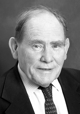 Sydney Brenner合作论文数Crick-Jacobs Center Brenner Laboratory2Byrappa Venkatesh合作论文数2Chee Hong Wong合作论文数2
 Heinz Höfler合作论文数Institut für Allgemeine Pathologie und Pathologische Anatomie der Technischen Universität München2
 Seng Gee Lim合作论文数Division of Gastroenterology and Hepatology, Department of Medicine, National University Hospital;National University Centre for Organ Transplantation, National University Hospital;Department of Medicine, Yong Loo Lin School of Medicine, National University of Singapore1




