IntroductionHuman fetal neural stem cells (hfNSCs) from the subventricular zone (SVZ) are employed in clinical trials for neurodegenerative diseases, yet their bioelectrical properties remain largely unexplored. Molecular markers alone do not reliably correlate with functional state, highlighting the need for complementary functional descriptors.MethodsWe performed whole-cell patch-clamp recordings on hfNSCs from three independent SVZ donors (15–16 weeks of gestation) to characterize resting membrane potential (Vm) and voltage-gated currents, assessing inter-donor reproducibility and differentiation dynamics over 30 days in vitro.ResultshfNSCs exhibited a highly reproducible bioelectrical signature across all donors, characterized by a depolarized resting membrane potential (∼−30 mV), a non-excitable profile, and a stereotyped composition of outward K+ currents. The two current components were resolved using combined biophysical and pharmacological approaches, while Western blot analysis confirmed the expression of Kv4.2 and Kv1.1 channel subtypes functionally consistent with IA and IK, respectively. Remarkably, inter-donor variability in bioelectrical parameters was minimal despite independent cell line derivation. During differentiation, Vm underwent rapid hyperpolarization within 24 h, representing the earliest detectable functional transition. IA showed progressive reduction detectable as early as 24 h and more pronounced by day 15, while IK remained stable throughout. By day 30, inward voltage-gated currents emerged in approximately 60% of cells, consistent with progression toward more differentiated neuro-glial electrophysiological states; however, cells remained non-excitable under our recording conditions. This late-stage divergence highlights heterogeneity in maturation trajectories and completes a temporally ordered sequence of electrical remodeling.ConclusionSVZ-derived hfNSCs possess a reproducible bioelectrical signature within the 15–16-week gestational window across independent donors, supporting electrophysiological profiling as a quantitative functional benchmark for identity and standardization. During in vitro differentiation, Vm hyperpolarization and IA/IK remodeling track early functional progression, whereas the late emergence of inward currents in only a subset of cells indicates increased heterogeneity at later stages. Overall, these findings support bioelectrical profiling as a quantitative functional biomarker with potential utility for standardization and quality assessment in hfNSC-based regenerative therapies.
Neural stem cell (NSC) transplantation holds promising therapeutic potential for neurodegenerative disorders like amyotrophic lateral sclerosis (ALS). However, pre-clinical studies and early-phase clinical trials have faced challenges hindering the effective clinical translation of this approach. Crucial hurdles include the side-effects of prolonged immunosuppression, concerns regarding cell origin and transplantation dosage, identification of the most appropriate therapeutic window, and invasiveness of surgical procedures. Here, we assessed the safety and efficacy of intracerebroventricular (ICV) hNSC transplantation as a novel and possibly more effective experimental approach for ALS. We evaluated the safety of administering up to 1 × 10⁶ hNSCs in immunodeficient mice and assessed their potential efficacy in reducing ALS hallmarks employing the SOD1G93A mouse model. Both transient (15 days) and prolonged immunosuppression regimens, at low (15 mg/kg) and high (30 mg/kg) doses, were tested along with two different cell dosages (3 × 10⁵ and 1 × 10⁶). Our study suggests that: (i) a bilateral ICV transplantation of 1 × 10⁶ hNSCs is safe and non-tumorigenic in immunodeficient hosts; (ii) sustained and high-dose immunosuppression is essential for ensuring cell survival in immunocompetent SOD1G93A mice; and (iii) hNSCs may delay motor symptom progression and reduce spinal cord microgliosis in SOD1G93A mice when administered in the lateral ventricles under prolonged high-dose (30 mg/kg) immunosuppression. ICV transplantation of hNSCs emerges as a safe and promising strategy for ALS, demonstrating potential to delay motor decline and reduce spinal cord microgliosis. However, sustained high-dose immunosuppression is crucial for therapeutic efficacy, emphasizing the need for further optimization to overcome translational challenges and achieve durable clinical benefits.
Background and aims: Neural stem cell (NSC) transplantation holds promising therapeutic potential for neurodegenerative disorders like amyotrophic lateral sclerosis (ALS). However, pre-clinical studies and early-phase clinical trials have faced challenges hindering the effective clinical translation of this approach. Crucial hurdles include the side-effects of prolonged immunosuppression, concerns regarding cell origin and transplantation dosage, identification of the most appropriate therapeutic window, and invasiveness of surgical procedures. Here, we show challenges and promises in optimizing a non-clinical protocol to assess safety and efficacy of human NSC (hNSC) intracerebroventricular (ICV) transplantation for ALS. Methods: We evaluated the safety of administering up to 1x10⁶ hNSCs in immunodeficient mice and assessed their potential efficacy in reducing ALS hallmarks employing the SOD1G93A mouse model. Both, transient (15 days) and prolonged immunosuppression regimens, at low (15 mg/kg) and high (30 mg/kg) doses, were tested along with two different cell dosages (3x10⁵ and 1x10⁶). Results: Bilateral ICV injection of up to 1x10⁶ hNSCs proved to be safe, with no evidence of tumor formation. At 40 days post-transplantation, hNSCs induced a trend toward delaying motor decline and reducing spinal cord (SC) microgliosis when transplanted under prolonged high-dose (30 mg/kg) immunosuppression. Conclusions: Our study suggests that: (i) a bilateral ICV transplantation of 1x10⁶ hNSCs is safe and non-tumorigenic in immunodeficient hosts; (ii) sustained high-dose immunosuppression is essential for ensuring cell survival in immunocompetent mice; and (iii) hNSC transplantation may provide therapeutic benefits in ALS by delaying motor decline and reducing microgliosis. This study also highlights persisting hurdles that need to be further addressed, such as the aggressive murine immune response to exogenous cells. ### Competing Interest Statement The authors have declared no competing interest.
This is an open-label, first-in-human, dose-escalation phase 1 study (NCT03282760, EudraCT2015‐004855‐37) to determine the feasibility, safety, and tolerability of the transplantation of allogeneic human neural stem/progenitor cells (hNSCs) for the treatment of progressive multiple sclerosis. We report the analysis on 1 year of data from the first cohort of 15 patients from two trial sites that received increasing numbers of allogeneic hNSCs delivered via intracerebroventricular injection in combination with an immunosuppressive regimen. No treatment-related deaths nor serious adverse events (AEs) were observed over the 12-month follow-up. Participants displayed stability of clinical and laboratory parameters, and lesion loads and activity at the brain MRIs, compared to study entry and run in. A significant inverse correlation was found between the number of injected hNSCs and the parenchymal brain volume change (PBVC; Spearman’s rho=-0.7, p= 0.0302). The absence of AEs and the stability of functional and structural outcomes suggest a neutral balance between benefits and risks for this intervention and represent a main milestone to rigorously address the challenges for the safe translation of key principles of stem cell biology into effective regenerative medicines.
We report the analysis of 1 year of data from the first cohort of 15 patients enrolled in an open-label, first-in-human, dose-escalation phase I study (ClinicalTrials.gov: NCT03282760, EudraCT2015-004855-37) to determine the feasibility, safety, and tolerability of the transplantation of allogeneic human neural stem/progenitor cells (hNSCs) for the treatment of secondary progressive multiple sclerosis. Participants were treated with hNSCs delivered via intracerebroventricular injection in combination with an immunosuppressive regimen. No treatment-related deaths nor serious adverse events (AEs) were observed. All participants displayed stability of clinical and laboratory outcomes, as well as lesion load and brain activity (MRI), compared with the study entry. Longitudinal metabolomics and lipidomics of biological fluids identified time- and dose-dependent responses with increased levels of acyl-carnitines and fatty acids in the cerebrospinal fluid (CSF). The absence of AEs and the stability of functional and structural outcomes are reassuring and represent a milestone for the safe translation of stem cells into regenerative medicines.
Translation of cell therapies into clinical practice requires the adoption of robust production protocols in order to optimize and standardize the manufacture and cryopreservation of cells, in compliance with good manufacturing practice regulations. Between 2012 and 2020, we conducted two phase I clinical trials (EudraCT 2009-014484-39, EudraCT 2015-004855-37) on amyotrophic lateral sclerosis secondary progressive multiple sclerosis patients, respectively, treating them with human neural stem cells. Our production process of a hNSC-based medicinal product is the first to use brain tissue samples extracted from fetuses that died in spontaneous abortion or miscarriage. It consists of selection, isolation and expansion of hNSCs and ends with the final pharmaceutical formulation tailored to a specific patient, in compliance with the approved clinical protocol. The cells used in these clinical trials were analyzed in order to confirm their microbiological safety; each batch was also tested to assess identity, potency and safety through morphological and functional assays. Preclinical, clinical and in vitro nonclinical data have proved that our cells are safe and stable, and that the production process can provide a high level of reproducibility of the cultures. Here, we describe the quality control strategy for the characterization of the hNSCs used in the above-mentioned clinical trials.
Intraventricular hemorrhage is a common cause of morbidity and mortality in premature infants. The rupture of the germinal zone into the ventricles entails loss of neural stem cells and disturbs the normal cytoarchitecture of the region, compromising late neurogliogenesis. Here we demonstrate that neural stem cells can be easily and robustly isolated from the hemorrhagic cerebrospinal fluid obtained during therapeutic neuroendoscopic lavage in preterm infants with severe intraventricular hemorrhage. Our analyses demonstrate that these neural stem cells, although similar to human fetal cell lines, display distinctive hallmarks related to their regional and developmental origin in the germinal zone of the ventral forebrain, the ganglionic eminences that give rise to interneurons and oligodendrocytes. These cells can be expanded, cryopreserved, and differentiated in vitro and in vivo in the brain of nude mice and show no sign of tumoral transformation 6 months after transplantation. This novel class of neural stem cells poses no ethical concerns, as the fluid is usually discarded, and could be useful for the development of an autologous therapy for preterm infants, aiming to restore late neurogliogenesis and attenuate neurocognitive deficits. Furthermore, these cells represent a valuable tool for the study of the final stages of human brain development and germinal zone biology.
The main objective of this phase I trial was to assess the feasibility and safety of microtransplanting human neural stem cell (hNSC) lines into the spinal cord of patients with amyotrophic lateral sclerosis (ALS). Eighteen patients with a definite diagnosis of ALS received microinjections of hNSCs into the gray matter tracts of the lumbar or cervical spinal cord. Patients were monitored before and after transplantation by clinical, psychological, neuroradiological, and neurophysiological assessment. For up to 60 months after surgery, none of the patients manifested severe adverse effects or increased disease progression because of the treatment. Eleven patients died, and two underwent tracheotomy as a result of the natural history of the disease. We detected a transitory decrease in progression of ALS Functional Rating Scale Revised, starting within the first month after surgery and up to 4 months after transplantation. Our results show that transplantation of hNSC is a safe procedure that causes no major deleterious effects over the short or long term. This study is the first example of medical transplantation of a highly standardized cell drug product, which can be reproducibly and stably expanded ex vivo, comprising hNSC that are not immortalized, and are derived from the forebrain of the same two donors throughout this entire study as well as across future trials. Our experimental design provides benefits in terms of enhancing both intra- and interstudy reproducibility and homogeneity. Given the potential therapeutic effects of the hNSCs, our observations support undertaking future phase II clinical studies in which increased cell dosages are studied in larger cohorts of patients. Stem Cells Translational Medicine 2019;8:887&897
Stem cells are emerging as a therapeutic option for incurable diseases, such as Amyotrophic Lateral Sclerosis (ALS). However, critical issues are related to their origin as well as to the need to deepen our knowledge of the therapeutic actions exerted by these cells. Here, we investigate the therapeutic potential of clinical-grade human neural stem cells (hNSCs) that have been successfully used in a recently concluded phase I clinical trial for ALS patients (NCT01640067). The hNSCs were transplanted bilaterally into the anterior horns of the lumbar spinal cord (four grafts each, segments L3–L4) of superoxide dismutase 1 G93A transgenic rats (SOD1 rats) at the symptomatic stage. Controls included untreated SOD1 rats (CTRL) and those treated with HBSS (HBSS). Motor symptoms and histological hallmarks of the disease were evaluated at three progressive time points: 15 and 40 days after transplant (DAT), and end stage. Animals were treated by transient immunosuppression (for 15 days, starting at time of transplantation). Under these conditions, hNSCs integrated extensively within the cord, differentiated into neural phenotypes and migrated rostro-caudally, up to 3.77 ± 0.63 cm from the injection site. The transplanted cells delayed decreases in body weight and deterioration of motor performance in the SOD1 rats. At 40DAT, the anterior horns at L3–L4 revealed a higher density of motoneurons and fewer activated astroglial and microglial cells. Accordingly, the overall survival of transplanted rats was significantly enhanced with no rejection of hNSCs observed. We demonstrated that the beneficial effects observed after stem cell transplantation arises from multiple events that counteract several aspects of the disease, a crucial feature for multifactorial diseases, such as ALS. The combination of therapeutic approaches that target different pathogenic mechanisms of the disorder, including pharmacology, molecular therapy and cell transplantation, will increase the chances of a clinically successful therapy for ALS.
Clinical trials for Parkinson's disease, which used primary brain fetal tissue, have demonstrated that neural stem cell therapy could be suitable for neurodegenerative diseases. The use of fetal tissue presents several issues that have hampered the clinical development of this approach. In addition to the ethical concerns related to the required continuous supply of fetal specimen, the necessity to use cells from multiple fetuses in a single graft greatly compounded the problem. Cell viability and composition vary in different donors, and, further, the heterogeneity in the donor cells increased the probability of immunological rejection or contamination. An ideal cell source for cell therapy is one that is renewable, thus eliminating the need for transplantation of primary fetal tissue, and that also allows for viability, sterility, cell composition, and cell maturation to be controlled, while being inherently not tumorigenic. The availability of continuous and standardized clinical grade normal human neural cells, able to combine the plasticity of fetal tissue with an extensive proliferating capacity and functional stability, would be of paramount importance for the translation of cell therapy for central nervous system (CNS) disorders into the clinic. Here we describe a well-established protocol to produce human neural stem cells following GMP guidelines that allows us to obtain "clinical grade" cell lines.
Establishing specific cell lineages from human induced pluripotent stem cells (hiPSCs) is vital for cell therapy approaches in regenerative medicine, particularly for neurodegenerative disorders. While neural precursors have been induced from hiPSCs, the establishment of hiPSC-derived human neural stem cells (hiNSCs), with characteristics that match foetal hNSCs and abide by cGMP standards, thus allowing clinical applications, has not been described. We generated hiNSCs by a virus-free technique, whose properties recapitulate those of the clinical-grade hNSCs successfully used in an Amyotrophic Lateral Sclerosis (ALS) phase I clinical trial. Ex vivo, hiNSCs critically depend on exogenous mitogens for stable self-renewal and amplification and spontaneously differentiate into astrocytes, oligodendrocytes and neurons upon their removal. In the brain of immunodeficient mice, hiNSCs engraft and differentiate into neurons and glia, without tumour formation. These findings now warrant the establishment of clinical-grade, autologous and continuous hiNSC lines for clinical trials in neurological diseases such as Huntington's, Parkinson's and Alzheimer's, among others.
We report the initial results from a phase I clinical trial for ALS. We transplanted GMP-grade, fetal human neural stem cells from natural in utero death (hNSCs) into the anterior horns of the spinal cord to test for the safety of both cells and neurosurgical procedures in these patients. The trial was approved by the Istituto Superiore di Sanità and the competent Ethics Committees and was monitored by an external Safety Board.
NSCs have been demonstrated to be very useful in grafts into the mammalian central nervous system to investigate the exploitation of NSC for the therapy of neurodegenerative disorders in animal models of neurodegenerative diseases. To push cell therapy in CNS on stage of clinical application, it is necessary to establish a continuous and standardized, clinical grade (i.e., produced following the good manufacturing practice guidelines) human neural stem cell lines. In this chapter, we illustrate some of the protocols routinely used into our GMP cell bank for the production of "clinical grade" human neural stem cell lines.
 Maurizio Gelati合作论文数14
 Vescovi Angelo Luigi合作论文数11Daniela Ferrari合作论文数10Muzi Gianmarco合作论文数7
 Massimilano Copetti合作论文数6Claudia Ricciolini合作论文数6Sgaravizzi Giada合作论文数5
 Zalfa Cristina合作论文数4
 Elena Binda合作论文数Clinical Research Center for Rare Diseases, From the Mario Negri Institute for Pharmacological Research;Unit of Nephrology and Dialysis, Azienda Ospedaliera;and Clinica Universitaria, Universidad de Navarra;and Clinica Universitaria Facultad de Medicina, Universidad de Navarra4Laura Bernardini合作论文数3Domenico Frondizi合作论文数3Alberto Visioli合作论文数3Laura Rota Nodari合作论文数3
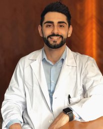 Ivan Lombardi合作论文数Dept Biotechnol & Biosci, Univ Milano Bicocca3Torres Barbara合作论文数3Spera Cristina合作论文数3Massimo Projetti Pensi合作论文数3
 Marina Boido合作论文数3
 Alessandro Vercelli合作论文数3Jessica Rosati合作论文数IBPM CNR, Università La Sapienza3
 Letizia Mazzini合作论文数3Francesco Petruzzelli合作论文数2Sandro Carletti合作论文数2Maurilio Massara合作论文数2Laura Cajola合作论文数2Gianni Sorarù合作论文数2
 Annamaria Maglione合作论文数2Francesca Pinos合作论文数2Elisa Perciballi合作论文数2
 Domenico Trombetta合作论文数2
 Stefano Masiero合作论文数2Carlo Cisari合作论文数2
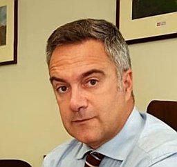 Alessandro Stecco合作论文数Radiol Dept, Maggiore Carita Hosp Novara2
 Maurizio Leone合作论文数Department of Medical Sciences, Fondazione IRCCS Casa Sollievo della Sofferenza2
 Maria Svelto合作论文数Dept Biosci Biotechnol & Biopharmaceut, Univ Bari2
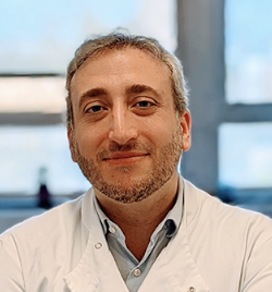 Luca Peruzzotti-Jametti合作论文数Department of Clinical Neurosciences, University of Cambridge;King's College, University of Cambridge;Imperial College London2
 Emanuele Pravata合作论文数Neuroradiology Department, Neurocenter of Italian Switzerland;Department of Neuroradiology, Catholic University of Rome;Rome, Italy - emanuele.pravata@gmail.com.;Neurocenter of Italian Switzerland, Catholic University of Rome2
 Lidia De Filippis合作论文数2Querin Giorgia合作论文数2Gaiani Alessandra合作论文数2Bersano Enrica合作论文数2Claudio Gobbi合作论文数2Giorgi Cesare合作论文数2Sarnelli Maria Francesca合作论文数2
 Roberto Cantello合作论文数Department of Neurology, Università del Piemonte Orientale “A. Avogadro”,2
 Nicholas M Boulis合作论文数Department of Psychiatry, Yale University School of Medicine2Emilio Ciusani合作论文数2
 Massimo Carella合作论文数2
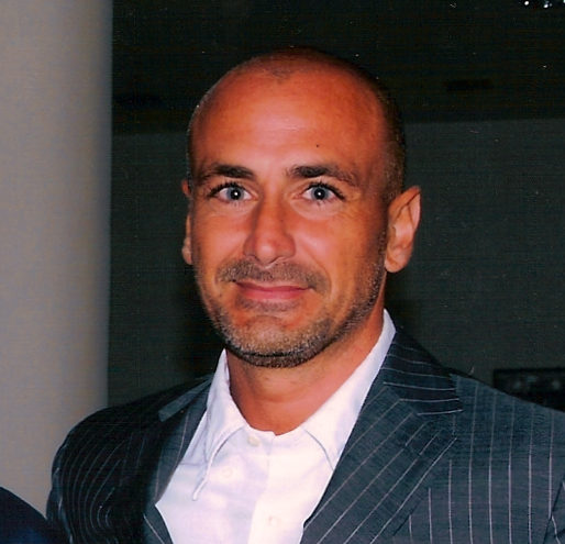 Stefano Pluchino合作论文数Department of Clinical Neurosciences, School of Clinical Medicine, University of Cambridge2




