Introduction:Brain metastases (BrMs) are poorly studied and likely underreported in diffuse pleural mesothelioma (DPM), limiting understanding of risk factors associated with their development. We evaluated the genomic, histologic, and clinical landscapes of patients with DPM and BrMs. Methods:We retrospectively reviewed all patients with a mesothelioma diagnosis treated at the Memorial Sloan Kettering Cancer Center between January 1, 2010, and May 1, 2025, with cross-sectional brain imaging. Clinicopathologic data and treatment outcomes were annotated for patients with BrMs. Results:Among 194 patients with mesothelioma and brain imaging, 16 (8%) had BrMs. Half (n = 8) had tumors of epithelioid histology, 44% (n = 7) biphasic, and 6% (n = 1) sarcomatoid. Compared with our DPM cohort regardless of BrMs (n = 194) or a DPM cohort from The Cancer Genome Atlas (n = 74), patients with DPM and BrMs had a higher prevalence of LATS2 alterations (46% versus 7% versus 12%, respectively [p < 0.00001]). BrMs were typically (88%, n = 14) diagnosed after neurologic symptoms prompting imaging. Among patients with BrMs, the median overall survival from initial mesothelioma diagnosis was 35.4 (95% confidence interval, 19.3-not reached) months and 3.7 (95% confidence interval, 3.5-not reached) months from BrM diagnosis. Conclusions:Our findings suggest that BrM develops relatively late in DPM, is more common than previously reported, and may be enriched in patients with LATS2 alterations. Prospective, multi-institutional studies with standardized brain imaging are needed to further characterize the incidence of BrMs in DPM and associated risk factors. Routine brain surveillance at diagnosis and with symptoms should be considered for patients with DPM.
On the basis that T-cell acute lymphoblastic leukemia (T-ALL) cells overexpress IL7 receptor (IL7R), which promotes resistance to chemotherapy and disease relapse, here we develop IL7Rα (CD127)-targeted chimeric antigen receptor (CAR) T cells with low- and high-affinity single-chain variable fragments. We establish the antitumor efficacy of CD127 CAR against T-ALL cells in vitro, in female mouse models of T-ALL, and against blasts from patients with T-ALL using the patients' own T cells transduced with CD127 CAR. Antitumor efficacy is higher with low-affinity CAR T cells than high-affinity CAR T cells, albeit with fratricide of CAR T cells following eradication of CD127-overexpressing blasts. CRISPR-Cas9 knockout of CD127 eliminates fratricide at the risk of prolonged lymphopenia in vivo. To overcome fratricide, we investigate short-term ( < 7 days) co-culture with or without dasatinib, a tyrosine kinase inhibitor, versus a natural selection method (10 days) and demonstrate that co-culturing with dasatinib facilitates higher CAR T-cell yield, improved fitness, and preserved functionality. In vivo, dasatinib can be used to temporarily and reversibly suppress CAR T-cell activity. With this supporting translational data, we are initiating a trial with low-affinity CD127 CAR T cells for adult and pediatric patients with relapsed or refractory T-ALL.
PURPOSE:One recurring challenge in cell therapy for solid tumors is poor tumor infiltration of adoptively transferred T cells. We previously showed that a subablative dose of tumor-targeted radiation generates a chemokine gradient that promotes infiltration, proliferation, and a memory phenotype of chimeric antigen receptor (CAR) T cells in solid tumors. However, radiation is cytotoxic to infiltrating CAR T cells, limiting its repeated use. EXPERIMENTAL DESIGN:We hypothesized that irreversible electroporation could generate a chemokine gradient that promotes CAR T-cell infiltration into solid tumors and that selective irreversible electroporation (sIRE) tuned for selective cancer cell lysis (thus sparing infiltrating CAR T cells) can be used in a repeated fashion. Using experimental screening and simulation models, we optimized sIRE parameters to kill cancer cells while sparing T cells. RESULTS:Using 3D tumor mimics and mouse models of malignant pleural mesothelioma, we confirmed the therapeutic benefit of repeated sIRE. Chemokine secretion by cancer cells injured by sIRE promoted migration and tumor infiltration of systemically administered CAR T cells and facilitated sustained immunity in a tumor-rechallenge model. CONCLUSIONS:By leveraging a dual-purpose translational strategy-through direct cancer cell-targeted cytotoxicity and augmented CAR T-cell infiltration-sIRE can reduce cancer burden while preserving and enhancing CAR T-cell function.
BACKGROUND Micropapillary and solid subtypes of lung adenocarcinoma are associated with high rates of recurrence in early-stage non-small cell lung cancer and may help guide the extent of resection if known preoperatively. We examined the accuracy of CT-guided core needle biopsy in determining high-risk subtypes in small (≤2 cm) tumors. METHODS A retrospective review was performed to identify patients with clinical T1a-T1bN0M0 (≤2 cm) lung adenocarcinoma who underwent CT-guided core needle biopsy before surgical resection from 2010 to 2024. The accuracy, sensitivity, and specificity of preoperative biopsy were determined for each histologic subtype. Multivariable logistic regression identified preoperatively known clinicopathologic risk factors for micropapillary/solid subtypes on surgical pathology. RESULTS We identified 658 patients with early-stage lung adenocarcinoma who underwent preoperative biopsy, with 372 (57%) patients undergoing core-needle biopsy resulting in a pathology specimen with histologic subtype reported. Preoperative biopsy was highly specific (96.9%) but not sensitive (37.1%) for the presence of solid or micropapillary subtypes. Of the 288 patients without a high-risk subtype identified on preoperative biopsy, 134 (47%) had a high-risk subtype identified on surgical pathology. Multivariable analysis identified male sex, current/former smoking history, tumor maximum standardized uptake value (SUVmax), and solid tumor CT texture as independently associated with greater risk of having micropaillary/solid features on surgical pathology. CONCLUSIONS Preoperative biopsy is effective at ruling in, but not ruling out, high-risk lung adenocarcinoma subtypes. The presence of high-risk features on biopsy may be helpful in treatment planning.
Supplemental Figure 4. Intratumoral T cell viability after sIRE or IRE in an immunocompetent syngeneic flank tumor model.
Purpose Although we typically consider diffuse pleural mesothelioma (DPM) as a disease of elderly males with a history of asbestos exposure, young individuals are also affected. Given the increasing incidence of cancer in young individuals, we investigated the characteristics and underlying biology of young patients with DPM. Methods We identified patients with DPM age <= 50 years from Memorial Sloan Kettering Cancer Center (1990-2023). Clinicopathologic and genetic characteristics were compared between very young (<= 35 years) and young (36-50 years) patients. Results We identified 273 patients with DPM age <= 50 years; 39% were female, 40% had a history of self-reported occupational asbestos exposure (very young, 16% v young, 45%; P < .01), 16% had a personal history of cancer (very young, 7% v young, 18%; P = .07), and 70% had a family history of cancer (most commonly breast, lung, and colon). Most frequently altered somatic genes (51/273 tested; 46/51 positive) included BAP1 (22/47, 47%), NF2(18/47, 38%), CDKN2A/B (12/48, 25%), and TP53 (10/48, 21%). Very young patients had less somatic alterations in BAP1 (13% v 63%, P < .01) and a lower tumor mutational burden (0.9 v 1.8 mut/mB, P < .01); they were less frequently carriers of germline alterations (0% [0/8] v 43% [6/14], P = .05). Somatic alterations in CDKN2B were more frequent among females (females 42% v males 12%, P = .03). Conclusion Young patients with DPM have strong personal and family histories of cancer, and heterogeneous somatic and germline alterations, indicating divergent underlying biology. With the increasing prevalence of young adults with cancers, mesotheliomas, although uncommon, should be on the differential for patients even without asbestos exposure history in this age group.
Supplemental Figure 7. sIRE promotes infiltration of CAR T cells with minimal killing of preexisting intratumoral T cells.
Objective:We evaluated the feasibility and long-term outcomes of minimally invasive surgery (MIS) after neoadjuvant immunotherapy in patients with resectable non-small cell lung cancer (NSCLC). Methods:Patients with stage IB-IIIB NSCLC who underwent induction immunotherapy followed by surgery (2015-2022) were included. Overall and event-free survival were estimated using the Kaplan-Meier approach. Cox regression was used to quantify associations between clinical and pathologic variables and survival outcomes. Results:In total, 69 patients met the inclusion criteria (MIS, n = 38; open surgery, n = 31). Patients who underwent open surgery had larger tumors (P < .001), more-frequent extended resection (P = .007), more major complications (P = .03), and longer in-hospital stay (P = .002), compared with patients who underwent MIS. The rate of conversion to open surgery in the MIS group was 16% (6/38). Histologic subtype (P = .057), completeness of resection (P > .9), response to treatment (P > .9), and stage (P = .6) were not statistically different between groups. Five-year event-free and overall survival in the MIS patients were 72% and 80%, respectively. Among all patients, pathologic N2 disease (hazard ratio [HR], 3.04; 95% CI, 1.1-8.38; P = .031) and upstaging (HR, 2.57; 95% CI, 1.05-6.34; P = .04) were associated with a greater risk of recurrence. Major pathologic response was associated with longer event-free survival (HR, 0.22; 95% CI, 0.05-0.93; P = .039). Programmed death-ligand 1 level (HR, 1.02; 95% CI, 1.01-1.04; P = .007) and tumor dimensional reduction after treatment (HR, 0.96; 95% CI, 0.93-0.99; P = .016) were associated with major pathologic response. Conclusions:MIS after induction immunotherapy is feasible and does not compromise long-term oncologic outcomes.
A 52-year-old man presented with sarcomatoid diffuse pleural mesothelioma that had relapsed at an isolated site after a complete response to dual-immune checkpoint inhibition (ICI). Targeted sequencing exhibited amplification of chromosome 9p24, encompassing JAK2, PD-L1, PD-L2, and PTPRD in the relapsed (post-ICI) tumor, compared with baseline (pre-ICI). On multiplex immunofluorescence, tumor-associated macrophages (TAMs) and CD8+ cytotoxic T lymphocytes (CTLs) made up most of the cells in baseline and relapsed tumor (59% and 47%, respectively). Baseline tumor cells expressed genes linked to extracellular matrix remodeling and epithelial-mesenchymal transition, intermixed with M2-like TAMs and tissue-resident, effector-like CTLs. Relapsed tumor cells shifted to a growth factor-driven phenotype (NT5E, NOD1, GATA2, FN1, PDCD1LG2) that is known to cause functional impairment of CTLs, which then transitioned to an exhausted state (FCRL3, CST7, GPR171, TRAT1, LAG3); exhausted CD8+ and CD4+ T cells are seen in the peripheral blood at relapse. TAMs were enriched in antigen-presentation (CD80, CD86, CXCL10), extracellular matrix-degradation (MMP9, CTSL), and CTL-suppression (ARG1, PLA2G7) pathways. Our analyses revealed that regional immunosuppression mediated by adaptive reprogramming of tumor-cell and immune-cell (TAMs, CTLs)-intrinsic changes-rather than by immune evasion or stromal exclusion-served as a mechanism of acquired resistance to dual-ICI therapy.
Supplemental Figure 1. Mesothelioma and T cell viability at 24 hours post-IRE in vitro. Mesothelioma cell lines.
BACKGROUND:Investigating the biological mechanisms of acquired resistance to immunotherapy remains a necessity for effective treatment of solid tumors. METHODS:Diffuse pleural mesothelioma (DPM) tumors, from immunocompetent and immunodeficient mouse models with acquired resistance to chimeric antigen receptor (CAR) T cell therapy were analyzed using flow cytometry, immunofluorescence, and ELISA. The interplay between genetic alterations in the cancer cell and site-specific tumor immune microenvironment (TIME) was characterized; therapies to reverse immunotherapy resistance were investigated. FINDINGS:Analysis of tumors with acquired resistance to CAR T cell therapy demonstrated lower expression of tumor suppressor NF2. Enforced depletion of NF2 promoted resistance to CAR T cell and anti-PD-1 therapy in DPM but not in heterotopic subcutaneous tumors. Loss of NF2 was associated with accumulation of immunosuppressive complement receptor of immunoglobulin superfamily (CRIg)+ macrophages in pleural tumors, which was dependent on tumor-secreted chemokines and retinoic acid. Ablation of chemokines or pharmacologic inhibition of retinoic acid or the Hippo pathway by use of TEAD inhibitors restored sensitivity to CAR T cell therapy. Correspondingly, in two independent cohorts of patients, DPM tumors with NF2 copy-number loss, but not mutations, were associated with higher infiltration of M2 macrophages, lower infiltration and function of CD8 T cells, and an anti-PD-1-resistant gene signature. CONCLUSIONS:Our study uncovers a previously unknown mechanism of resistance to immunotherapy by identifying the dynamic interplay between cancer cell genetic alterations and the TIME. FUNDING:This research was funded in part through the NCI UG3CA290241, R01CA292664, R01CA235667, R01CA236615, and Department of War CA200437.
Background Guidelines recommend invasive mediastinal nodal staging (IMNS) for patients with synchronous primary lung cancer (SPLC) who are candidates for curative resection. We investigated the prevalence of occult nodal disease and selective use of IMNS in patients with SPLC. Methods Patients with SPLC suspected on preoperative imaging who underwent resection from 2000 to 2022 were included. Exclusion criteria included metachronous and recurrent lung cancer, cStage III-IV, carcinoid histology, absence of preoperative PET/CT, and receipt of neoadjuvant therapy. Univariable analysis identified clinicopathologic risk factors for occult nodal disease. Proposed criteria for identifying a patient at low risk for occult nodal disease were as follows: all primary tumors are peripheral, subsolid, ≤3 cm on preoperative CT, and have a maximum standardized uptake value (SUVmax) < 3.6, and lymph nodes must be PET negative. Results Of 484 eligible patients, only 19% (n = 90) underwent IMNS. After excluding patients with clinical nodal disease on IMNS and/or imaging, 472 patients remained, of which 14% (68/472) had occult nodal disease, with 5.9% (28/472) having occult N2 disease. Patients with occult nodal disease had dominant tumors with a higher SUVmax (median, 6.0 vs. 3.2; p < 0.001), central location (31% vs. 17%; p = 0.009) and solid appearance (76% vs. 46%; p < 0.001). Our proposed criteria categorized 21% of patients with complete data (96/449)as low risk for occult nodal disease(sensitivity = 24.4%, specificity = 96.9%, positive predictive value = 97.9%, negative predictive value = 17.6%). Conclusions The decision to pursue IMNS in patients with SPLC should be individualized using imaging characteristics including central location, solid-appearance, tumor size, and SUVmax.
Supplemental Figure 3. Fluorescent imaging of IRE and sIRE treatment effects on (A) MSTO-GM, (B) untransduced, and (C) CAR T cells embedded in 3D collagen mimics that were treated with various parameters of sIRE or IRE 24 hours post-treatment in vitro. 4x magnification images were scanned and stitched to illustrate PEF treatment area of effect within the whole well of a 24-well using a 2-pin probe electrodes with 4 mm spacing. Dead cells were identified by propidium iodide (red) and live cells identified with Calcein AM (green). (D–F) Area of cell death were quantified using script on FIJI ImageJ. Scale bar = 4 mm.
Introduction Brain metastases (BrMs) are poorly studied and likely underreported in diffuse pleural mesothelioma (DPM), limiting understanding of risk factors associated with their development. We evaluated the genomic, histologic, and clinical landscapes of patients with DPM and BrMs. Methods We retrospectively reviewed all patients with a mesothelioma diagnosis treated at Memorial Sloan Kettering Cancer Center between January 1, 2010, and May 1, 2025, with cross sectional brain imaging. Clinicopathologic data and treatment outcomes were annotated for patients with BrMs. Results Among 194 patients with mesothelioma and brain imaging, 16 (8%) had BrMs. Half (n=8) had tumors of epithelioid histology, 44% (n=7) biphasic, and 6% (n=1) sarcomatoid. Compared with our DPM cohort regardless of BrMs (n=194) or a DPM cohort from The Cancer Genome Atlas (n=74), patients with DPM and BrMs had a higher prevalence of LATS2 alterations (46% vs. 7% vs. 12%, respectively [p < 0.00001]). BrMs were typically (88%, n=14) diagnosed after neurological symptoms prompting imaging. Among patients with BrMs, the median overall survival (OS) from initial mesothelioma diagnosis was 35.4 (95% CI, 19.3–NA) months and 3.7 (95% CI, 3.5–NA) months from BrM diagnosis. Conclusions Our findings suggest BrM develops relatively late in DPM, is more common than previously reported, and may be enriched in patients with LATS2 alterations. Prospective, multi-institutional studies with standardized brain imaging are needed to further characterize the incidence of BrMs in DPM and associated risk factors. Routine brain surveillance at diagnosis and with symptoms should be considered for patients with DPM.
Supplemental Figure 6. Flow cytometry analysis of T cell populations in blood samples was carried out from mice treated with intravenous CAR T cells, sIRE, or combinational therapy (sIRE + CAR & sIRE + CAR + sIRE).
 Valerie W. Rusch合作论文数Department of Surgery, Memorial Sloan-Kettering Cancer Center;Weill Cornell Medical College180
 David R. Jones合作论文数Department of Surgery, Memorial Sloan-Kettering Cancer Center169
 William D. Travis合作论文数Memorial Sloan Kettering Cancer Center148
 Kay See Tan合作论文数Department of Epidemiology & Biostatistics, Memorial Sloan Kettering Cancer Center129
 Takashi Eguchi合作论文数Department of Surgery, Shinshu University School of Medicine78
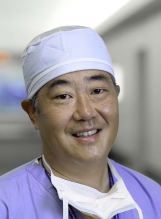 Bernard J. Park合作论文数Memorial Sloan Kettering Cancer Center77
 Matthew Bott合作论文数Department of Pathology;Memorial Sloan-Kettering Cancer Center;Department of Pathology, Memorial Sloan-Kettering Cancer Center75
 Kyuichi Kadota合作论文数68
 James Z Huang合作论文数65
 Manjit S. Bains合作论文数Memorial Sloan Kettering Cancer Center;Weill Cornell Medical College64
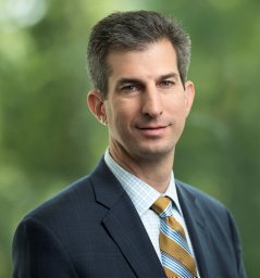 James M Isbell合作论文数Virginia Cardiac Surgery Quality Initiative, University of Virginia64
 Zauderer Marjorie G合作论文数Mem Sloan Kettering Canc Ctr62
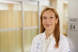 Molena Daniela合作论文数Department of Surgery, Johns Hopkins University60
 Gaetano Rocco合作论文数Memorial Sloan Kettering Cancer Center58
 Yuman Fong合作论文数Department of Surgery, City of Hope;Center for International Medicine, City of Hope;Center for Surgical Innovation, City of Hope51
 Natasha Rekhtman合作论文数51Camelia S. Sima合作论文数49
 Smita Sihag合作论文数Memorial Sloan Kettering Cancer Center46
 Andreas Rimner合作论文数Department of Radiation Oncology, University of Freiburg45Sauter Jennifer L合作论文数39Nabil Rizk合作论文数38
 Robert J. Downey合作论文数Memorial Sloan-Kettering Cancer Center36Michael D. Offin合作论文数35
 Navin Chintala合作论文数School of Pharmacy, Texas Tech University32Stephen Solomon合作论文数30Rania G. Aly合作论文数Faculty of Medicine, University of Alexandria28
 Marc Ladanyi合作论文数Memorial Sloan Kettering Cancer Center27
 Jun-ichi Nitadori合作论文数26
 Brendon M. Stiles合作论文数Memorial Sloan-Kettering Cancer Center25Shaohua Lu (卢韶华)合作论文数22Kei Suzuki合作论文数Matsusaka Chuo General Hospital Internal Medicine21
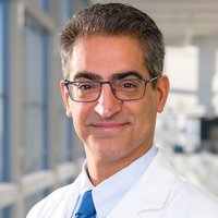 Inderpal S. Sarkaria合作论文数Harold C. Simmons Comprehensive Cancer Center, The University of Texas Southwestern Medical Center;Department of Cardiovascular and Thoracic Surgery, The University of Texas Southwestern Medical Center19
 Michael Hezel合作论文数Karolinska Institutet19
 Joseph Montecalvo合作论文数Dept Pathol, Mem Sloan Kettering Canc Ctr18
 Christos Colovos合作论文数Mem Sloan Kettering Canc Ctr18
 Mark G. Kris合作论文数David H. Koch Center for Cancer Care, Memorial Sloan Kettering Cancer Center18Villena-Vargas Jonathan合作论文数18Elliot L Servais合作论文数17
 Abraham J. Wu合作论文数Memorial Sloan Kettering Cancer Center17
 Takahashi Yusuke合作论文数Department of General Thoracic Surgery, Tokyo Metropolitan Cancer and Infectious Diseases Center16Michelle S. Ginsberg合作论文数16David P. Eisenberg合作论文数15Daniel Gomez合作论文数15Katherine D Gray合作论文数Department of Surgery, New York Presbyterian Hospital–Weill Cornell Medical Center15
 Katsura Emoto合作论文数15Mei-Ki Chan合作论文数15Joseph Dycoco合作论文数15Brooke Mastrogiacomo合作论文数14
 Charles M. Rudin合作论文数David H. Koch Center for Cancer Care, Memorial Sloan Kettering Cancer Center14




