The success of in vitro fertilization (IVF) at many clinics relies on the accurate morphological assessment of day 5 blastocysts, a process that is often subjective and inconsistent. While artificial intelligence can help standardize this evaluation, models require large, diverse, and balanced datasets, which are often unavailable due to data scarcity, natural class imbalance, and privacy constraints. Existing generative embryo models can mitigate these issues but face several limitations, such as poor image quality, small training datasets, non-robust evaluation, and lack of clinically relevant image generation for effective data augmentation. Here, we present the Diffusion Based Imaging Model for Artificial Blastocysts (DIA) framework, a set of latent diffusion models trained to generate high-fidelity, novel day 5 blastocyst images. Our models provide granular control by conditioning on Gardner-based morphological categories and z-axis focal depth. We rigorously evaluated the models using FID, a memorization metric, an embryologist Turing test, and three downstream classification tasks. Our results show that DIA models generate realistic images that embryologists could not reliably distinguish from real images. Most importantly, we demonstrated clear clinical value. Augmenting an imbalanced dataset with synthetic images significantly improved classification accuracy (p < 0.05). Also, adding synthetic images to an already large, balanced dataset yielded statistically significant performance gains, and synthetic data could replace up to 40
Embryo assessment in in vitro fertilization (IVF) involves multiple tasks-including ploidy prediction, quality scoring, component segmentation, embryo identification, and timing of developmental milestones. Existing methods address these tasks individually, leading to inefficiencies due to high costs and lack of standardization. Here, we introduce FEMI (Foundational IVF Model for Imaging), a foundation model trained on approximately 18 million time-lapse embryo images. We evaluate FEMI on ploidy prediction, blastocyst quality scoring, embryo component segmentation, embryo witnessing, blastulation time prediction, and stage prediction. FEMI attains area under the receiver operating characteristic (AUROC) > 0.75 for ploidy prediction using only image data-significantly outpacing benchmark models. It has higher accuracy than both traditional and deep-learning approaches for overall blastocyst quality and its subcomponents. Moreover, FEMI has strong performance in embryo witnessing, blastulation-time, and stage prediction. Our results demonstrate that FEMI can leverage large-scale, unlabelled data to improve predictive accuracy in several embryology-related tasks in IVF.
(Abstracted from Nat Commun 2024;15(1):7756 Assisted reproductive technology, including in vitro fertilization, has become an important treatment for individuals who desire familial expansion but are unable to conceive. A crucial part of this process is the determination of embryo viability and the selection of the highest quality embryos for transfer into the uterus.
Background: In recent times, various algorithms have been developed to assist in the selection of embryos fortransfer based on artificial intelligence (AI). Nevertheless, the majority of AI models employed in this context werecharacterized by a lack of transparency. To address these concerns, we aim to design an interpretable tool to automatehuman embryo evaluation by combining artificial neural networks (ANNs) and genetic algorithms (GA).Materials and Methods: This retrospective cohort study included 223 human blastocyst time-lapse (TL) imagestaken at 110 hours post-injection. All the images were evaluated by five embryologists from different clinics in termsof blastocyst expansion (BE), quality of the inner cell mass (ICM), and trophectoderm (TE). The embryo databasewas used to develop an AI system (70% training, 15% validation, and 15% test) for automate blastocyst assessment.The entire set of images underwent a standardization process, followed by processing and segmentation using Matlabsoftware. The resulting quantified variables were utilized in AI techniques (ANN and GA). Finally, the accuracy andperformance of the automation tool was assessed with the area under the receiver operating characteristic (ROC)curve (AUC). Then, the level of agreement among embryologists and between embryologists and the AI system wascompared with Kappa Index.Results: The overall agreement among embryologists was low (Kappa: 0.4 for BE; and 0.3 for TE and ICM). The AItool achieved higher consistency (Kappa 0.7 for BE and ICM; and 0.4 for TE). The AI exhibited high accuracy in classifyingBE (test 81.5%), ICM (test 78.8%), and TE (test 78.3%) and better performance for BE (AUC 0.888-0.956)than for ICM (AUC 0.605-0.854) and TE (AUC 0.726-0.769) assessment.Conclusion: Our AI tool highlighted the superior consistency of AI compared to human operators in grading blastocystmorphology. This research represents an important step towards fully automating objective embryo evaluation.
Previous studies have demonstrated equivocal euploidy rates between blastocysts that have undergone direct unequal cleavage (DUC) and non-DUC blastocysts.1 Even among euploid embryos, non-DUC embryos are chosen over DUC embryos at our institution although a difference in pregnancy outcomes between the groups have not been demonstrated.
Assessing fertilized human embryos is crucial for in vitro-fertilization (IVF), a task being revolutionized by artificial intelligence and deep learning. Existing models used for embryo quality assessment and chromosomal abnormality (ploidy) detection could be significantly improved by effectively utilizing time-lapse imaging to identify critical developmental time points for maximizing prediction accuracy. Addressing this, we developed and compared various embryo ploidy status prediction models across distinct embryo development stages. We present BELA (Blastocyst Evaluation Learning Algorithm), a state-of-the-art ploidy prediction model surpassing previous image- and video-based models, without necessitating subjective input from embryologists. BELA uses multitask learning to predict quality scores that are used downstream to predict ploidy status. By achieving an AUC of 0.76 for discriminating between euploidy and aneuploidy embryos on the Weill Cornell dataset, BELA matches the performance of models trained on embryologists' manual scores. While not a replacement for preimplantation genetic testing for aneuploidy (PGT-A), BELA exemplifies how such models can streamline the embryo evaluation process, reducing time and effort required by embryologists.
Background One challenge in the field of in-vitro fertilisation is the selection of the most viable embryos for transfer. Morphological quality assessment and morphokinetic analysis both have the disadvantage of intra-observer and interobserver variability. A third method, preimplantation genetic testing for aneuploidy (PGT-A), has limitations too, including its invasiveness and cost. We hypothesised that differences in aneuploid and euploid embryos that allow for model-based classification are reflected in morphology, morphokinetics, and associated clinical information. Methods In this retrospective study, we used machine-learning and deep-learning approaches to develop STORK-A, a non-invasive and automated method of embryo evaluation that uses artificial intelligence to predict embryo ploidy status. Our method used a dataset of 10 378 embryos that consisted of static images captured at 110 h after intracytoplasmic sperm injection, morphokinetic parameters, blastocyst morphological assessments, maternal age, and ploidy status. Independent and external datasets, Weill Cornell Medicine EmbryoScope+ (WCM-ES+; Weill Cornell Medicine Center of Reproductive Medicine, NY, USA) and IVI Valencia (IVI Valencia, Health Research Institute la Fe, Valencia, Spain) were used to test the generalisability of STORK-A and were compared measuring accuracy and area under the receiver operating characteristic curve (AUC). Findings Analysis and model development included the use of 10 378 embryos, all with PGT-A results, from 1385 patients (maternal age range 21-48 years; mean age 36.98 years [SD 4.62]). STORK-A predicted aneuploid versus euploid embryos with an accuracy of 69.3% (95% CI 66.9-71.5; AUC 0.761; positive predictive value [PPV] 76.1%; negative predictive value [NPV] 62.1%) when using images, maternal age, morphokinetics, and blastocyst score. A second classification task trained to predict complex aneuploidy versus euploidy and single aneuploidy produced an accuracy of 74.0% (95% CI 71.7-76.1; AUC 0.760; PPV 54.9%; NPV 87.6%) using an image, maternal age, morphokinetic parameters, and blastocyst grade. A third classification task trained to predict complex aneuploidy versus euploidy had an accuracy of 77.6% (95% CI 75.0-80.0; AUC 0.847; PPV 76.7%; NPV 78.0%). STORK-A reported accuracies of 63.4% (AUC 0.702) on the WCM-ES+ dataset and 65.7% (AUC 0.715) on the IVI Valencia dataset, when using an image, maternal age, and morphokinetic parameters, similar to the STORK-A test dataset accuracy of 67.8% (AUC 0.737), showing generalisability. Interpretation As a proof of concept, STORK-A shows an ability to predict embryo ploidy in a non-invasive manner and shows future potential as a standardised supplementation to traditional methods of embryo selection and prioritisation for implantation or recommendation for PGT-A.
To assess the biological relevance of inner cell mass (ICM) surface area with regards to embryo grading and ranking by experienced embryologists, and ploidy, clinical and live birth outcome.
To compare biomarkers automatically annotated by CHLOE EQTM (Fairtility) with human annotations, and to better understand their biological relevance.
Germline mutations in the BRCA genes are associated with a higher risk of carcinogenesis, which is linked to an increased mutation rate and loss of the second unaffected BRCA allele (loss of heterozygosity, LOH). However, the mechanisms triggering mutagenesis are not clearly understood. The BRCA genes contain high numbers of repetitive DNA sequences. We detected replication forks stalling, DNA breaks, and deletions at these sites in haploinsufficient BRCA cells, thus identifying the BRCA genes as fragile sites. Next, we found that stalled forks are repaired by error-prone pathways, such as microhomology-mediated break-induced replication (MMBIR) in haploinsufficient BRCA1 breast epithelial cells. We detected MMBIR mutations in BRCA1 tumor cells and noticed deletions-insertions (>50 bp) at the BRCA1 genes in BRCA1 patients. Altogether, these results suggest that under stress, error-prone repair of stalled forks is upregulated and induces mutations, including complex genomic rearrangements at the BRCA genes (LOH), in haploinsufficient BRCA1 cells.
The aim of this study was to identify if the combined age of a couple undergoing infertility treatment is associated with the probability of a full chromosome mosaic (FCM) or partial chromosome mosaic (PCM) preimplantation genetic testing (PGT) diagnosis.
1. To determine the impact of two different culture media on embryo development. 2. To develop machine learning (ML) models to predict the probability of blast formation based on morphokinetic and clinical parameters accessible at 66 hours of incubation in both culture media. 3. To implement an accessible ML-supported tool to assist ART decisions that can be easily adapted to the ever-evolving technological advances of the IVF laboratory. A total of 31,040 embryos were cultured in two different media (Media-A, single step, N=11128/33.7%; Media-B, sequential, N=19912/66.3%) over a 2-year period using the Embryoscope (Vitrolife, Sweden) time-lapse system, with developmental features annotated manually. A retrospective analysis of the morphokinetic timings and the inner cell mass (ICM), trophectoderm (TE), and expansion (Ex) grades of the blastocyst was performed. Differences in the development rates of embryos cultured were evaluated by logistic regression adjusting for maternal age. A logistic regression model was developed to estimate the probability of the embryo reaching the blastocyst stage at 110 hours. The model used maternal age and morphokinetic parameters observed earlier than 66 hours of incubation as inputs. Model features were selected using LASSO L-1 regularization with an 80% training/20% cross-validation split. The two different culture media employed in this study had a significant (p<0.01) impact both on the dynamics of post-fertilization development and embryo quality, as assessed by subjective morphological parameters. Media-A had a significantly higher proportion of embryos reaching the blastocyst stage at <110 hours and a higher proportion of top-quality blastocysts. The area under the receiver operating curve obtained from the ML models was 80.9% for Media-A and 80.5% for Media-B. The models were deployed as a web-based service running a Python script to extract the relevant morphokinetic and clinical parameters of embryos in real-time to provide the predicted probability of 110-hour blast formation. There were significant differences in the rate of embryo development and the proportion of top-graded blastocysts between the two culture media. The models developed to estimate the probability of reaching the blastocyst stage in <110 hours had a similar ROC-AUC but different structures to account for the different dynamics of development induced by the culture media.
During in vitro fertilization (IVF), the timing of cell divisions in early human embryos is a key predictor of embryo viability. Recent developments in time-lapse microscopy (TLM) have allowed us to observe cell divisions in much greater detail than previously possible. However, it is a time-consuming process that relies on a highly trained staff and subjective observations. We describe an automated method based on a convolutional neural network to detect and classify cell divisions from original (unprocessed) TLM images. Here, we used two embryo TLM image datasets to evaluate our method: a public dataset with mouse embryos up to the 4-cell stage and a private dataset with human embryos up to the 8-cell stage. Compared to embryologists’ annotations, our results were almost 100% accurate for the mouse embryo images and accurate within five frames in 93.9% of cell stage transitions for the human embryos. Our approach can be used to improve the consistency and quality of the existing annotations or as part of a platform for fully automated embryo assessment. The code is available at http://github.com/JonasEMalmsten/CellDivision .
The current method of preimplantation genetic testing for aneuploidy (PGT-A) involves invasive trophectoderm (TE) biopsy. Although PGT-A has improved the success rate per embryo transfer, it has notable limitations. These include the cost of sequencing, mosaicism, the skill required to biopsy TE cells, and the fact that only a select number of blastocysts (BLs) can be tested. Embryologists rely on morphological assessment and clinical information to select BLs for PGT-A. The development of noninvasive methods of embryo screening, as an alternative to PGT-A, is essential. We propose an embryo selection method that leverages the power of deep learning classification methods trained on spatial and temporal information stored in time-lapse images (TLM) that capture embryo development along with clinical parameters; this method can be used to select embryos for PGT-A and to predict embryo ploidy (euploid vs. aneuploid) without a biopsy. In our study, we used a retrospective dataset consisting of 10,872 embryos of known ploidy status (euploid or aneuploid) to train and validate several deep learning models. We developed deep learning models for embryo image analysis based on pre-trained ResNet18. The models utilized images of human embryos captured using time-lapse microscopy (EmbryoScopeTM) at 110 hours post-ICSI and known PGT-A results (aneuploid n = 6,443; euploid n = 4,429) as ground truth labels. The developed models use several clinical features, including maternal age, morphokinetics, BL grade, and BL score. Using an 80/20 training-validation split of the data, performances were measured by validation accuracy and the AUC. Class activation mapping (CAM) was employed to identify which areas within embryo images were used to predict ploidy. Several models were trained and validated, each with varying features.Tabled 1ModelFeaturesAUCAccuracyAImage0.621462.87%BImage, Age0.729368.06%CImage, Morphokinetics0.609462.55%DImage, BL Score0.690164.07%EImage, BL Grade0.687665.51%FImage Age, Morphokinetics0.734168.29%GImage, Age, BL Score0.755870.23%HImage, Age, BL Grade0.759569.54%IImage, Age, Morphokinetics, BL Score0.761469.68%JImage, Age, Morphokinetics, BL Grade0.75669.03% Open table in a new tab The implementation of deep learning image analysis permits a more objective assessment of BLs and, with the addition of oocyte age, morphokinetics, and BL grade or score, improves the model’s ability to predict embryo ploidy in a noninvasive manner. CAM results from BL images suggest that the model focuses on the presence and absence of cavitation to predict embryo ploidy at 110 hours.
Objective: To convert blastocyst (BL) morphological grade and BL day into a numeric blastocyst score (BS). Design: Retrospective cohort study. Setting: Academic center. Patient(s): A total of 5,653 BL of known implantation (fetal heart, FH) and 11,348 biopsied BL. Intervention(s): Based on their FH rates and/or significance, a score (1-4) was assigned to each BL grade component. The BL morphological score (BMS) is the sum (BS = BMS on day 5; BS = BMS + 2 on day 6). Main Outcome Measure(s): Statistics characterized the FH and euploidy odds with BS. Result(s): All three morphology grade components and BL day were associated with implantation and euploidy probability. The FH rate and euploidy odds decrease with larger BS. The BS was the most important factor (odds ratio [OR] per unit change = 0.807, 95% confidence interval [CI] 0.784, 0.831) for untested and euploid BL implantation, and the sole one for euploid BL (OR/unit change = 0.845, 95% CI 0.803, 0.889). The BS is the second most significant factor after maternal age for euploidy probability (OR/unit change = 0.808, 95% CI 0.795, 0.822). In training and validation sets (75:25), the BS can predict implantation with similar area under the curve [AUC] (training = 0.628, 95% CI 0.613, 0.643; validation = 0.606, 95% CI 0.581, 0.631). The BS has better euploidy prediction ability (training AUC = 0.683, 95% CI 0.673, 0.693; validation AUC = 0.698, 95% CI 0.681, 0.715). The BS can stratify BL into good (3-5), fair (6-9), and poor (10-14) groups, reflecting their FH, live birth rates, and ploidy status. Advanced maternal age was associated with lower untested BL implantation and lower euploidy odds across all groups. Conclusion(s): The BS is a predictor of BL ploidy and FH implantation. (Fertil Steril Rep (R) 2020;1:133-41. (c) 2020 by American Society for Reproductive Medicine.)
Visual morphology assessment is routinely used for evaluating of embryo quality and selecting human blastocysts for transfer after in vitro fertilization (IVF). However, the assessment produces different results between embryologists and as a result, the success rate of IVF remains low. To overcome uncertainties in embryo quality, multiple embryos are often implanted resulting in undesired multiple pregnancies and complications. Unlike in other imaging fields, human embryology and IVF have not yet leveraged artificial intelligence (AI) for unbiased, automated embryo assessment. We postulated that an AI approach trained on thousands of embryos can reliably predict embryo quality without human intervention. We implemented an AI approach based on deep neural networks (DNNs) to select highest quality embryos using a large collection of human embryo time-lapse images (about 50,000 images) from a high-volume fertility center in the United States. We developed a framework (STORK) based on Google's Inception model. STORK predicts blastocyst quality with an AUC of >0.98 and generalizes well to images from other clinics outside the US and outperforms individual embryologists. Using clinical data for 2182 embryos, we created a decision tree to integrate embryo quality and patient age to identify scenarios associated with pregnancy likelihood. Our analysis shows that the chance of pregnancy based on individual embryos varies from 13.8% (age ≥41 and poor-quality) to 66.3% (age <37 and good-quality) depending on automated blastocyst quality assessment and patient age. In conclusion, our AI-driven approach provides a reproducible way to assess embryo quality and uncovers new, potentially personalized strategies to select embryos.
To assess whether the duration of recovery following the warming of cryopreserved oocytes affects the morphological and morphokinetic properties of the developing embryo. This is a retrospective data analysis examining the effect of the post-warming recovery duration of vitrified oocytes prior to ICSI. Frozen oocytes from 36 patients (July 2018 to March 2019), either autologous or donor, were included in the study. The study examined cycles in which the oocytes were injected within a 2-hour recovery period and compared them to those in which the oocytes were injected within 3 hours or more within the same patient oocyte cohort. All injected oocytes were cultured individually in time-lapse incubators (TLM; EmbryoScope, Vitrolife, Sweden). Embryo transfers occurred on D3 or D5 regardless of the study groups. Standard oocyte freezing protocol included vitrification 2 hours after retrieval using a Kitazato-based (Japan) vitrification media. Oocyte warming was performed using Kitazato thaw media. ICSI was performed in the standard fashion, and embryos were annotated daily for their developmental hallmarks. Embryo selection for ET or freezing was performed by standard laboratory protocols. No significant differences were observed in 2PN and/or abnormal fertilization rates between the groups. Similarly, on day 3, no significant differences were found in the average cell numbers or in the average number of good (≥8 cells, <20% frag.), fair (5-7 cells, any abnormal cleavages), and poor (≤4 cells) embryos. Equally, the incidence of abnormal cleavage patterns was similar among the cohort. More embryos were selected for transfer from the 3-hour group than from the 2-hour group (51.1% vs. 48.9%; p=0.78). There were no significant differences in the utilization rate (the number of embryos transferred fresh plus blastocysts frozen) between the 3- and 2-hour groups (49.6% vs. 49.6%, respectively). The time-lapse morphokinetic data indicated faster-growing embryos in the 3- versus 2-hour group. This difference is apparent in late-stage morphokinetic parameters (t9 to tsB). The 3-hour group produced significantly better quality blastocysts in cycles that were either frozen upfront, fresh D5 ET, or PGT (p=0.03). The duration of the recovery time post-warming showed no significant differences in overall embryology outcomes prior to blastocyst formation. However, at the BL stage, the 3-hour group demonstrated improvement in morphokinetic parameters and in overall BL quality.
To determine whether convolutional neural network (CNN) can be used to predict whether an embryo capable of achieving a pregnancy will ultimately miscarry or lead to live birth based on Artificial Intelligence (AI) analysis of time-lapse (TLM) embryo images.
During in-vitro fertilization, the timings of cell divisions in early human embryos are important predictors of embryo viability. Recent developments in time-lapse microscopy (TLM) allows for observing cell divisions in much greater detail than before. However, it is a time-consuming process relying on highly trained staff and subjective observations. We present an automated method based on a convolutional neural network to predict cell divisions from original (unprocessed) TLM images. Our method was evaluated on two embryo TLM image datasets: a public dataset with mouse embryos and a private dataset with human embryos up to 4-cell stage. Compared to embryologists' annotations, our results were almost 100% accurate for mouse embryos and accurate within five frames in 93% of cell stage transitions for human embryos. Our approach can be used to improve consistency and quality of existing annotations or as part of a platform for fully automatic embryo assessment.
 Nikica Zaninovic合作论文数29
 Zev Rosenwaks合作论文数Weill Cornell Medicine;NewYork-Presbyterian Hospital27
 Jonas Malmsten合作论文数Ronald O Perelman and Claudia Cohen Center for Reproductive Medicine, Weill Cornell Medicine14
 Iman Hajirasouliha合作论文数Weill Cornell Medicine9Marcos Meseguer合作论文数9
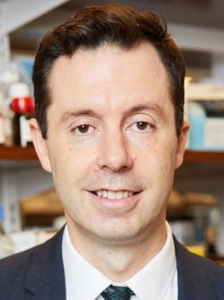 Olivier Elemento合作论文数Englander Institute for Precision Medicine, Weill Cornell Medical College;Institute for Computational Biomedicine , Weill Cornell Medical College;Clinical & Translational Science Center, Weill Cornell Medical College;Department of Physiology and Biophysics, Weill Cornell Medical College;Laboratory of Cancer Systems Biology, Weill Cornell Medical College;Institute for Computational Medicine, Weill Cornell Medical College;Meyer Cancer Center, Weill Cornell Medical College9Cristina Hickman合作论文数6Zhen Ye合作论文数5Josue Barnes合作论文数4Pantelis Zisimopoulos合作论文数Institute for Computational Biomedicine, Weill Cornell Medicine4
 Suraj Rajendran合作论文数Institute for Computational Biomedicine, Weill Cornell Medicine4
 Alexandros Sigaras合作论文数4RN Clarke合作论文数4Marco Toschi合作论文数4Kathleen A. Miller合作论文数3Matthew Brendel合作论文数Institute for Computational Biomedicine, Weill Cornell Medicine3
 Pegah Khosravi合作论文数Department of Biological Sciences, New York City College of Technology;The City University of New York Graduate Center;Biomind AI Lab3
 Jeannine Gerhardt合作论文数Department of Cell Biology, Albert Einstein College of Medicine3Adriana Brualla Mora合作论文数2Jose Sierra合作论文数2Iris Har Vardi合作论文数2David Hoffman合作论文数2
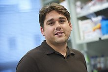 Christopher C Kloss合作论文数Dept Med, Weill Cornell Med Coll2Zi Yan合作论文数2
 Shan Juan合作论文数Department of Computer Science, Seidenberg School of Computer Science and Information Systems, Pace University2Rocha José Celso合作论文数2
 Advaitha Madireddy合作论文数Department of Cell Biology, Albert Einstein College of Medicine2
 Kazemi Ehsan合作论文数Google DeepMind2
 Shahin Rafii合作论文数Cell & Developmental Biology, Weill Cornell Graduate School of Medical Sciences, Cornell University2
 Daylon James合作论文数Department of Genetic Medicine, Weill Cornell Medical College2
 Assaf Ben-Meir合作论文数Department of Obstetrics and Gynecology, Hebrew University of Jerusalem;Hadassah Hebrew-University Medical Center;Fairtility2
 Carl L Schildkraut合作论文数Department of Cell Biology;Einstein College of Medicine, Albert;Department of Cell Biology, Albert Einstein College of Medicine2Richard J. Bodine合作论文数2R. Bodine合作论文数1Theodore Paniza合作论文数1Jason Park合作论文数1Anna Malkova合作论文数1Ryan Miller合作论文数1Qianzi Li合作论文数1Andrea Cabassa Miskimen合作论文数1Rishabh Hariharan合作论文数1Jerzy Twarowski合作论文数1Vianne R. Gao合作论文数1
 Eva R. Hoffmann合作论文数Department of Cellular and Molecular Medicine, Faculty of Health and Medical Sciences, University of Copenhagen1Nina Vyas合作论文数1C L O'Neill合作论文数1Christian S. Ottolini合作论文数1
 Junbum Kim合作论文数Institute for Computational Biomedicine, Weill Cornell Medicine1




