The Society for Craniofacial Genetics and Developmental Biology (SCGDB) held its 48th Annual Meeting at the University of Minnesota in Minneapolis on September 29-October 1, 2025. On the first day of the meeting, Drs. Timothy Cox and Jennifer Fish were honored with Excellence in Craniofacial Research Awards for their exceptional contributions to the field of craniofacial biology. The following 2 days of the meeting featured five sessions that highlighted new discoveries in human genetics, systems biology of craniofacial development and disease, evolutionary connections, signaling mechanisms, and a special session on clinical and patient perspectives. The meeting also featured workshops on scientific writing and the role of artificial intelligence in advancing research and care. A poster session facilitated dynamic and insightful interactions among the 113 attendees, who represented diverse career stages and research backgrounds in developmental biology and genetics, further strengthening the SCGDB community.
Humans with pathogenic loss of function alanyl-tRNA synthetase 1 (AARS1) variants have a range of congenital brain phenotypes, including a high prevalence of microcephaly. The molecular mechanisms for this are unclear but zebrafish mutants in aars1 have reduced neurogenesis and increased apoptosis. Here, we report two individuals with compound heterozygous AARS1 variants. We created two mouse models to study the role of Aars1 in embryonic brain development. We provide evidence from these mouse models and in vitro splicing assays that both human AARS1 alleles are pathogenic. Mice homozygous for either a missense allele or an indel allele are both lethal very early in embryonic development. Aars1G80S/WT heterozygous animals show reduced Purkinje cell immunoreactivity at 8 months of age but no gross morphological cerebellar phenotypes or impaired performance in a motor coordination assay. We conclude these are pathogenic alleles in AARS1 but lethality in mice preclude a detailed study of neural development.
Tumor Suppressor Candidate 3 (TUSC3) is an integral component of the oligosaccharyltransferase complex and is required for the N-glycosylation of proteins. While TUSC3 has been implicated in cancer and autosomal recessive non-syndromic intellectual disability, some patients also exhibit distinct facial features. However, the role of Tusc3 in craniofacial development is unknown. Here, we report a patient presenting with cleft lip and palate who was found to carry a homozygous variant in the TUSC3 promoter region in addition to a maternally inherited chromosomal duplication. In order to evaluate a potential role for TUSC3 in craniofacial development, we analyzed Tusc3 deletion mouse mutants from the International Mouse Phenotyping Consortium (IMPC). Initial IMPC phenotyping data suggested that a subset of Tusc3 deletion mice could develop cleft palate, micrognathia, and aglossia. We further identified that Tusc3 is expressed in the craniofacial and brain regions during embryonic development. Upon characterizing the Tusc3 deletion line, we observed that most homozygous mutants exhibit preweaning lethality. While craniofacial defects occur at a very low frequency in the deletion mutants, we identified a modest reduction in cortical area. Furthermore, RNA-seq analysis surprisingly revealed that no other genes in the developing brain or face were significantly affected upon loss of Tusc3. These findings suggest that Tusc3 can contribute to congenital malformations in both craniofacial and cortical development.
Glycosylphosphatidylinositol (GPI) anchors are a post-translational modification made to over 150 proteins. These GPI-anchored proteins are enriched in lipid rafts in the plasma membrane and serve a variety of functions. Human pathogenic variants in GPI biosynthesis pathway enzymes are collectively called inherited GPI deficiencies and lead to several brain anomalies, but we still lack a deep understanding of GPI-anchor functions in brain development. PIGA and PGAP2 are two enzymes in the GPI-anchor biosynthesis pathway. A Nestin-Cre mediated deletion of Piga in the mouse led to early postnatal death and significant structural brain malformations. There are no studies on Pgap2 loss of function in the brain to date. We extended these studies with a series of genetic ablations to further determine the role of Piga and Pgap2 in the forebrain, oligodendrocytes, and cerebellum. We find Piga expression in the hindbrain is absolutely required for survival while Pgap2 ablations were much less deleterious.
Primary cilia play a pivotal role in cellular signaling and development. Human primary microcephaly is strongly associated with pathogenic variants in primary cilia genes. Here, we examine the role of Ttc21b, a component of the intraflagellar transport-A complex, during mouse forebrain development by using a Ttc21balien null allele. Our findings reveal that significant microcephaly in homozygous mutants is caused by disrupted neural progenitor proliferation and differentiation. Histological and immunohistochemical analyses show an enlarged ventricular zone and reduced cortical plate thickness accompanied by altered mitotic spindle angles, suggesting defects in symmetric versus asymmetric cell divisions. Embryonic expression patterns suggest that perdurant TTC21B protein underlies these phenotypes. Progenitor proliferation kinetics were disrupted along with changes in TBR2positive intermediate progenitors and TBR1-positive early-developing neurons. Neuronal processes in the cortical plate were significantly shortened. Our findings support a model in which early expression of Ttc21b in neural precursor cells destined for the forebrain is critical to ensure TTC21B protein levels to sustain subsequent neural progenitor proliferation and differentiation. These results advance our understanding of the role primary cilia have in cortical development.
Germline ZFX variants are associated with an X-linked neurodevelopmental disorder, with 14 males and 16 females reported to date. We describe a 20-year-old female with a heterozygous ZFX frameshift variant, p.(Met666Valfs*2), identified by genome sequencing, previously reported in an affected male. She exhibited motor and speech delays with hypotonia in early childhood, and was later diagnosed with congenital heart defects, autism spectrum disorder, mild intellectual disability, and absence seizures. She further developed sensorineural hearing loss, skin hyperpigmentation, and ophthalmoplegia. Novel phenotypic features included inferior cerebellar vermian hypoplasia, hypoplastic right vertebral artery, aberrant subclavian artery, long palpebral fissures, ophthalmoplegia, skin hyperpigmentation, and a short uvula, expanding the known clinical spectrum. Female carriers of pathogenic ZFX variants demonstrate highly variable expressivity, ranging from apparently unaffected individuals to syndromic presentations. Individuals with heterozygous missense variants often exhibit hyperparathyroidism, suggesting a genotype-phenotype correlation. Reanalysis of published RNA-sequencing data identified 15 ZFX target genes involved in neurodevelopment, suggesting a role for these genes in disease pathogenesis. These findings confirm the pathogenicity of the p.(Met666Valfs*2) variant in the proband and highlight the phenotypic heterogeneity of the disorder in females. Clinical care should include cardiac and endocrine monitoring, with endocrine testing offered to unaffected females carrying missense variants.
The Society for Craniofacial Genetics and Developmental Biology (SCGDB) hosted its 47th Annual Meeting on September 9-10, 2024, at the Stowers Institute for Medical Research and Children's Mercy Research Institute in Kansas City, Missouri. On the opening day, Drs. Jean-Pierre Saint-Jeannet and Elizabeth Leslie received the SCGDB Distinguished Scientist Awards in recognition of their exceptional contributions to craniofacial biology. Additionally, Dr. Daniel Jensen discussed his unique perspective on Treacher Collins syndrome, speaking as both a physician and a patient. Over the next 2 days, five sessions showcased groundbreaking research on cell signaling and genomic mechanisms regulating craniofacial development, human genetics, translational and regenerative approaches, and clinical management of craniofacial differences. The meeting also featured interactive workshops on engaging with journal editors during the manuscript review process and empowering mentees to take an active role in maximizing their mentorship experience. A poster session further fostered meaningful interactions among the attendees, who represented diverse career stages and research backgrounds in developmental biology and genetics, further strengthening the SCGDB community.
A recurrent de novo germline variant in the MAX gene, p.(Arg60Gln), has recently been associated with polydactyly-macrocephaly syndrome in six unrelated individuals. Affected individuals presented with progressive macrocephaly, post-axial polydactyly, developmental delay, autistic features and a series of craniofacial, brain, cardiac, ocular, and renal anomalies. Here, we describe two unrelated female probands with the known recurrent MAX variant, c.179G>A p.(Arg60Gln), who presented with the emerging phenotypes of the MAX-associated syndrome. We also propose that genitourinary abnormalities, including Mayer-Rokitanski-Kuster-Hauser syndrome in one individual, may constitute an expansion of the known phenotype. These findings contribute to the current knowledge regarding the phenotypic spectrum of MAX-associated polydactyly-macrocephaly syndrome.
Glycosylphosphatidylinositol (GPI) anchors are a class of post-translational modifications observed on over 150 proteins. Pathogenic variants in the GPI biosynthesis enzyme, PIGA, in humans are associated with several brain anomalies such as hypomyelination, cerebellar hypoplasia, ataxic gait, and can lead to premature mortality. We previously genetically deleted Piga from the embryonic mouse brain which led to early postnatal death and significant structural brain malformations similar to those observed in humans with PIGA variants. The current treatment options for PIGA patients only manage symptoms and provides palliative care, demonstrating a need for new therapeutic options. We employed an AAV9-mediated PIGA gene-replacement (AAV9-hPIGA) strategy to assess the efficacy of gene therapy in the brain. We show that a single intracerebroventricular treatment on the first day of life successfully rescued survival rates, structural brain anomalies, and neurological impairments. Additionally, we used mass spectrometry to identify and quantify GPI-anchored proteins in untreated and treated mutant mice. We found that AAV9-hPIGA treatment restored GPI-anchored protein levels in mutant animals. These investigations enhance our understanding of GPI-anchored protein production during brain development and contribute to the development of a more effective intervention for PIGA-related symptoms.
Cleft lip and cleft palate are among the most common congenital anomalies and are the result of incomplete fusion of embryonic craniofacial processes or palatal shelves, respectively. We know that genetics play a large role in these anomalies but the list of known causal genes is far from complete. As part of a larger sequencing effort of patients with congenital craniofacial anomalies, we identified a rare candidate variant in transforming growth factor beta receptor 2 (TGFBR2). This variant alters a highly conserved amino acid and is predicted to be pathogenic by a number of metrics. The family history and population genetics suggest that this specific variant would be incompletely penetrant, but this gene has been convincingly implicated in craniofacial development. In order to test the hypothesis this might be a causal variant, we used genome editing to create the orthologous variant in a new mouse model. Surprisingly, Tgfbr2V387M mice did not exhibit craniofacial anomalies or have reduced survival, suggesting Tgfbr2V387M is not a causal variant for cleft palate/ micrognathia. The discrepancy between in silico predictions and mouse phenotypes highlights the complexity of translating human genetic findings to mouse models. We expect these findings will aid in interpretation of future variants seen in TGFBR2 from ongoing sequencing of patients with congenital craniofacial anomalies.
Human congenital anomalies account for twice the mortality of childhood cancer. Despite advancements in genome sequencing and transgenic mouse models that have aided in understanding their pathogenesis, significant gaps remain. Through a forward genetics approach, we previously discovered the hypo-morphic anteater allele of Cse1l which displayed variable craniofacial phenotypes. To circumvent the variability seen in this model, we generated a conditional allele of Cse1l and genetically ablated it in the dorsal midline giving rise to portions of the nervous system and the cranial neural crest cells using the Wnt1-Cre 2 driver. Our analysis revealed that Wnt1-Cre2; Cse1l CRISPR/flox embryos exhibited severe malformations in the forebrain, midbrain, and hindbrain, accompanied by a dramatic hypoplasia of the frontonasal, maxillary, and mandibular processes, and the second pharyngeal arch. Wnt1-Cre2; Cse1l CRISPR/flox embryos were embryonic lethal by E11.5 likely due to defects in the ventricular myocardium. Wnt1-Cre2; Cse1l CRISPR/flox embryos exhibited consistently increased apoptosis at E9.5 in the affected tissues along with an increase in p53 expression. These data together show a previously unknown critical function of CSE1L in neural crest cell survival during development. Summary Statement:Cse1l is critical for neural crest cell survival and genetic ablation of Cse1l in neural crest cells resulted in dramatic apoptosis with increase in p53 expression.
Ribosome biogenesis is a key cellular function and disruptions in this process can lead to congenital anomalies or "ribosomopathies" with varying phenotypes including craniofacial malformations and neurodevelopment symptoms. Classically, the mouse is a robust model to understand the molecular mechanisms underlying ribosomopathies to further elucidate human pathogenesis. We identified novel compound heterozygous missense variants in the TATA-box binding protein associated factor, RNA polymerase I subunit C (TAF1C) locus in a patient with some phenotypes consistent with ribosomopathies. TAF1C encodes a subunit of the SL1 complex which is critical for the RNA PolI complex to initiate ribosomal RNA transcription. We hypothesized that functional TAF1C is required at developmental stages critical for craniofacial and neurodevelopment. To test this hypothesis, we created mouse Taf1c variants orthologous to the human variants using CRISPR-CAS9 technology (Taf1cR202Q and Taf1cS428A). We also created an 11bp deletion to complement the missense variants (Taf1c11bpdel). We created multiple allelic combinations to determine the roles for Taf1c in survival and craniofacial development. Homozygous mice for any of these novel variants were underrepresented at organogenesis stages. We did not observe craniofacial anomalies in any surviving mice. Our results suggest that these specific TAF1C variants are not the cause of any human phenotype present in the patient motivating the study. However, we showed that Taf1c is required for embryonic survival and our studies contribute to knowledge about the role of ribosome biogenesis machinery throughout organogenesis.
The primary cilium is a small organelle that plays key roles in cellular signaling. Defects in primary cilia formation, morphology, and function cause a heterogeneous group of developmental syndromes termed ciliopathies. The inturned planar cell polarity protein (INTU) gene acts in the CPLANE complex to facilitate ciliogenesis and support cilia signaling. Bi-allelic genetic variants in INTU have previously been reported in seven patients with pleiotropic disorders, but a core set of phenotypes from these patients has not been codified and functional studies into these variants have failed to fully demonstrate mechanistic perturbations caused by INTU dysfunction. Here, we report on a person with cardiac abnormalities, distinctive craniofacial features, developmental delays, tongue hamartomas, bilateral clinodactyly, and polydactyly of the left great toe. Trio whole-exome sequencing identified compound heterozygous variants in the INTU gene. Functional studies provide evidence that these INTU variants confer human disease through altered ciliogenesis and/or cilia signaling. Furthermore, we suggest that this study along with previous reports sufficiently establishes an association between a pleiotropic disorder and variants in the INTU gene to enhance clinical interpretation of INTU variants in future studies.
Microcephaly affects 1 in 2,500 babies per year. Primary microcephaly results from aberrant neurogenesis leading to a small brain at birth. This is due to altered patterns of proliferation and/or early differentiation of neurons. Premature differentiation of neurons is associated with defects in the centrosome and/or primary cilia. In this study, we report on the first patients identified with NUBP2 -deficiency and utilize a conditional mouse model to ascertain the molecular mechanisms associated with NUBP2 -deficient primary microcephaly. We identified homozygous NUBP2 variants in these patients who displayed profound primary microcephaly in addition to intrauterine growth restriction, cervical kyphosis, severe contractures of joints, and facial dysmorphia. We then generated a mouse model using Emx1-Cre to ablate Nubp2 from the forebrain. The mice presented with severe microcephaly starting at E18.5. Neurospheres generated from the forebrain of Emx1-Cre; Nubp2 flox/flox conditional deletion mice were used to support the pathogenicity of the patient variants. We show that loss of Nubp2 increases both canonical and non-canonical cell death, but that loss of p53 fails to rescue microcephaly in the mouse model. Examination of neurogenesis in Emx1-Cre; Nubp2 flox/flox mice revealed distinct alterations in proliferation and cellular migration accompanied by supernumerary centrosomes and cilia. We therefore propose that NUBP2 is a novel primary microcephaly-related gene and that the role of Nubp2 in centrosome and cilia regulation is crucial for proper neurogenesis.
Human congenital anomalies account for twice the mortality of childhood cancer. Despite advancements in genome sequencing and transgenic mouse models that have aided in understanding their pathogenesis, significant gaps remain. Through a forward genetics approach, we previously discovered the hypo-morphic anteater allele of Cse1l which displayed variable craniofacial phenotypes. To circumvent the variability seen in this model, we generated a conditional allele of Cse1l and genetically ablated it in the dorsal midline giving rise to portions of the nervous system and the cranial neural crest cells using the Wnt1-Cre2 driver. Our analysis revealed that Wnt1-Cre2; Cse1lCRISPR/flox embryos exhibited severe malformations in the forebrain, midbrain, and hindbrain, accompanied by a dramatic hypoplasia of the frontonasal, maxillary, and mandibular processes, and the second pharyngeal arch. Wnt1-Cre2; Cse1lCRISPR/flox embryos were embryonic lethal by E11.5 likely due to proliferative defects in the ventricular myocardium. Wnt1-Cre2; Cse1lCRISPR/flox embryos exhibited consistently increased apoptosis at E9.5 in the affected tissues along with an increase in p53 expression. These data together show a previously unknown critical function of CSE1L in neural crest cell survival during development.
Primary cilia play a pivotal role in cellular signaling and development and disruptions in ciliary form and/or function leads to human ciliopathies. Here, we examine the role of Ttc21b , a key component of the intraflagellar transport-A complex, in mouse forebrain development using a Ttc21b alien null allele. Our findings reveal significant microcephaly in homozygous mutants is caused by disrupted neural progenitor proliferation and differentiation. Histological and immunohistochemical analyses show an enlarged ventricular zone and reduced cortical plate thickness, accompanied by altered mitotic spindle angles, suggesting defects in symmetric versus asymmetric cell divisions. Despite low Ttc21b expression in the forebrain epithelium, early embryonic expression patterns imply that perdurant TTC21B protein may underlie these phenotypes. Progenitor proliferation kinetics were disrupted, with fewer cells re-entering the cell cycle, correlating with reduced TBR2-positive intermediate progenitors and altered neurogenesis dynamics. Neuronal processes in the cortical plate were significantly shortened, suggesting cytoskeletal defects specific to terminal differentiation stages. Our findings support a model where early Ttc21b expression in precursors destined for the forebrain is critical for sustaining later neural progenitor proliferation and differentiation. These results advance our understanding of primary cilia in cortical development and provide a framework for exploring cytoskeletal contributions to ciliopathies.
Cleft lip and cleft palate are among the most common congenital anomalies and are the result of incomplete fusion of embryonic craniofacial processes or palatal shelves. Genetic factors are known to play a large role in these anomalies, but the list of known causal genes is far from complete. As part of a larger effort to sequence patients with micrognathia and cleft palate, we identified candidate pathogenic variants in dmx-like 1 (DMXL1). We used genome editing to create an allelic series of Dmxl1 in the mouse: a small deletion and the two orthologous missense variants. We do not find evidence that either missense allele is pathogenic, but we do see that loss of Dmxl1 leads to very early embryonic lethality. This confirms and extends two recent findings about Dmxl1, suggesting this gene has crucial basal functions in the cell and should be further considered in human disease genetics.
Despite advances in next generation sequencing (NGS), genetic diagnoses remain elusive for many patients with neurologic syndromes. Long-read sequencing (LRS) and optical genome mapping (OGM) technologies improve upon existing capabilities in the detection and interpretation of structural variation in repetitive DNA, on a single haplotype, while also providing enhanced breakpoint resolution. We performed LRS and OGM on two patients with known chromosomal rearrangements and inconclusive Sanger or NGS. The first patient, who had epilepsy and developmental delay, had a complex translocation between two chromosomes that included insertion and inversion events. The second patient, who had a movement disorder, had an inversion on a single chromosome disrupted by multiple smaller inversions and insertions. Sequence level resolution of the rearrangements identified pathogenic breaks in noncoding sequence in or near known disease-causing genes with relevant neurologic phenotypes (MBD5, NKX2-1). These specific variants have not been reported previously, but expected molecular consequences are consistent with previously reported cases. As the use of LRS and OGM technologies for clinical testing increases and data analyses become more standardized, these methods along with multiomic data to validate noncoding variation effects will improve diagnostic yield and increase the proportion of probands with detectable pathogenic variants for known genes implicated in neurogenetic disease.
There are currently over 7,000 rare diseases estimated to affect nearly 30 million Americans, according to the National Organization for Rare Disease (NORD). A variety of genetic, biochemical, and other diagnostic tests are available to such patients. While microarrays and gene panels are often considered standard of care, clinicians are increasingly ordering exome sequencing on such patients as a first-tier diagnostic assay. Large cohort studies have shown that exome sequencing has a consistent diagnostic yield of 30-50% depending on the patient’s condition, but this still fails to provide an answer for more than half of individuals tested.
Genetic variants in multiple sphingolipid biosynthesis genes cause human brain disorders. A recent study looked at people from 12 unrelated families with variants in the gene SMPD4, a neutral sphingomyelinase that metabolizes sphingomyelin into ceramide at an early stage of the biosynthesis pathway. These individuals have severe developmental brain malformations, including microcephaly and cerebellar hypoplasia. The disease mechanism of SMPD4 was not known and so we pursued a new mouse model. We hypothesized that the role of SMPD4 in producing ceramide is important for making primary cilia, a crucial organelle mediating cellular signaling. We found that the mouse model has cerebellar hypoplasia due to failure of Purkinje cell development. Human induced pluripotent stem cells lacking SMPD4 exhibit neural progenitor cell death and have shortened primary cilia, which is rescued by adding exogenous ceramide. SMPD4 production of ceramide is crucial for human brain development.
 David R. Beier合作论文数13Samantha A. Brugmann合作论文数12Weaver K Nicole合作论文数9
 John Klingensmith合作论文数The Graduate School, Duke University;School of Medicine, Duke University8
 Ching-Fang Chang合作论文数8Ashley M. Driver合作论文数University of Wisconsin Madison, University of Wisconsin-Madison8Chelsea Menke合作论文数7
 Cynthia A. Prows合作论文数Division of Human Genetics, Cincinnati Children's Hospital Medical Center7
 Robert J. Hopkin合作论文数Department of Pediatrics, University of Cincinnati;Division of Human Genetics, Cincinnati Children's Hospital Medical Center, University of Cincinnati College of Medicine7
 Andrew Distasio合作论文数6Marshall Lukacs合作论文数6
 Sund Kristen L合作论文数Cincinnati Children's Hospital, University of Cincinnati College of Medicine6Pamela Tran合作论文数Departments of Human Genetics, Pediatrics and Biology, McGill University—Montreal Children's Hospital Research Institute6
 Howard Saal合作论文数Division of Human Genetics, Cincinnati Children's Hospital Medical Center6
 Zakia I Abdelhamed合作论文数Leeds Institute of Molecular Medicine, St James’s University Hospital6D R Beier合作论文数5
 Amy E Merrill合作论文数David Geffen School of Medicine, University of California5
 Choi Murim合作论文数Coll Med, Seoul Natl Univ5Liegel Ryan合作论文数5
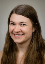 Schock Elizabeth N合作论文数Department of Surgery, Cincinnati Children's Hospital Medical Center;Department of Pediatrics, Cincinnati Children's Hospital Medical Center5
 Robert B. Hufnagel合作论文数Ophthalmic Genomics Laboratory, National Eye Institute;Medical Genetics and Ophthalmic Genomics Unit, National Eye Institute5
 Ammar Husami合作论文数4Milene Donlin合作论文数4
 Lauren Blizzard合作论文数4David Beier合作论文数4J Klingensmith合作论文数4Bonnie R Sullivan合作论文数4Cionni Megan合作论文数4Attia Aria C合作论文数4Cox Timothy C合作论文数Center for Developmental Biology &Regenerative Medicine, Seattle Children's Research Institute;Department of Pediatrics (Craniofacial Medicine),, University of Washington;Department of Oral Health Sciences, University of Washington;Department of Anatomy &Developmental Biology, Monash University;University of Washington, Monash University4
 Matthew P. Harris合作论文数Department of Anatomy, University of Wisconsin4Bimal P Chaudhari合作论文数Biomedical Science, Creighton University School of Medicine4David E Clouthier合作论文数Department of Cranio. Biol., Univ. of Colorado Denver4John Snedeker合作论文数3Erin Finnerty合作论文数3Katherine A Inskeep合作论文数3Brandon Stone合作论文数3
 Elizabeth Bittermann合作论文数3Jesse M Hunter合作论文数3
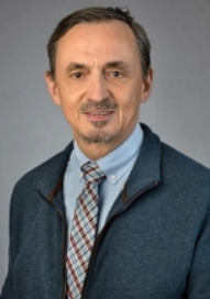 Jean-Pierre Saint-Jeannet合作论文数Department of Molecular Pathobiology, College of Dentistry, New York University3Turbe-Doan Annick合作论文数3
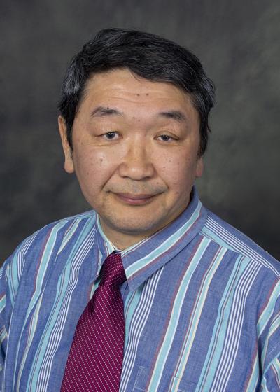 Yuji Mishina合作论文数Department of Biologic and Materials Sciences & Prosthodontics, School of Dentistry, University of Michigan3Knudson Luke合作论文数3
 William Dobyns合作论文数Department of Pediatrics, Medical School, University of Minnesota;Division of Genetics and Metabolism, University of Minnesota3
 Ben Pode-Shakked合作论文数Institute for Rare Diseases, Sheba Medical Center3
 Dan Koboldt合作论文数Institute for Genomic Medicine, Nationwide Children's Hospital;The Ohio State University3Beth M. Kline-Fath合作论文数Cincinnati Children’s Hospital Medical Center, University of Cincinnati Medical Center3
 Fowzan S. Alkuraya (Fowzan Alkuraya)合作论文数College of Medicine, Alfaisal University;Department of Translational Genomics, King Faisal Specialist Hospital and Research Center3
 Katherine A. Fantauzzo合作论文数Department of Genetics and Development, Columbia University3




