Mammalian lung development requires coordinated gene regulation to drive lung bud formation, branching morphogenesis, proximal-distal patterning, and epithelial specification. While key transcriptional and signaling regulators are known, the epigenetic regulators are less well studied. Here, we identify the canonical BAF complex as essential for lung epithelial development. Complete loss of BAF complex function causes failure of lung formation, and selective deletion of ARID1A leads to loss of distal patterning and reduced alveolar type 1 (AT1) cell differentiation, with emergence of a highly proliferative cell state defined by joint activation of YAP and WNT signaling and loss of BMP response. Epigenomic analyses demonstrate broad failure of cell type-specific enhancer activation. Notably, exogenous BMP4 rescues distal differentiation in embryonic murine lung organoids, while YAP and WNT signaling require functional BAF complex. These data demonstrate a requirement for BAF complex activity during lung epithelial development and reveal a surprising differential specificity between signaling pathways.
Neural crest cells (NCCs) are a population of multipotent cells that undergo specification, epithelial-to-mesenchymal transition, migration and differentiation into a plethora of cell types. A wealth of studies across various embryonic model systems have established a dogma as to the molecular mechanisms and signaling cascades that contribute to NCC development. While Wnt, FGF and BMP signaling pathways have well-established and essential roles in several aspects of NCC development, the Hedgehog (HH) signaling pathway has received limited attention for any specific role in this process. Herein, we propose two distinct, temporal roles for the transcription factor GLI3 in NCC development. Gli3, and other members of the HH pathway, were robustly co-expressed with established NCC induction and specification markers in chick, mouse and human embryonic stem cell-derived NCCs. Early knockdown of GLI3 reduced expression of key markers of NCC specification and conditional knockout of Gli3 post-specification specifically impaired the ability of cranial NCCs to differentiate into ectomesenchymal derivatives. Together, these results demonstrate dual roles for GLI3 in early NCC specification and later in cranial NCC differentiation.
Pathogenic missense variants in PRKACA cause craniofacial, skeletal, and cardiac defects similar to Ellis-van Creveld syndrome. We report an individual with a previously unreported, de novo 3-amino-acid deletion in PRKACA, identified on trio genome sequencing, and phenotypic features including severe neonatal hypotonia, appendicular skeletal abnormalities, osteopenia, aortic dilation, coronary dilation, and vascular tortuosity. To assess this variant's effects, we performed in vitro and in vivo studies, generated an in silico model, and assessed cell ciliation in induced pluripotent stem cells from the patient. Although the protein product of the PRKACA (Protein kinase A [PKA]-Cα) 3-amino-acid-deletion variant is catalytically active, it shows reduced interaction with the regulatory subunits of PKA (particularly type II), resulting in overactivation of the PKA pathway and/or an inability to initiate Hedgehog signaling. The deletion affects a key portion of PKA-C important for substrate tethering. Patient-derived induced pluripotent stem cells (iPSCs) have reduced ciliation compared to controls. Collectively, this supports that the PRKACA variant is pathogenic, and we propose that it is causal for our patient's unique skeletal dysplasia and vasculopathy phenotypes. This expands the phenotypic spectrum of pathogenic variants in PRKACA and suggests that affected individuals may require periodic screening for aortic and coronary dilation as well as osteopenia.
Animal models have demonstrated a critical role of the homeodomain transcription factor Genetic-Screened Homeobox 2 (Gsx2) in the developing basal ganglia. Moreover, recent clinical genetic studies have shown that GSX2 patient variants are associated with severe neurological symptoms and basal ganglia dysgenesis. Unfortunately, technical limitations with existing animal models, such as progenitor heterogeneity and limited temporal control, have impeded the investigation of direct regulatory targets. In this study, we engineered a Dox-inducible human embryonic stem cell (hESC) line to investigate the function of GSX2 in directed differentiation cultures that model developing lateral ganglionic eminence-like (LGE-like) progenitors. Transcriptomic, chromatin accessibility, and genomic binding studies revealed that GSX2: (1) binds both high- and low-accessibility chromatin using varying binding site preferences; (2) alters chromatin accessibility largely through indirect mechanisms; (3) functions primarily as a transcriptional repressor; and (4) regulates key conserved target genes that impact both neuronal progenitor maturation and regional specification. These results provide insight into the key regulatory roles and targets of GSX2, thereby establishing a new tractable experimental system to investigate basal ganglia development.
Nuclear receptors are iteratively deployed during neural crest development, from pre-induction through differentiation stages. NR2F1 and NR2F2 in particular have been proposed as broad regulators of early neural crest gene expression in mammals, but the timing, extent, and redundancy of their developmental requirement has remained unclear, as Nr2f1 and Nr2f2 single mouse mutants present only minimal craniofacial phenotypes. Here we report the dynamic expression patterns of Nr2f1 and Nr2f2 in the mouse cranial neural crest from specification through post-migratory stages. Combined conditional knockout of both Nr2f1 and Nr2f2 in the neural crest with Wnt1-Cre or Pax3Cre caused severe midfacial clefting, loss of the maxilla and palate, and hypoplasticity of all other facial skeletal elements except the distal mandible. These perinatal phenotypes were rooted in a major shortage of pharyngeal arch mesenchyme at mid-gestation. This in turn traced to a deficiency of migrating neural crest cells, first evident in the trailing part of the first arch migratory stream at embryonic day 8.75. RNAseq at a slightly earlier stage revealed downregulation of many migratory neural crest genes, including a possible direct target, the phospholipase Plcg2. These findings reveal a vital requirement for NR2F1/2 within the later-forming cranial neural crest.
A recurrent de novo germline variant in the MAX gene, p.(Arg60Gln), has recently been associated with polydactyly-macrocephaly syndrome in six unrelated individuals. Affected individuals presented with progressive macrocephaly, post-axial polydactyly, developmental delay, autistic features and a series of craniofacial, brain, cardiac, ocular, and renal anomalies. Here, we describe two unrelated female probands with the known recurrent MAX variant, c.179G>A p.(Arg60Gln), who presented with the emerging phenotypes of the MAX-associated syndrome. We also propose that genitourinary abnormalities, including Mayer-Rokitanski-Kuster-Hauser syndrome in one individual, may constitute an expansion of the known phenotype. These findings contribute to the current knowledge regarding the phenotypic spectrum of MAX-associated polydactyly-macrocephaly syndrome.
Cleft lip and cleft palate are among the most common congenital anomalies and are the result of incomplete fusion of embryonic craniofacial processes or palatal shelves, respectively. We know that genetics play a large role in these anomalies but the list of known causal genes is far from complete. As part of a larger sequencing effort of patients with congenital craniofacial anomalies, we identified a rare candidate variant in transforming growth factor beta receptor 2 (TGFBR2). This variant alters a highly conserved amino acid and is predicted to be pathogenic by a number of metrics. The family history and population genetics suggest that this specific variant would be incompletely penetrant, but this gene has been convincingly implicated in craniofacial development. In order to test the hypothesis this might be a causal variant, we used genome editing to create the orthologous variant in a new mouse model. Surprisingly, Tgfbr2V387M mice did not exhibit craniofacial anomalies or have reduced survival, suggesting Tgfbr2V387M is not a causal variant for cleft palate/ micrognathia. The discrepancy between in silico predictions and mouse phenotypes highlights the complexity of translating human genetic findings to mouse models. We expect these findings will aid in interpretation of future variants seen in TGFBR2 from ongoing sequencing of patients with congenital craniofacial anomalies.
Bacterial artificial chromosome transgenic models, including most Cre-recombinases, enable potent interrogation of gene function in vivo but require rigorous validation as limitations emerge. Due to its high relevance to metabolic studies, we perform comprehensive analysis of the Ucp1-CreEvdr line which is widely used for brown fat research. Hemizygotes exhibit major brown and white fat transcriptomic dysregulation, indicating potential altered tissue function. Ucp1-CreEvdr homozygotes also show high mortality, tissue specific growth defects, and craniofacial abnormalities. Mapping the transgene insertion site reveals insertion in chromosome 1 accompanied by large genomic alterations disrupting several genes expressed in a range of tissues. Notably, Ucp1-CreEvdr transgene retains an extra Ucp1 gene copy that may be highly expressed under high thermogenic burden. Our multi-faceted analysis highlights a complex phenotype arising from the presence of the Ucp1-CreEvdr transgene independently of intended genetic manipulations. Overall, comprehensive validation of transgenic mice is imperative to maximize discovery while mitigating unexpected, off-target effects.
Orofacial clefts are the most common form of congenital craniofacial malformation worldwide. The etiology of these birth defects is multifactorial, involving genetic and environmental factors. However, in most cases, the underlying causes remain unexplained, precluding a molecular understanding of disease mechanisms. Here, we integrated genome-wide association data, targeted resequencing of case and control cohorts, tissue- and cell-type-specific epigenomic profiling, and genome architecture analyses to molecularly dissect a genomic locus associated with an increased risk of non-syndromic orofacial cleft. We found that common and rare risk variants associated with orofacial cleft intersect with an enhancer (e2p24.2) that is active in human embryonic craniofacial tissue. We mapped e2p24.2 long-range interactions to a topologically associated domain harboring MYCN, DDX1, and CYRIA. We found that MYCN and DDX1, but not CYRIA, are required during craniofacial development in chicken embryos. We investigated the role of DDX1, a key component of the tRNA splicing complex, in cranial neural crest cells (cNCCs). The loss of DDX1 in cNCCs resulted in the accumulation of unspliced tRNA fragments, depletion of mature intron-containing tRNAs, and ribosome stalling at codons decoded by these tRNAs. This was accompanied by defects in both global protein synthesis and cNCC migration. We further showed that the induction of tRNA fragments is sufficient to disrupt craniofacial development. Together, these results uncovered a molecular mechanism in which impaired tRNA splicing affects cNCCs and craniofacial development and positioned MYCN, DDX1, and tRNA processing defects as risk factors in the pathogenesis of orofacial clefts.
Primary cilia are microtubule based extensions on the surface of most cells that play a crucial role in cellular signaling during development, tissue homeostasis, and organ function. Defective cilia result in a wide variety of clinical manifestations affecting multiple organ systems, collectively termed ciliopathies. Ciliopathies are rare, exhibit tremendous genetic diversity and an overlap of clinical features, making diagnosis and treatment challenging. Identifying and characterizing novel ciliary variants is critical to gain an improved understanding of ciliopathic pathologies. To address this need, we performed a forward genetic screen using N-ethyl-N-nitrosourea (ENU) mutagenesis and subsequent complementation analysis. We found a novel variant in Pibf1, a gene essential for ciliogenesis and previously linked to the ciliopathy, Joubert syndrome. Pibf1(m1Bei/Null) embryos exhibited a collection of craniofacial anomalies associated with ciliopathies including midline defects, maxillary hyperplasia, micrognathia, and high arched palate. Interestingly, Pibf1(m1Bei/Null) embryos also presented with semilobar holoprosencephaly, a phenotype not typically associated with ciliopathies. Molecular analysis revealed aberrant Shh expression and GLI3 processing concomitant with an expansion of Fgf8 and Lhx6 expression across structures in the face, brain, and oral cavity. In summary, these data suggest a role for PIBF1 and cilia in establishing proper SHH/FGF8 signaling axes across the embryo and suggest that holoprosencephaly is a part of the ciliopathic phenotypic spectrum associated with Joubert syndrome.
The Society for Craniofacial Genetics and Developmental Biology (SCGDB) held its 46th Annual Meeting at Cincinnati Children's Hospital Medical Center in Cincinnati, Ohio on October 10th-12th, 2023. On the first day of the meeting, Drs. Sally Moody and Justin Cotney were each honored with the SCGDB Distinguished Scientist Awards for their exceptional contributions to the field of craniofacial biology. The following two days of the meeting featured five sessions that highlighted new discoveries in signaling and genomic mechanisms regulating craniofacial development, human genetics, translational and regenerative approaches, and clinical management of craniofacial differences. Interactive workshops on spatial transcriptomics and scientific communication, as well as a poster session facilitated meaningful interactions among the 122 attendees representing diverse career stages and research backgrounds in developmental biology and genetics, strengthened the SCGDB community.
The BAF chromatin remodeler regulates lineage commitment including cranial neural crest cell (CNCC) specification. Variants in BAF subunits cause Coffin-Siris syndrome (CSS), a congenital disorder characterized by coarse craniofacial features and intellectual disability. Approximately 50% of individuals with CSS harbor variants in one of the mutually exclusive BAF subunits, ARID1A/ARID1B. . While Arid1a deletion in mouse neural crest causes severe craniofacial phenotypes, little is known about the role of ARID1A in CNCC specification. Using CSS-patient-derived ARID1A+/- +/- induced pluripotent stem cells to model CNCC specification, we discovered that ARID1A-- haploinsufficiency impairs epithelial-to-mesenchymal transition (EMT), a process necessary for CNCC delamination and migration from the neural tube. Furthermore, wild-type ARID1A-BAF regulates enhancers associated with EMT genes. ARID1A-BAF binding at these enhancers is impaired in heterozygotes while binding at promoters is unaffected. At the sequence level, these EMT enhancers contain binding motifs for ZIC2, and ZIC2 binding at these sites is ARID1A-dependent. When excluded from EMT enhancers, ZIC2 relocates to neuronal enhancers, triggering aberrant neuronal gene activation. In mice, deletion of Zic2 impairs NCC delamination, while ZIC2 over- expression in chick embryos at post-migratory neural crest stages elicits ectopic delamination from the neural tube. These findings reveal an essential ARID1A-ZIC2 axis essential for EMT and CNCC delamination.
The mandible is composed of several musculoskeletal tissues including bone, cartilage, and tendon that require precise patterning to ensure structural and functional integrity. Interestingly, most of these tissues are derived from one multipotent cell population called cranial neural crest cells (CNCCs). How CNCCs are properly instructed to differentiate into various tissue types remains nebulous. To better understand the mechanisms necessary for the patterning of mandibular musculoskeletal tissues we utilized the avian mutant talpid2 (ta2) which presents with several malformations of the facial skeleton including dysplastic tendons, mispatterned musculature, and bilateral ectopic cartilaginous processes extending off Meckel’s cartilage. We found an ectopic epithelial BMP signaling domain in the ta2 mandibular prominence (MNP) that correlated with the subsequent expansion of SOX9+ cartilage precursors. These findings were validated with conditional murine models suggesting an evolutionarily conserved mechanism for CNCC-derived musculoskeletal patterning. Collectively, these data support a model in which cilia are required to define epithelial signal centers essential for proper musculoskeletal patterning of CNCC-derived mesenchyme.
Craniofacial abnormalities account for approximately one third of birth defects. The regulatory programs that build the face require precisely controlled spatiotemporal gene expression, achieved through tissue-specific enhancers. Clusters of coactivated enhancers and their target genes, known as superenhancers, are important in determining cell identity but have been largely unexplored in development. In this study we identified superenhancer regions unique to human embryonic craniofacial tissue. To demonstrate the importance of such regions in craniofacial development and disease, we focused on an ~600 kb noncoding region located between NPVF and NFE2L3 . We identified long range interactions with this region in both human and mouse embryonic craniofacial tissue with the anterior portion of the HOXA gene cluster. Mice lacking this superenhancer exhibit perinatal lethality, and present with highly penetrant skull defects and orofacial clefts phenocopying Hoxa2-/- mice. Moreover, we identified two cases of de novo copy number changes of the superenhancer in humans both with severe craniofacial abnormalities. This evidence suggests we have identified a critical noncoding locus control region that specifically regulates anterior HOXA genes and copy number changes are pathogenic in human patients.
Generation and characterization of FA-deficient (HPV positive and HPV negative) HNSCC cell models.
Summary Bacterial artificial chromosome transgenic models, including most Cre-recombinases , enable potent interrogation of gene function in vivo but require rigorous validation as limitations emerge. Due to its high relevance to metabolic studies, we performed comprehensive analysis of the Ucp1-Cre Evdr line which is widely used for brown fat research. Hemizygotes exhibited major brown and white fat transcriptomic dysregulation, indicating potential altered tissue function. Ucp1-Cre Evdr homozygotes also show high mortality, growth defects, and craniofacial abnormalities. Mapping the transgene insertion site revealed insertion in chromosome 1 accompanied by large genomic alterations disrupting several genes expressed in a range of tissues. Notably, Ucp1-Cre Evdr transgene retains an extra Ucp1 gene copy that may be highly expressed under high thermogenic burden. Our multi-faceted analysis highlights a complex phenotype arising from the presence of the Ucp1-Cre Evdr transgene independently of the intended genetic manipulations. Overall, comprehensive validation of transgenic mice is imperative to maximize discovery while mitigating unexpected, off-target effects. Highlights Hemizygous Ucp1-Cre Evdr mice exhibit substantial brown and white fat tissue dysregulation. Homozygous Ucp1-Cre Evdr mice display high mortality, growth defects, and craniofacial abnormalities. The Ucp1-Cre Evdr transgene integration resulted in major genomic disruptions affecting multiple genes. The Ucp1-Cre Evdr transgene retains a possibly functional extra Ucp1 copy.
The genes encoding proteins involved in cilia formation and function are thought to be well conserved, but ciliopathies are associated with a broad range of tissue-specific phenotypes. A new paper in Development investigates differences in ciliary gene expression across different tissues and developmental stages. To hear more about the story, we caught up with first author Kelsey Elliott and her doctoral supervisor Samantha Brugmann, Professor at Cincinnati Children's Hospital Medical Center.
ABSTRACT Primary cilia are nearly ubiquitous organelles that transduce molecular and mechanical signals. Although the basic structure of the cilium and the cadre of genes that contribute to ciliary formation and function (the ciliome) are believed to be evolutionarily conserved, the presentation of ciliopathies with narrow, tissue-specific phenotypes and distinct molecular readouts suggests that an unappreciated heterogeneity exists within this organelle. Here, we provide a searchable transcriptomic resource for a curated primary ciliome, detailing various subgroups of differentially expressed genes within the ciliome that display tissue and temporal specificity. Genes within the differentially expressed ciliome exhibited a lower level of functional constraint across species, suggesting organism and cell-specific function adaptation. The biological relevance of ciliary heterogeneity was functionally validated by using Cas9 gene-editing to disrupt ciliary genes that displayed dynamic gene expression profiles during osteogenic differentiation of multipotent neural crest cells. Collectively, this novel primary cilia-focused resource will allow researchers to explore longstanding questions related to how tissue and cell-type specific functions and ciliary heterogeneity may contribute to the range of phenotypes associated with ciliopathies.
 Ching-Fang Chang合作论文数21
 Schock Elizabeth N合作论文数Department of Surgery, Cincinnati Children's Hospital Medical Center;Department of Pediatrics, Cincinnati Children's Hospital Medical Center16
 Jill Helms合作论文数Department of Surgery, School of Medicine, Stanford University;Ankasa Regenerative Therapeutics, Inc.14
 Rolf W Stottmann合作论文数Department of Cell Biology;Medical Center, Duke University;Medical Center, Duke University12
 Kelsey H. Elliott合作论文数10
 Christian Louis Bonatto Paese合作论文数7
 Aaron James合作论文数School of Medicine, Johns Hopkins University;The Johns Hopkins Hospital7
 Michael T. Longaker合作论文数Department of Materials Science & Engineering, Stanford University6
 Praneet Chaturvedi合作论文数<institution>Division of Developmental Biology, Department of Pediatrics, University of Cincinnati College of Medicine</institution>, Cincinnati, OH, <country>USA</country>5Mary E. Delany合作论文数5
 Marion Brusadelli合作论文数4Elizabeth L Virts合作论文数4Grethel Millington合作论文数4
 Ya-Ting Chang合作论文数Department of Pediatrics, Cincinnati Children's Hospital Medical Center4
 S. Steven Potter合作论文数Department of Pediatrics, Division of Developmental Biology, Cincinnati Children's Hospital Medical Center4
 Sally A. Moody合作论文数Moody Lab, Department of Anatomy and Regenerative Biology, School of Medicine and Health Sciences, The George Washington University4Attia Aria C合作论文数4Foglesong Grant D合作论文数4
 Privette Vinnedge Lisa M合作论文数Cancer and Blood Diseases Institute, Cincinnati Children's Hospital Medical Center4Susanne I. Wells合作论文数4Lindsey E. Romick-Rosendale合作论文数4
 Maura L. Gillison合作论文数Department of Thoracic/Head and Neck Medical Oncology, Division of Cancer Medicine, The University of Texas MD Anderson Cancer Center4
 Elizabeth Hoskins合作论文数4Nancy C. Allen合作论文数3Jaime N Struve合作论文数3
 Evan Brooks合作论文数3
 Gijs W E Santen合作论文数Department of Clinical Genetics, Leiden University Medical Center, Leiden, The Netherlands.3
 Chiara Scopa合作论文数THOMAS JEFFERSON UNIVERSITY3Kari Brown合作论文数3
 Matthew T Weirauch合作论文数Cincinnati Childrens Hosp Med Ctr, Ctr Autoimmune Genom & Etiol, Cincinnati, OH 45229 USA3
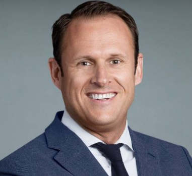 Philipp Leucht合作论文数Department of Cell Biology, Grossman School of Medicine, New York University3
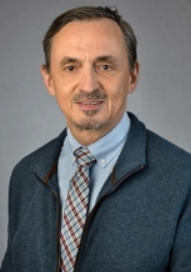 Jean-Pierre Saint-Jeannet合作论文数Department of Molecular Pathobiology, College of Dentistry, New York University3
 Kevin J. Peterson合作论文数Department of Biological Sciences, Dartmouth College3
 L. Henry Goodnough合作论文数Department of Pathology, Case Western Reserve University3Salomone Joseph R合作论文数3Emily R. Nelson合作论文数3
 Marco Trizzino合作论文数Dipartimento di Biologia Animale e Dell’Uomo, Università di Roma3
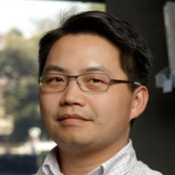 Howard Y. Chang合作论文数Chang Lab, Stanford University3Brian Gebelein合作论文数3Harald Mikkers合作论文数3Benjamin Levi合作论文数3David E Clouthier合作论文数Department of Cranio. Biol., Univ. of Colorado Denver3
 Ruchi Bajpai合作论文数Centre for Cellular and Molecular Biology, Uppal Road, Hyderabad, 500 007, India3
 Ammar Husami合作论文数2John Snedeker合作论文数2David A Billmire合作论文数2Milene Donlin合作论文数2Megan Aarnio-Peterson合作论文数2




