Many patients with urinary obstruction have an accelerated decline in renal function despite early urologic interventions. They also have defects in urinary concentrating capacity that may predispose them to acute kidney injury and chronic kidney disease. Because urinary concentrating capacity depends on having intact solute concentrating mechanisms in the renal medulla, it is likely that this results from obstruction-induced abnormalities in renal medullary structure, function, or both. This review focuses on findings from a recent study characterizing the long-term effects of reversible unilateral ureteral obstruction in mice that address these questions. These findings show that there is delayed long-term growth of the inner medulla, which is initially shrunken, that results in complete restoration of inner medullary size and gross tissue architecture 3 months after reversal of the obstruction. More detailed analysis shows that despite relatively normal histologic appearances, there are permanent defects in the cellular organization and function of the medulla that account for the loss of urinary concentrating capacity. We discuss the extent to which regenerative versus nonregenerative repair mechanisms may contribute to the growth and functional recovery of the renal medulla and consider how these findings shed light on the mechanisms of tissue repair and dysfunction after reversal of urinary obstruction.
Inflammatory diseases contribute to secondary osteoporosis. Hypertension is a highly prevalent inflammatory condition that is clinically associated with reduced bone mineral density and increased risk of fragility fracture. In this study, we showed that a significant loss in bone mass and strength occurs in two preclinical models of hypertension. This accompanied increases in immune cell populations, including monocytes, macrophages, and IL-17A-producing T cell subtypes in the bone marrow of hypertensive mice. Neutralizing IL-17A in angiotensin II-infused mice blunted hypertension-induced loss of bone mass and strength as a result of decreased osteoclastogenesis. Likewise, the inhibition of the CSF1 receptor blunted loss of bone mass and prevented loss of bone strength in hypertensive mice. In an analysis of UK Biobank data, circulating bone remodeling markers exhibited striking associations with blood pressure and bone mineral density in more than 27,000 humans. These findings illustrate a potential mechanism by which hypertension activates immune cells in the bone marrow, encouraging osteoclastogenesis and eventual loss in bone mass and strength.
Urinary obstruction causes injury to the renal medulla, impairing the ability to concentrate urine and increasing the risk of progressive kidney disease. However, the regenerative capacity of the renal medulla after reversal of obstruction is poorly understood. To investigate this, we developed a mouse model of reversible urinary obstruction. Despite robust regeneration and complete histological recovery of the renal medulla, these mice exhibited a permanent defect in urinary concentrating capacity. However, there were lasting changes in the composition, organization, and transcriptional profiles of epithelial, endothelial, and interstitial cells. Persistent inflammatory responses were also seen in patients with renal stone disease, but there were also adaptive responses to the increasingly hypoxic environment of the renal medulla that occurred only after reversal of obstruction. These findings indicate that while partial repair occurs after reversal of urinary obstruction, there are lasting structural and functional changes across all major cellular compartments of the renal medulla. These changes reflect shared and distinct responses to different renal medullary injuries in humans and mice.
Urinary obstruction causes injury to the renal papilla and leads to defects in the ability to concentrate urine which predisposes to progressive kidney injury. However, the regenerative capacity of the papilla after reversal of obstruction is poorly understood. To address this, we developed a mouse model of reversible urinary obstruction which is characterized by extensive papillary injury, followed by a robust regeneration response and complete histological recovery over a 3- month period. However, these mice have a pronounced defect in urinary concentrating capacity. We now show that this is due to permanent changes in the composition, organization, and transcriptional signatures of epithelial, endothelial, and interstitial cell lineages in the papilla. There are persistent inflammatory responses that are also seen in patients with renal stone disease but are associated with cell-specific adaptive responses to the increasingly hypoxic environment of the papilla after reversal of obstruction. Taken together, our analysis of a new model of reversible urinary obstruction reveals that partial repair leads to permanent changes in the structure and function of all of the major cellular compartments in the papilla that include both shared and distinct responses to different types of renal papillary injury in humans and mice. Summary:Partial repair after reversal of urinary obstruction leads to permanent changes in structure and function of all major cellular compartments in the renal papilla.
Kidney tubules use fatty acid oxidation (FAO) to support their high energetic requirements. Carnitine palmitoyltransferase 1A (CPT1A) is the rate-limiting enzyme for FAO, and it is necessary to transport long-chain fatty acids into mitochondria. To define the role of tubular CPT1A in aging and injury, we generated mice with tubule-specific deletion of Cpt1a (Cpt1aCKO mice), and the mice were either aged for 2 years or injured by aristolochic acid or unilateral ureteral obstruction. Surprisingly, Cpt1aCKO mice had no significant differences in kidney function or fibrosis compared with wild-type mice after aging or chronic injury. Primary tubule cells from aged Cpt1aCKO mice had a modest decrease in palmitate oxidation but retained the ability to metabolize long-chain fatty acids. Very-long-chain fatty acids, exclusively oxidized by peroxisomes, were reduced in kidneys lacking tubular CPT1A, consistent with increased peroxisomal activity. Single-nuclear RNA-Seq showed significantly increased expression of peroxisomal FAO enzymes in proximal tubules of mice lacking tubular CPT1A. These data suggest that peroxisomal FAO may compensate in the absence of CPT1A, and future genetic studies are needed to confirm the role of peroxisomal β-oxidation when mitochondrial FAO is impaired.
BACKGROUND: Renal ischemia and reperfusion (IR) contribute to perioperative acute kidney injury, and oxygen is a key regulator of this process. We hypothesized that oxygen administration during surgery and renal IR would impact postoperative kidney function and injury in mice. METHODS: Mice were anesthetized, intubated, and mechanically ventilated with a fraction of inspired oxygen (Fio2) 0.10 (hypoxia), 0.21 (normoxia), 0.60 (moderate hyperoxia), or 1.00 (severe hyperoxia) during 67 minutes of renal IR or sham IR surgery. Additional mice were treated before IR or sham IR surgery with 50 mg/kg tempol, a superoxide scavenger. At 24 hours, mice were sacrificed, and blood and kidney collected. We assessed and compared kidney function and injury across groups by measuring blood urea nitrogen (BUN, primary end point), renal histological injury, renal expression of neutrophil gelatinase–associated lipocalin (NGAL), and renal heme oxygenase 1 (Ho-1), peroxisome proliferator–activated receptor gamma coactivator 1-α (Pgc1-α), and glutathione peroxidase 4 (Gpx-4) transcripts, to explore potential mechanisms of any effect of oxygen. RESULTS: Hyperoxia and hypoxia during renal IR surgery decreased renal function and increased kidney injury compared to normoxia. Baseline median (interquartile range) BUN was 22.2 mg/dL (18.4–26.0), and 24 hours after IR surgery, BUN was 17.5 mg/dL (95% confidence interval [CI], 1.3–38.4; P = .034) higher in moderate hyperoxia–treated animals, 51.8 mg/dL (95% CI, 24.9–74.8; P < .001) higher in severe hyperoxia–treated animals, and 64.9 mg/dL (95% CI, 41.2–80.3; P < .001) higher in hypoxia-treated animals compared to animals treated with normoxia (P < .001, overall effect of hyperoxia). Hyperoxia-induced injury, but not hypoxia-induced injury, was attenuated by pretreatment with tempol. Histological injury scores, renal NGAL staining, and renal transcription of Ho-1 and suppression of Pgc1-α followed the same pattern as BUN, in relation to the effects of oxygen treatment. CONCLUSIONS: In this controlled preclinical study of oxygen treatment during renal IR surgery, hyperoxia and hypoxia impaired renal function, increased renal injury, and impacted expression of genes that affect mitochondrial biogenesis and antioxidant response. These results might have implications for patients during surgery when high concentrations of oxygen are frequently administered, especially in cases involving renal IR.
Retinoic acid receptor (RAR) signaling is essential for mammalian kidney development but, in the adult kidney, is restricted to occasional collecting duct epithelial cells. We now show that there is widespread reactivation of RAR signaling in proximal tubular epithelial cells (PTECs) in human sepsis-associated acute kidney injury (AKI) and in mouse models of AKI. Genetic inhibition of RAR signaling in PTECs protected against experimental AKI but was unexpectedly associated with increased expression of the PTEC injury marker Kim1. However, the protective effects of inhibiting PTEC RAR signaling were associated with increased Kim1-dependent apoptotic cell clearance, or efferocytosis, and this was associated with dedifferentiation, proliferation, and metabolic reprogramming of PTECs. These data demonstrate the functional role that reactivation of RAR signaling plays in regulating PTEC differentiation and function in human and experimental AKI.
Pretreatment with 3-deacyl 6-acyl phosphorylated hexaacyl disaccharide; a novel synthetic Toll-like receptor 4 agonist, preserves kidney function during ischemia-reperfusion injury-induced acute kidney injury.
Tubular atrophy and fibrosis are pathological changes that determine the prognosis of kidney disease induced by acute kidney injury (AKI). We aimed to evaluate multiple magnetic resonance imaging (MRI) parameters, including pool size ratio (PSR) from quantitative magnetization transfer, relaxation rates, and measures from spin-lock imaging ( R 1 ρ and S ρ ), for assessing the pathological changes associated with AKI-induced kidney disease. Eight-week-old male C57BL/6 J mice first underwent unilateral ischemia reperfusion injury (IRI) induced by reperfusion after 45 min of ischemia. They were imaged using a 7T MRI system 56 days after the injury. Paraffin tissue sections were stained using Masson trichrome and picrosirius red to identify histopathological changes such as tubular atrophy and fibrosis. Histology detected extensive tubular atrophy and moderate fibrosis in the cortex and outer stripe of the outer medulla (CR + OSOM) and more prominent fibrosis in the inner stripe of the outer medulla (ISOM) of IRI kidneys. In the CR + OSOM region, evident decreases in PSR, R 1 , R 2 , R 1 ρ , and S ρ showed in IRI compared with contralateral kidneys, with PSR and S ρ exhibiting the most significant changes. In addition, the exchange parameter S ρ dropped by the largest degree among all the MRI parameters, while R 2 * increased significantly. In the ISOM of IRI kidneys, PSR increased while S ρ kept decreasing. R 2 , R 1 ρ , and R 2 * all increased due to more severe fibrosis in this region. Among MRI measures, PSR and R 1 ρ showed the highest detectability of renal changes no matter whether tubular atrophy or fibrosis dominated. R 2 * and S ρ could be more specific to a single pathological event than other MRI measures because only R 2 * increased and S ρ decreased consistently when either fibrosis or tubular atrophy dominated, and their correlations with fibrosis scores were higher than other MRI measures. Multiparametric MRI may enable a more comprehensive analysis of histopathological changes following AKI.
Acute kidney injury (AKI) is a strong independent predictor of mortality and often results in incomplete recovery of renal function, leading to progressive chronic kidney disease (CKD). Many clinical trials have been conducted on the basis of promising preclinical data, but no therapeutic interventions have been shown to improve long-term outcomes after AKI. This is partly due to the failure of preclinical studies to accurately model clinically relevant injury and long-term outcomes on CKD progression. Here, we evaluated the long-term effects of AKI on CKD progression in three animal models reflecting diverse etiologies of AKI: repeat-dose cisplatin, rhabdomyolysis, and ischemia-reperfusion injury. Using transdermal measurement of glomerular filtration rate as a clinically relevant measure of kidney function and quantification of peritubular capillary density to measure capillary rarefaction, we showed that repeat-dose cisplatin caused capillary rarefaction and decreased renal function in mice without a significant increase in interstitial fibrosis, whereas rhabdomyolysis-induced AKI led to severe interstitial fibrosis, but renal function and peritubular capillary density were preserved. Furthermore, long-term experiments in mice with unilateral ischemia-reperfusion injury showed that restoration of renal function 12 wk after a contralateral nephrectomy was associated with increasing fibrosis, but a reversal of capillary rarefaction was seen at 4 wk. These data demonstrate that clear dissociation between kidney function and fibrosis in these models of AKI to CKD progression and suggest that peritubular capillary rarefaction is more strongly associated with CKD progression than renal fibrosis.
Severe acute kidney injury has a high mortality and is a risk factor for progressive chronic kidney disease. None of the potential therapies that have been identified in preclinical studies have successfully improved clinical outcomes. This failure is partly because animal models rarely reflect the complexity of human disease: most preclinical studies are short term and are commonly performed in healthy, young, male mice. Therapies that are effective in preclinical models that share common clinical features seen in patients with acute kidney injury, including genetic diversity, different sexes, and comorbidities, and evaluate long-term outcomes are more likely to predict success in the clinic. Here, we evaluated susceptibility to chronic kidney disease after ischemia-reperfusion injury with delayed nephrectomy by monitoring long-term functional and histological responses to injury. We defined conditions required to induce long-term postinjury renal dysfunction and fibrosis without increased mortality in a reproducible way and evaluate effect of mouse strains, sexes, and preexisting diabetes on these responses.
 Mark de Caestecker合作论文数16
 Hai-Chun Yang合作论文数Fu Dan University Medical Center, Shanghai Medical University8
 Maya Brewer合作论文数7Anna Menshikh合作论文数5Lauren Scarfe合作论文数4
 Gewin Leslie合作论文数4Lauren N Lopez合作论文数3Yuantee Zhu合作论文数3
 Lauren Himmel合作论文数3
 Julia K. Bohannon合作论文数Vanderbilt University Medical Center3Patil Naeem K合作论文数M.) and Department of Pathology (N.G.),, University of Arkansas for Medical Sciences3
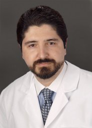 Hernandez Antonio合作论文数Division of Critical Care Medicine and Cardiothoracic Anesthesiology Department of Anesthesiology, Vanderbilt University Medical Center3
 Alan Davidson合作论文数Department of Molecular Medicine and Pathology, School of Medical Sciences, Faculty of Medical and Health Sciences, The University of Auckland3
 Veronika Sander合作论文数Department of Molecular Medicine and Pathology, School of Medical Sciences, Faculty of Medical and Health Sciences, The University of Auckland3Thitinee Vanichapol合作论文数3
 Edward R. Sherwood合作论文数Department of Anesthesiology;Medical Branch, University of Texas;Shriners Hospital for Children;Shriners Hospital for Children, University of Texas3
 Min Yang (杨敏)合作论文数Guangdong Provincial People's Hospital3Yosuke Osaki合作论文数2Charlene Finney合作论文数2Louise Lantier合作论文数2
 Raymond C. Harris合作论文数Vanderbilt Institute for Infection, Immunology and Inflammation, Vanderbilt University Medical Center;Division of Nephrology, Vanderbilt University Medical Center2
 Mark P. De Caestecker合作论文数Department of Medicine, Vanderbilt University2Chris R Kennedy合作论文数1Jane Tonello合作论文数1Melissa J Kimlinger合作论文数1Eric H. Mace合作论文数1Michael D. McDaniels合作论文数1Olga M Viquez合作论文数1Justin T Kern合作论文数1
 Jams Tom合作论文数1
 Feng Wang合作论文数1
 Allison M Owen合作论文数1Nidia C Messias合作论文数1Elizabeth Hennen合作论文数Dept Biomed Engn, Vanderbilt Univ1
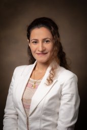 Fatemeh Adelnia合作论文数Vanderbilt University Institute of Imaging Science, Vanderbilt University Medical Center;Department of Radiology and Radiological Sciences, Vanderbilt University Medical Center;Vanderbilt University Medical Center, Vanderbilt University Medical Center1Keith Long合作论文数1Wei Chen合作论文数1
 Dongliang Lu合作论文数1Mateusz Siedlinski合作论文数1
 Nestor de la Visitacion合作论文数1
 Alla V Ivanova合作论文数1
 Nyman Jeffry S合作论文数Dept Orthopaed Surg & Rehabil, Vanderbilt Univ1
 Tadashi Otsuka合作论文数1
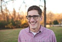 Harkins Kevin D合作论文数1
 Frederic Billings合作论文数Department of Anesthesiology, Vanderbilt University1
 John C. Gore合作论文数Department of Radiology, Vanderbilt University Medical Center;Department of Molecular Physiology and Biophysics, School of Medicine, Vanderbilt University1
 Irfan J Lodhi合作论文数Division of Endocrinology, Metabolism & Lipid Research, Washington University School of Medicine1
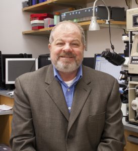 David H. Wasserman合作论文数Department of Molecular Physiology & Biophysics, School of Medicine, Vanderbilt University;Mouse Metabolic Phenotyping Center, School of Medicine, Vanderbilt University1
 Uppuganti Sasidhar合作论文数Department of Orthopaedic Surgery & Rehabilitation, Vanderbilt University Medical Center1




