The macromolecular crystallography (MX) beamline I04 [ref 1] at Diamond has evolved over time through various upgrade projects that aimed at increasing scientific capability but at the same time aiming for increased stability so that the best possible data can be obtained by the user. We have implemented an optical concept for beam delivery combining simplicity with stability. Variable focus beam is provided through the combination of a double crystal monochromator (DCM) with a F-switch which houses compound refractive lenses (CRL) that can be brought individually into the beam path, providing maximum flexibility. Both devices were designed inhouse and this combination allows delivery of a very stable beam from the microfocus regime (8 μm x 5 μm (h x v)) to larger beam sizes (up to 110 μm x 100 μm). Beam delivery within 3% RMS of the beamsize is achieved by making use of a dedicated feedback system using X-ray beam position monitors (XBPMs) [ref 2]. The original X-ray source has been replaced by a 17.6 mm period CPMU in June 2022 and has resulted in a significant flux increase over the whole energy range (6-18 keV) thereby generating new scientific opportunities.Optimal data collection in MX strongly depends on setting up the right data collection parameters which should be defined by the experimental aim or scientific question that is being asked. With high intensity beamlines radiation damage has become the primary limitation and therefore the total exposure of the sample plays a key role. It became soon clear that with the combination of the high variability of the flux profile from the source combined with the large variability of beam sizes, that the concept of exposure per data collection frame is no longer feasible. Therefore, we have implemented the concept of dose aware data collection [ref 3] where the user is given the option to dial a dose per data set (instead of an exposure time per frame) and this dose should of course be compatible with the experimental aim. We use the programme RADDOSE-3D [ref 4, 5] which takes known information from the beamline (energy, flux, beam size) and currently assumes a standard macromolecular crystal which means that the sample only contains lighter elements and combines this information in the dose calculation to produce optimal exposure times per frame and adjustment of transmission if required. Future improvements will consider better information about sample composition and size. In most cases, we aim for the shortest possible exposure time to take advantage of the Eiger2 XE 16M detector capabilities which allows acquisition rates up to 500 Hz. Depending on the experimental aim we usually implement a multi-sweep (and often multi-crystal) approach [ref 6] using different crystal orientations which can be realised with the SmarGon multi-axis goniometer. The dose-based approach is also fully implemented in our unattended data collection (UDC) protocols. Apart from the UDC and remote interactive modes we also strongly encourage in person visits to train users in best practice dose aware data collection and enabling them to make the best choices using the available tools in the data collection software. We constantly aim to streamline the user experience further by addition of new functionality and tools.
Optimal data collection in macromolecular crystallography (MX) strongly depends on setting the appropriate data collection parameters which should be guided by the experimental aim and scientific question being asked. While the goal is to maximize signal-to-noise, this must be weighed against the risk of radiation damage to the sample. Due to the diverse optical setups resulting in varying flux with energy and beam size, determining optimal exposure times can be a complex task, particularly for inexperienced users. Here we present the concept and implementation of dose aware data collection, its aim to facilitate the choice of data collection parameters for MX experiments and how transformative this approach is particularly for a beamline where changing beam size is standard operation and regularly used down to microfocus level.
Aflatoxin B1 (AFB1) is one of the most potent carcinogens and a widespread food and feed contaminant. As for other toxins, many efforts are devoted to find efficient and environmentally-friendly methods to degrade AFB1, such as enzymatic treatments, thus improving the safety of food and feed products. In this regard, the dye decolorizing peroxidase of type B (DypB) can efficiently degrade AFB1. The molecular mechanism, which is required to drive protein optimization in view of the usage of DypB as a mycotoxin reduction agent in large scale application, is unknown. Here, we focused on the role of four DypB residues in the degradation of AFB1 by alanine-scanning (residues 156, 215, 239 and 246), which were identified from biochemical assays to be kinetically relevant for the degradation. As a result of DypB degradation, AFB1 is converted into four products. Interestingly, the relative abundancy of these products depends on the replaced residues. Molecular dynamics simulations were used to investigate the role of these residues in the binding step between protein and manganese, a metal ion which is expected to be involved in the degradation process. We found that the size of the haem pocket as well as conformational changes in the protein structure could play a role in determining the kinetics of AFB1 removal and, consequently, guide the process towards specific degradation products.
Diamond Light Source currently operates seven beamlines for macromolecular crystallography [1]. I04 [2,3] is an energy (6-18 keV) and beam size (5-100 microns) tuneable microfocus beamline suitable for projects ranging from large scale high throughput ligand screening to difficult single or multiple anomalous dispersion experiments. These large spectrum capabilities are built on top state of the art hardware equipment and software developments. The hardware highlights are a multi-axis goniometer for crystal alignment and X-ray centring, a large and very fast frame rate pixel array detector for diffraction data collection and the use of compound refractive index lenses for focusing. The software stack includes the diamond wide data acquisition and GUI software GDA [4-5] as well as the SyncWeb interface [6] for ISPYB [7]. Collecting at its full potential I04 can load a sample, centre it automatically using X-ray diffraction at two different orientations and collect a 7.2 second rotation dataset of 360 degrees in less than 2 minutes providing on average over 32 samples throughput per hour. I04 is also part of the recent Diamond MX beamline developments on the use of Unattended Data Collection (UDC) [8] where the only interaction of the users with the beamline is done prior to the beamtime on defining how do they want to collect their data based on a series of pre-set recipes (unpublished). In addition to UDC, I04 provides onsite and remote access capabilities and has special tools like built-in Raddose3D [9] calculator to help design experiments for optimal data. Here we present a brief overview of the beamline automation in data collection including the recent enhancements on using dose to increase efficiency on both the unattended data collection as well as driving queues of experiments dialling dose instead of an exposure (Figure 1, Figure 2). The latter enhancement was critical
The macromolecular crystallography beamline I04 at Diamond [1] is a versatile variable and microfocus beamline aiming to provide the best quality diffraction data from crystals of macromolecules mainly at cryo temperatures.Beam delivery is achieved through the combination of a double crystal monochromator (DCM) with a F-switch which houses compound refractive lenses (CRL) that can be brought individually into the beam path.Both devices were designed inhouse and this combination allows variable focus of a very stable beam from the microfocus regime (8 µm x 5 µm (h x v)) to larger beam sizes (up to 110 µm x 100 µm) over the whole energy range of 6-18 keV.Beam delivery within 3% RMS of the beam size is achieved by making use of a dedicated feedback system using X-ray beam position monitors (XBPMs).In June 2022 the original U23 insertion device has been replaced with a cryocooled permanent magnet undulator (CPMU) and this has resulted in a significant flux increase which has opened up new opportunities, including faster data collection and the ability to address more challenging data collections, in particular for microcrystallography. Related to the insertion device upgrade we are carefully monitoring any heat load effects on the optics and are further characterising the performance of our F-switch device with the aim to optimise beam delivery.
Diamond Light Source currently operates seven beamlines for macromolecular crystallography [1].I04 [2,3] is an energy (6-18 keV) and beam size (5-100 microns) tuneable microfocus beamline suitable for projects ranging from large scale high throughput ligand screening to difficult single or multiple anomalous dispersion experiments.These large spectrum capabilities are built on top state of the art hardware equipment and software developments.The hardware highlights are a multi-axis goniometer for crystal alignment and X-ray centring, a large and very fast frame rate pixel array detector for diffraction data collection and the use of compound refractive index lenses for focusing.The software stack includes the diamond wide data acquisition and GUI software GDA [4][5] as well as the SyncWeb interface [6] for ISPYB [7].Collecting at its full potential I04 can load a sample, centre it automatically using X-ray diffraction at two different orientations and collect a 7.2 second rotation dataset of 360 degrees in less than 2 minutes providing on average over 32 samples throughput per hour.I04 is also part of the recent Diamond MX beamline developments on the use of Unattended Data Collection (UDC) [8] where the only interaction of the users with the beamline is done prior to the beamtime on defining how do they want to collect their data based on a series of pre-set recipes (unpublished).In addition to UDC, I04 provides onsite and remote access capabilities and has special tools like built-in Raddose3D [9] calculator to help design experiments for optimal data.Here we present a brief overview of the beamline automation in data collection including the recent enhancements on using dose to drive data collection.The latter allows both the unattended data collection as well as queues of experiments dialling dose instead of an exposure (Figure 1, Figure 2).This enhancement is vital to open the automation avenue but also training of new generations on how to collect data without using an urban legend type of knowledge that passes from older to younger generation orally.Its major benefit is that it permits, driven from a mix of prior known and live data, not to compromise on radiation damage as well as not under exposing a diffracting crystal.
Raw diffraction data for mpro-x1187 / PDB ID 5RFA (see: https://www.ebi.ac.uk/pdbe/entry/pdb/5RFA) - SARS-CoV-2 main protease in complex with Z2643472210 (SMILES:CN1C=CC(=N1)C(=O)NC[C@@H]2CCCO2) collected as part of an XChem crystallographic fragment screening campaign on beamline i04-1 at Diamond Light Source. The deposited structure was automatically processed with standard Diamond tools and PanDDA, however the raw data are being made available to allow reanalysis by any interested party. For more details see: https://www.diamond.ac.uk/covid-19/for-scientists/Main-protease-structure-and-XChem.html
Raw diffraction data for mpro-x1374 / PDB ID 5RFM (see: https://www.ebi.ac.uk/pdbe/entry/pdb/5RFM) - SARS-CoV-2 main protease in complex with PCM-0102539 (SMILES:Cc1ccc(cc1)N(C2CS(=O)(=O)C=C2)C(=O)CCl) collected as part of an XChem crystallographic fragment screening campaign on beamline i04-1 at Diamond Light Source. The deposited structure was automatically processed with standard Diamond tools and PanDDA, however the raw data are being made available to allow reanalysis by any interested party. For more details see: https://www.diamond.ac.uk/covid-19/for-scientists/Main-protease-structure-and-XChem.html
Raw diffraction data for mpro-x0749 / PDB ID 5REN (see: https://www.ebi.ac.uk/pdbe/entry/pdb/5REN) - SARS-CoV-2 main protease in complex with PCM-0102425 (SMILES:ClCC(=O)N1CCCC(C1)c2nc3ccccc3s2) collected as part of an XChem crystallographic fragment screening campaign on beamline i04-1 at Diamond Light Source. The deposited structure was automatically processed with standard Diamond tools and PanDDA, however the raw data are being made available to allow reanalysis by any interested party. For more details see: https://www.diamond.ac.uk/covid-19/for-scientists/Main-protease-structure-and-XChem.html
Raw diffraction data for mpro-x0305 / PDB ID 5R82 (see: https://www.ebi.ac.uk/pdbe/entry/pdb/5R82) - SARS-CoV-2 main protease in complex with Z219104216 (SMILES:CCNC=1C=CC(C#N)=CN1) collected as part of an XChem crystallographic fragment screening campaign on beamline i04-1 at Diamond Light Source. The deposited structure was automatically processed with standard Diamond tools and PanDDA, however the raw data are being made available to allow reanalysis by any interested party. For more details see: https://www.diamond.ac.uk/covid-19/for-scientists/Main-protease-structure-and-XChem.html
Raw diffraction data for mpro-x1336 / PDB ID 5RFI (see: https://www.ebi.ac.uk/pdbe/entry/pdb/5RFI) - SARS-CoV-2 main protease in complex with PCM-0102353 (SMILES:Cc1ccc(C)c(c1)S(=O)(=O)N2CCN(CC2)C(=O)CCl) collected as part of an XChem crystallographic fragment screening campaign on beamline i04-1 at Diamond Light Source. The deposited structure was automatically processed with standard Diamond tools and PanDDA, however the raw data are being made available to allow reanalysis by any interested party. For more details see: https://www.diamond.ac.uk/covid-19/for-scientists/Main-protease-structure-and-XChem.html
Raw diffraction data for mpro-x0769 / PDB ID 5RES (see: https://www.ebi.ac.uk/pdbe/entry/pdb/5RES) - SARS-CoV-2 main protease in complex with PCM-0102281 (SMILES:Fc1ccccc1S(=O)(=O)N2CCN(CC2)C(=O)CCl) collected as part of an XChem crystallographic fragment screening campaign on beamline i04-1 at Diamond Light Source. The deposited structure was automatically processed with standard Diamond tools and PanDDA, however the raw data are being made available to allow reanalysis by any interested party. For more details see: https://www.diamond.ac.uk/covid-19/for-scientists/Main-protease-structure-and-XChem.html
Raw diffraction data for mpro-x0752 / PDB ID 5REO (see: https://www.ebi.ac.uk/pdbe/entry/pdb/5REO) - SARS-CoV-2 main protease in complex with PCM-0102578 (SMILES:ClCC(=O)NCc1ccc2OCOc2c1) collected as part of an XChem crystallographic fragment screening campaign on beamline i04-1 at Diamond Light Source. The deposited structure was automatically processed with standard Diamond tools and PanDDA, however the raw data are being made available to allow reanalysis by any interested party. For more details see: https://www.diamond.ac.uk/covid-19/for-scientists/Main-protease-structure-and-XChem.html
Raw diffraction data for mpro-x1226 / PDB ID 5RFB (see: https://www.ebi.ac.uk/pdbe/entry/pdb/5RFB) - SARS-CoV-2 main protease in complex with Z1271660837 (SMILES:CCNCC1=CN(C)N=N1) collected as part of an XChem crystallographic fragment screening campaign on beamline i04-1 at Diamond Light Source. The deposited structure was automatically processed with standard Diamond tools and PanDDA, however the raw data are being made available to allow reanalysis by any interested party. For more details see: https://www.diamond.ac.uk/covid-19/for-scientists/Main-protease-structure-and-XChem.html
Raw diffraction data for mpro-x0376 / PDB ID 5REA (see: https://www.ebi.ac.uk/pdbe/entry/pdb/5REA) - SARS-CoV-2 main protease in complex with Z31432226 (SMILES:O=C(N1CCCCCC1)C=2C=CC=3OCOC3C2) collected as part of an XChem crystallographic fragment screening campaign on beamline i04-1 at Diamond Light Source. The deposited structure was automatically processed with standard Diamond tools and PanDDA, however the raw data are being made available to allow reanalysis by any interested party. For more details see: https://www.diamond.ac.uk/covid-19/for-scientists/Main-protease-structure-and-XChem.html
Raw diffraction data for mpro-x1119 / PDB ID 5RF8 (see: https://www.ebi.ac.uk/pdbe/entry/pdb/5RF8) - SARS-CoV-2 main protease in complex with Z271004858 (SMILES:NC=1C=CC(=CC1)S(=O)(=O)NC=2C=CC=CN2) collected as part of an XChem crystallographic fragment screening campaign on beamline i04-1 at Diamond Light Source. The deposited structure was automatically processed with standard Diamond tools and PanDDA, however the raw data are being made available to allow reanalysis by any interested party. For more details see: https://www.diamond.ac.uk/covid-19/for-scientists/Main-protease-structure-and-XChem.html
Raw diffraction data for mpro-x0194 / PDB ID 5RE6 (see: https://www.ebi.ac.uk/pdbe/entry/pdb/5RE6) - SARS-CoV-2 main protease in complex with Z54571979 (SMILES:CC(=O)NC=1C=CC(OC=2N=CC=CN2)=CC1) collected as part of an XChem crystallographic fragment screening campaign on beamline i04-1 at Diamond Light Source. The deposited structure was automatically processed with standard Diamond tools and PanDDA, however the raw data are being made available to allow reanalysis by any interested party. For more details see: https://www.diamond.ac.uk/covid-19/for-scientists/Main-protease-structure-and-XChem.html
Raw diffraction data for mpro-x1375 / PDB ID 5RFN (see: https://www.ebi.ac.uk/pdbe/entry/pdb/5RFN) - SARS-CoV-2 main protease in complex with PCM-0102868 (SMILES:Fc1ccc(cc1)N(C2CS(=O)(=O)C=C2)C(=O)CCl) collected as part of an XChem crystallographic fragment screening campaign on beamline i04-1 at Diamond Light Source. The deposited structure was automatically processed with standard Diamond tools and PanDDA, however the raw data are being made available to allow reanalysis by any interested party. For more details see: https://www.diamond.ac.uk/covid-19/for-scientists/Main-protease-structure-and-XChem.html
 Katherine McAuley合作论文数CCLRC Daresbury Laboratory37
 Winter Graeme合作论文数Diamond Light Source Ltd31
 Marco Mazzorana合作论文数Venetian Institute for Molecular Medicine–VIMM30
 Dave Hall合作论文数27David Aragão合作论文数26
 Peter Luger合作论文数Institut für Chemie und Biochemie, Freie Universität Berlin26
 Crawshaw Adam D合作论文数Institute for Cell and Molecular Biosciences, Newcastle University25Petra Lukacik合作论文数Structural Genomics Consortium, University of Oxford25Alexandre M.M. Dias合作论文数24Anna Carbery合作论文数24Conor Wild合作论文数24Efrat Resnick合作论文数Weizmann Inst Sci, Dept Organ Chem, IL-7610001 Rehovot, Israel24Claire Strain-Damerell合作论文数Diamond Light Source Ltd., Harwell Science and Innovation Campus24
 Martin de Porres Walsh合作论文数European Molecular Biology Laboratory, c/o DESY Notkestrasse 85, D-22603 Hamburg, Germany24
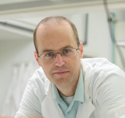 Nir London合作论文数Institute for Medical Research IMRIC, The Hebrew University24C David Owen合作论文数Univ St Andrews, Biomol Sci Bldg, St Andrews KY16 9ST, Fife, Scotland23R. Skyner合作论文数20Matt Snee合作论文数20D. Fearon合作论文数20A.J. Powell合作论文数20Louise Dunnett合作论文数Diamond Light Source Ltd, Harwell Sci & Innovat Campus, Didcot OX11 0QX, Oxon, England20
 Paul Gehrtz合作论文数Weizmann Inst Sci, Dept Organ Chem, IL-7610001 Rehovot, Israel20
 Reddi Rambabu N合作论文数Chemical Engineering and Process Development Division, National Chemical Laboratory (CSIR),20J. Brandao-Neto合作论文数17Dave Stuart合作论文数17Tibor S Koritsanszky合作论文数17
 Birger Dittrich合作论文数Faculty of Mathematics and Natural Sciences, University of Zurich12
 Wagner Armin合作论文数10
 D. R. Hall合作论文数9
 Thomas Lykke-Møller Sørensen合作论文数9Pierpaolo Romano合作论文数8Ashton Alun W合作论文数8
 Dino Moras合作论文数Department of Integrative Structural Biology, Institut de Génétique et de Biologie Moléculaire et Cellulaire8James Sandy合作论文数Department of Pharmacology, University of Oxford8
 Gwyndaf Evans合作论文数Center for Mechanistic Biology and Biotechnology;Argonne National Laboratory;Center for Mechanistic Biology and Biotechnology, Argonne National Laboratory8Holger Greschik合作论文数7Geoff Preece合作论文数7
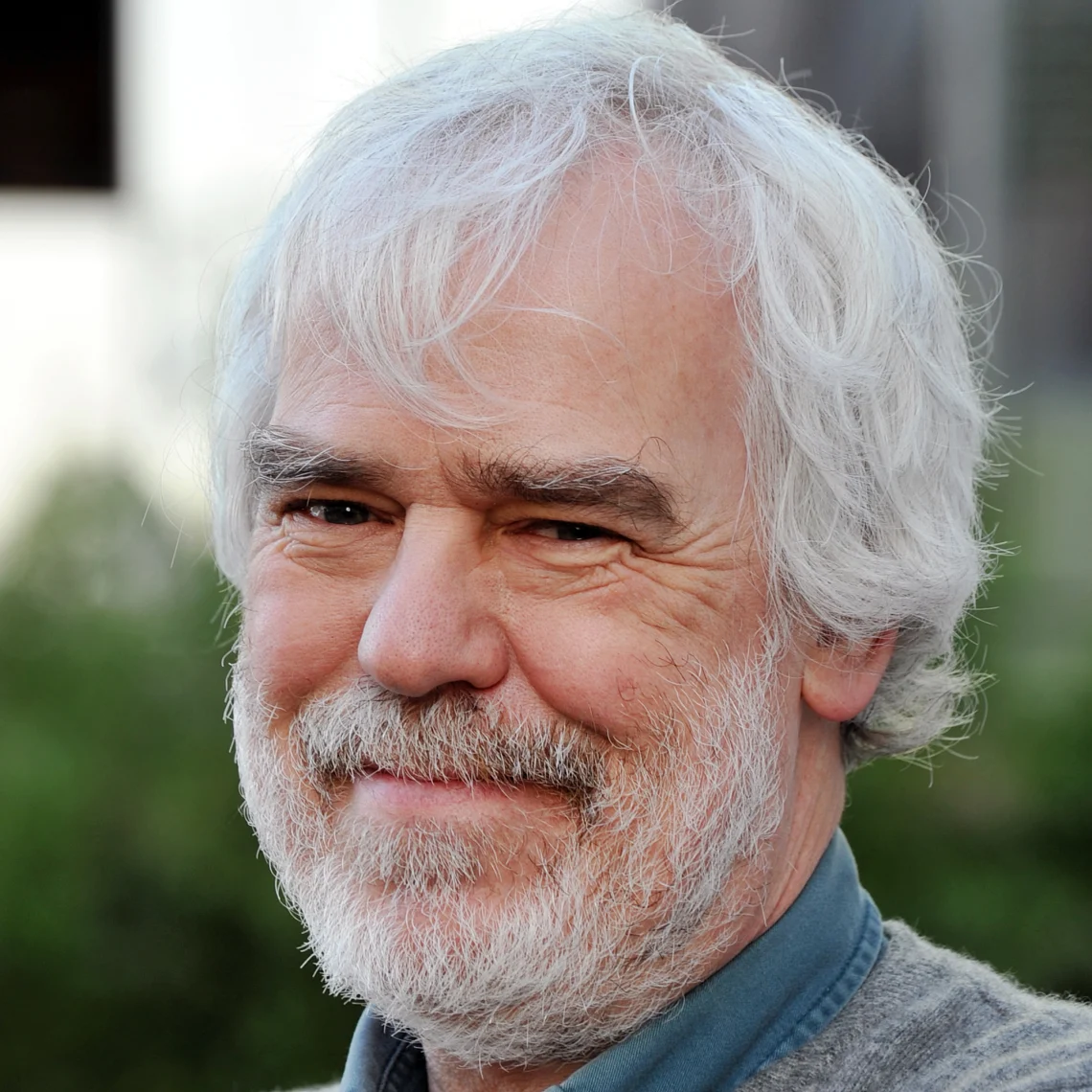 David Stuart合作论文数Division of Structural Biology, Nuffield Department of Medicine, University of Oxford7H. G. Krane合作论文数6
 Dieter Zobel合作论文数5D. Aragao合作论文数5
 David Geoffrey Waterman合作论文数York Structural Biology Laboratory, University of York5Mark Williams合作论文数4F. von Delft合作论文数4
 Jose Brandao-neto合作论文数4James O'Hea合作论文数4Fearn Richard合作论文数4Duke Elizabeth合作论文数4Carole Peluso-Iltis合作论文数4




