BRCA1 -linked cancer genomes contain abundant genome-wide ∼10 kb 'Group 1' tandem duplications (TDs) that are drivers of tumorigenesis. Group 1 TD formation is recapitulated at a chromosomal Tus/ Ter site-specific replication fork barrier in DNA end resection-defective mouse embryonic stem (mES) cells lacking Brca1 exon 11. To explore relationships between DNA end resection and Group 1 TD formation, we analyzed Brca1 coiled coil (CC) domain mutants-separation-of-function alleles that are impaired for homologous recombination but competent for DNA end resection. Notably, Brca1 CC mutants retain the ability to suppress Group 1 TDs in the Tus/ Ter system and in a mouse model of Brca1 -linked tumorigenesis. These data show that Brca1 CC domain mutant cancers follow a path of tumorigenesis distinct from that of other pathogenic Brca1 alleles. FANCM is a TD co-suppressor, the loss of which is synthetic lethal/sick in combination with Brca1 exon 11 mutation. In contrast, Fancm deletion is well-tolerated by Brca1 CC mutant mES cells. Thus, Group 1 TD formation and Fancm synthetic lethality are linked phenotypes related to defective BRCA1-mediated DNA end resection.
Pancreatic ductal adenocarcinomas (PDACs) are aggressive, stroma-rich tumours. They are unresponsive to treatments, and patients relapse quickly on DNA-damaging chemotherapies. PDAC stroma consists of extracellular matrix proteins (ECM), secreted by cancer-associated fibroblasts (CAFs). Here we show an unexpected link between CAF-secreted ECM proteins and enhanced DNA repair. We identify NDRG1 (N-myc downstream-regulated gene 1) as a key mediator that senses signals from the ECM via adhesion receptors and serum and glucocorticoid-activated kinase. We establish NDRG1 as a DNA repair factor that physically associates with replication forks, maintains DNA replication, resolves stalled forks caused by chemotherapies and is involved in reducing R-loops, RNA-DNA hybrids known to cause genomic instability. NDRG1 is highly expressed in PDAC tumours and its high expression correlates with poor disease-specific survival and poor response to chemotherapy. In conclusion, our data reveal an unexpected role for CAF-secreted ECM proteins in promoting DNA repair via NDRG1, mechanistically linking tumour stroma to replication fork homeostasis and R-loop regulation.
SUMMARY Structural variants (SVs) in cancer are associated with defects in DNA repair and replication stress, but the mechanisms generating common SV types remain unresolved. We propose that large (>100 kb) tandem duplications originate through a novel sister-fork breakage–fusion mechanism. To capture replication-related context beyond breakpoints, we developed an algorithm to characterize replication timing, origin density, and fork direction across SV-spanned regions, features that refine and differentiate previously defined SV signatures. Large tandem duplications frequently overlap replication origins from which forks proceed bidirectionally; combined with independent evidence from APOBEC strand asymmetry, this pattern is compatible uniquely with the proposed mechanism. Although tandem duplications in CCNE1 -amplified and CDK12 -mutant cancers also concentrate around origins and highly transcribed genes, they display distinct contexts: CDK12 -mutant SVs arise near later-firing origins, whereas those in CCNE1 -amplified tumors often coincide with genes in specific strand configurations, suggesting different causes of fork stalling. Incorporating replication features into signature analysis enabled the discovery of new SV signatures, which we used to build SVIG, a multi-class classifier of SV phenotypes. SV signatures attributed to replication stress may help guide therapies targeting this vulnerability.
In pancreatic ductal adenocarcinoma cancer (PDAC) drug resistance is a severe clinical problem and patients relapse within a few months after receiving the standard-of-care chemotherapy. One contributing factor to treatment resistance is the desmoplastic nature of PDAC; the tumours are surrounded by thick layers of stroma composing up to 90% of the tumour mass. This stroma, which is mostly comprised of extracellular matrix (ECM) proteins, is secreted by cancer-associated fibroblasts (CAFs) residing in the tumour microenvironment. However, the mechanistic basis by which the tumour stroma directly contributes to chemoresistance remains unclear. Here, we show that CAF-secreted ECM proteins induce chemoresistance by blunting chemotherapy-induced DNA damage. Mechanistically, we identify N-myc downstream regulated gene 1 (NDRG1) as a key protein required for stroma-induced chemoresistance that responds to signals from the ECM and adhesion receptors. We further show that NDRG1 is a novel DNA repair protein that physically interacts with replication forks, maintains DNA replication and functions to resolve stalled forks caused by chemotherapy. More specifically, NDRG1 reduces R-loops, RNA-DNA hybrids that are known to cause genomic instability. R-loops occur during replication-transcription conflicts in S-phase and after chemotherapy treatments, thus posing a major threat to normal replication fork homeostasis. We identify NDRG1 as highly expressed in PDAC tumours, and its high expression correlates with chemoresistance and poor disease-specific survival. Importantly, knock-out of NDRG1 or inhibition of its phosphorylation restores chemotherapy-induced DNA damage and resensitizes tumour cells to treatment. In conclusion, our data reveal an unexpected role for CAF-secreted ECM proteins in enhancing DNA repair via NDRG1, a novel DNA repair protein, directly linking tumour stroma to replication fork homeostasis and R-loop biology, with important therapeutic implications for restoring DNA damage response pathways in pancreatic cancer. Summary paragraph:Drug resistance is a severe clinical problem in stroma-rich tumours, such as pancreatic ductal adenocarcinoma (PDAC), and patients often relapse within a few months on chemotherapy 1-9 . The stroma, comprised of extracellular matrix (ECM) proteins, is secreted by cancer-associated fibroblasts (CAFs) residing in the tumour microenvironment 10-13 . Prior work show that ECM proteins provide survival benefits to cancer cells 14,15 . However, the precise role of CAF-secreted ECM in resistance to DNA damaging chemotherapies remains poorly understood. Here, we link ECM proteins to chemoresistance by enhanced DNA damage repair (DDR). Mechanistically, we identify N-myc downstream-regulated gene 1 (NDRG1) as a key effector downstream of ECM and the integrin-Src-SGK1-signalling axis that mediates enhanced DDR. We show that NDRG1 loss, mutation of conserved His194, or inhibition of NDRG1 phosphorylation by SGK1 lead to replication fork stalling, increased R-loops, and higher transcription-replication conflicts, resulting in genomic instability and sensitivity to chemotherapies. Our analysis of PDAC patient cohorts 16 found that high NDRG1 expression correlates with chemoresistance and poor patient survival. In conclusion, we uncover an unexpected role for CAF-secreted ECM proteins in promoting therapeutic resistance by enhancing DDR and establish NDRG1 as a novel DNA repair protein directly linking tumour stroma to DDR.
Replication fork collision with a DNA nick can generate a one-ended break, fostering genomic instability. The opposing fork’s collision with the nick could form a second DNA end, enabling conservative repair by homologous recombination (HR). To study mechanisms of nickase-induced HR, we developed the Flp recombinase “step arrest” nickase in mammalian cells. A Flp-nick induces two-ended, BRCA2/RAD51-dependent short tract gene conversion (STGC), BRCA2/RAD51-independent long tract gene conversion, and discoordinated two-ended invasions. HR pathways induced by a replication-independent break and the Flp-nickase differ in their dependence on BRCA1, MRE11, and CtIP. To determine the origin of the second DNA end during Flp-nickase-induced STGC, we blocked the opposing fork using a Tus/Ter replication fork barrier (RFB). Flp-nickase-induced STGC remained robust and two ended. Thus, a single replication fork’s collision with a Flp-nick triggers two-ended HR, possibly reflecting replicative bypass of lagging strand nicks. This response may limit genomic instability during replication of nicked DNA.
Cohesin folds genomes into chromatin loops, the roles of which are under debate. We found that double-strand breaks (DSBs) induce de novo formation of chromatin loops in human cells, with the loop base positioned at the DSB site. These loops form in the S and G2 phases of the cell cycle during homologous recombination repair, concomitantly with DNA end resection and radiation-sensitive protein 51 (RAD51) recruitment. RAD51 shows a broad (megabase-sized) chromatin domain reflective of the homology search. This domain is regulated by cohesin unloader and overlaps with chromatin regions reeled through the break-anchored loop, suggesting that loop extrusion regulates the homology search. Indeed, depletion of the loop-extruding cohesin subunit NIPBL lowers homologous recombination in mouse embryonic stem cells, and this effect is more pronounced when the homologous recombination donor is hundreds of kilobases from the DSB. Our data indicate that loop-extruding cohesin promotes the mammalian homology search by facilitating break-chromatin interactions.
Replisome collision with a nicked parental DNA template can lead to the formation of a replication-associated double strand break (DSB). How this break is repaired has implications for cancer initiation, cancer therapy and therapeutic gene editing. Recent work shows that collision of a replisome with a nicked DNA template can give rise to either a single-ended (se) or a double-ended (de)DSB, with potentially divergent effects on repair pathway choice and genomic instability. Emerging evidence suggests that the biochemical environment of the broken mammalian replication fork may be specialized in such a way as to skew repair in favor of homologous recombination at the expense of non-homologous end joining.
Tandem duplications (TD) are among the most frequent type of structural variant (SV) in the cancer genome. They are characterized by a single breakpoint junction that defines the boundaries and the size of the duplicated segment. Cancer-associated TDs often increase oncogene copy number or disrupt tumor suppressor gene function, and thus have important roles in tumor evolution. TDs in cancer genomes fall into three classes, defined by the size of duplications, and are associated with distinct genetic drivers. In this review, we survey key features of cancer-related TDs and consider possible underlying mechanisms in relation to stressed DNA replication and the 3D organization of the S phase genome.
This file includes Supplementary Figures S1-S9. Figure S1. PARP1 is a partner of the RAP80-BRCA1 complex and promotes BRCA1 PARsylation. Figure S2. In vitro PARP1-driven PARsylation of BRCA1 fragments. Figure S3. Identification of a BRCA1 sequence required for optimal mono- and poly- ADP-ribosylation of the BRCA1 F3.7 fragment. Figure S4. The BRCA1 D5 sequence is not required for IRIF localization of BRCA1 in S/G2 phase nuclei. Figure S5. Olaparib treatment leads to increased BRCA1 chromatin association at sites near an I-SceI induced DSB. Figure S6. (a) RAP80 depletion and/or Olaparib treatment does not interfere with the interaction between BARD1 and BACH1, CtIP or RAD51. (b) The BRCA1-D5 sequence is required for a stable interaction between BRCA1 and RAP80 after IR, but not for the interaction between BRCA1 and BACH1 or CtIP. (c) RAP80 is not required for BRCA1 PARsylation in cells. (d) The RAP80-BRCA1 complex contains both PARsylated and unmodified BRCA1 molecules. Figure S7. PID is required for efficient RAP80 binding to PAR and PARsylated BRCA1, but not required for RAP80 IRIF localization. Figure S8. PARP1 activity is required for normal HRR tuning. Figure S9. Effects of RAD51 or EXO1/DNA2L depletion on STGC of cells expressing endogenous BRCA1, BRCA1-WT or BRCA1-D5, or depleted of BRCA1.
PDF file - 71K, In vitro responses of BRCA1-mutant breast cancer cell lines to treatments with NVP-BKM120 (1 muM), Olaparib (10 muM) or their combination. Cells were seeded in quadruplicate in 96-well plates and treated for 7 days as indicated. A. HCC1937 parental cells, B. SUM149 cells, C. HCC1937 cells stably transfected with vector control or a PTEN expression construct (D)
PDF file - 159K, Erk phosphorylation after treatments with NVP-BKM120 in vivo. Tumor tissues were harvested from MMTV-CreBRCA1f/fp53+/- mice within 3 hours after the last treatment with NVP-BKM120. Tissues were fixed and processed for immunohistochemistry with anti-phospho ERK (Thr202/Tyr204) antibodies. 400 x magnification
PDF file - 71K, Standard Curve for the quantitation of NVP-BKM120 in tumor cell lysates. Counts were measured from the peaks of the total ion current for NVP-BKM-120, integrated using MultiQuant v2.0 software (AB/SCIEX). For the concentration curve data, BKM-120 was prepared at concentrations of 1 nM, 10 nM, 100 nM, 500 nM, 1 muM and 10 muM in 40% methanol
This file includes supplementary figure legends, supplementary methods, and supplementary references.
PDF file - 137K, Effects of the combination of NVP-BKM120 and Olaparib (left pair, treated with NVP-BKM120 at 30 mg/kg/day and Olaparib at 50 mg/kg/day) and of Olaparib alone (right pair, 50 mg/kg/day). Mice were imaged before and after 3 daily treatments, the repeat scan was obtained within 3 hours after the third treatment. The mouse on the right had multiple synchronous primary tumors, labeled in the before and after images with blue, orange and yellow arrows. Percent FDG-uptake was 65% (tumor with orange arrow), 55% (tumor with blue arrow) and 64% (tumor with yellow arrow). The mouse in the image pair on the right that was treated with Olaparib carried only one macroscopically detectable tumor (red arrow). Olaparib treatments increased FDG-uptake by 75% in this tumor. Note that in the after-Olaparib-treatment image (far right) several hotspots in the upper thorax are visible which upon necroscopy were found to be tumors of less than 2 mm in diameter
Vertebrate replication forks arrested at interstrand DNA cross-links (ICLs) engage the Fanconi anemia pathway to incise arrested forks, 'unhooking' the ICL and forming a double strand break (DSB) that is repaired by homologous recombination (HR). The FANCP product, SLX4, in complex with the XPF (also known as FANCQ or ERCC4)-ERCC1 endonuclease, mediates ICL unhooking. Whether this mechanism operates at replication fork barriers other than ICLs is unknown. Here, we study the role of mouse SLX4 in HR triggered by a site-specific chromosomal DNA-protein replication fork barrier formed by the Escherichia coli-derived Tus-Ter complex. We show that SLX4-XPF is required for Tus-Ter-induced HR but not for error-free HR induced by a replication-independent DSB. We additionally uncover a role for SLX4-XPF in DSB-induced long-tract gene conversion, an error-prone HR pathway related to break-induced replication. Notably, Slx4 and Xpf mutants that are defective for Tus-Ter-induced HR are hypersensitive to ICLs and also to the DNA-protein cross-linking agents 5-aza-2'-deoxycytidine and zebularine. Collectively, these findings show that SLX4-XPF can process DNA-protein fork barriers for HR and that the Tus-Ter system recapitulates this process.
EWS-FLI1, the fusion oncogene that drives aggressive pediatric Ewing sarcoma, is a strong inducer of replication stress. It accelerates G1/S phase transition, increases R-loop formation and increases DNA damage levels, thus leading to growth defects in normal cells. We previously demonstrated that gain of an additional copy of the cohesin subunit gene, RAD21, significantly mitigates EWS-FLI1-induced replication stress, promotes oncogenesis, and is an important contributor to chromosome 8 gain in Ewing sarcoma. In this study, we report that EWS-FLI1 expression in both normal euploid fibroblasts and Ewing sarcoma cancer cells impairs DNA replication progression and accumulates transcription-replication conflicts (TRCs). Importantly, we found that RAD21 is recruited to the stalled replication forks and enriched at the TRC regions. Moreover, overexpression of RAD21 significantly reduced stalled replication forks and the TRCs caused by EWS-FLI1 in primary euploid cells, and reduction of RAD21 levels in trisomy 8 cancer cells increases TRCs. Using a TurboID approach, we find that RAD21 has increased interactions with other cohesin subunits and several DNA damage repair initiation factors in primary cells experiencing oncogene-induced replication stress. These findings provide insights into how RAD21 promotes repair of oncogenic stress-induced DNA damage, and suggest targeting cohesin as a potential cancer treatment. Funding Information: This work was supported by NIH grant CA206157 and GM118066 to A.A., who was an investigator of the Howard Hughes Medical Institute, the Paul F. Glenn Center for the Biology of Aging Research at MIT and the Ludwig Center at MIT’s Koch Institute for Integrative Cancer Research. X.A.S. was supported through funding from the Virginia and D.K. Ludwig Fund for Cancer Research, Bridge Project and a Jane Coffin Childs Memorial Fellowship. R.W.W. was supported through Bridge Project and the Department of Biology. B.T.D. was supported by F30HL156404 from NHLBI and T32GM007753 from NIGMS. A.P. was supported by an AACR Fellowship (19-40-12-PAND). S.H. was supported by F30 CA260739 from NCI. This work was supported in part by the Koch Institute Support (core) Grant P30-CA14051 from the National Cancer Institute (NCI). We thank the Koch Institute's Robert A. Swanson (1969) Biotechnology Center for technical support, specifically IGB: Genomics and Bioinformatics, Biopolymers & Proteomics, High Throughput Sciences and Flow Cytometry core facilities. Declaration of Interests: The authors declare no conflicts of interests.
 Nicholas Adrian Willis合作论文数Howard Hughes Medical Institute, MIT27
 Anyong Xie (谢安勇)合作论文数Institute of Translational Medicine, Zhejiang University20
 David M. Livingston合作论文数Department of Cancer Biology, Dana-Farber Cancer Institute19
 John Asara合作论文数Harvard Medical School;Beth Israel Deaconess Medical Center13
 Lewis Cantley合作论文数Department of Cell Biology, Harvard Medical School;Department of Cell Biology, Dana-Farber Cancer Institute12
 Hai Hu (胡海)合作论文数Zhejiang Cancer Hospital;Hangzhou Institute of Medicine Chinese Academy of Sciences12
 Gerburg M. Wulf合作论文数Beth Israel Deaconess Medical Center;Medicine Department, Harvard Medical School;Dana-Farber Harvard Cancer Center12
 Rajendran Anbazhagan合作论文数10
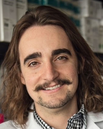 Costas A Lyssiotis合作论文数Rogel Cancer Center, Medical School, University of Michigan10Chandramouly Gurushankar合作论文数9Hartlerode Andrea合作论文数9
 Arvind Panday合作论文数School of Veterinary Medicine, Louisiana State University9Juvekar Ashish合作论文数9Amy Kwok合作论文数7Francesca Menghi合作论文数7
 Rajula Elango合作论文数7
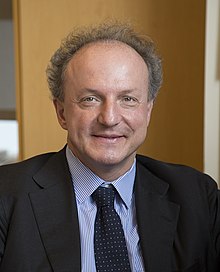 Pier Paolo Pandolfi合作论文数Institute of Cancer, Renown Health;Veneto Institute of Molecular Medicine7
 Ganesh Nagaraju合作论文数Department of Medicine, Harvard Medical School and Beth Israel Deaconess Medical Center7Nadine Puget合作论文数Department of Hematology/Oncology, Beth Israel Deaconess Medical Center and Harvard Medical School7
 Judith Balmana合作论文数6Katherine Spencer合作论文数6Elaine Lunsford合作论文数6
 Caterina Nardella合作论文数6
 Antonella Papa合作论文数Department of Biochemistry and Molecular Biology, School of Biomedical Sciences, Faculty of Medicine, Nursing and Health Sciences, Monash University6Burga Laura N合作论文数6
 Edison T. Liu合作论文数The JAX Cancer Center, The Jackson Laboratory6Shobu Odate合作论文数6
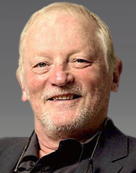 Frederick W. Alt合作论文数Department of Genetics, Blavatnik Institute, Harvard Medical School;Department of Pediatrics, Boston Children's Hospital6
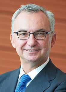 José Baselga合作论文数Weill Cornell Medical College6Yasir H. Ibrahim合作论文数University of Minnesota Cancer Center6Erin E Duffey合作论文数5
 Emilie Rass合作论文数4Yadegarynia Sina合作论文数4
 Andrew Kung合作论文数Department of Pediatrics, Memorial Sloan Kettering Cancer Center4
 Jean Feunteun合作论文数Centre National de la Recherche Scientifique, Institut Gustave-Roussy4T Ashley合作论文数3Thomas J. F. Nieland合作论文数3
 Sean T Bailey合作论文数3Yan Liu合作论文数School of Pharmacy, Lanzhou University3Zhao Chen (陈昭)合作论文数3
 Kwok-Kin Wong合作论文数Department of Medicine, Grossman School of Medicine, New York University3
 Peng Gao合作论文数Pulmonary and Critical Care, Department of Medicine, Feinberg School of Medicine, Northwestern University3
 Qingsong Liu (刘青松)合作论文数High Magnetic Field Laboratory, Hefei Institutes of Physical Science, Chinese Academy of Sciences;University of Science and Technology of China;PreceDo Pharmaceuticals Co.Ltd.3Vlasakova Katerina合作论文数3Altabef Abigail合作论文数3Kimmelman Alec C合作论文数3Tchaicha Jeremy H合作论文数3Choe Sung合作论文数Agios Pharmaceuticals3Cohoon Travis J合作论文数3




