Nearly all mitochondrial proteins are imported into mitochondria from the cytosol. How nascent mitochondrial precursors acquire and sustain import-competence in the cytosol under normal and stress conditions is incompletely understood. Here, we show that under normal conditions, the Hsc70 and Hsp90 systems interact with and redundantly minimize precursor degradation. During acute import stress, Hsp90 buffers precursor degradation, preserving proteins in an import-competent state until stress resolution. Unexpectedly, buffering by Hsp90 relies critically on a mitochondrial targeting signal (MTS), the absence of which greatly decreases precursor-Hsp90 interaction. Site-specific photo-crosslinking and biochemical reconstitution showed how the MTS directly engages co-chaperones of Hsc70 (St13 and Stip1) and Hsp90 (p23 and Cdc37) to facilitate chaperone retention on the mature domain. Thus, the MTS has a previously unappreciated role in regulating chaperone dynamics on mitochondrial precursors to buffer their degradation and maintain import competence, functions that may facilitate restoration of mitochondrial homeostasis after acute import stress.
Protein biogenesis at the endoplasmic reticulum requires translocons comprising the Sec61 protein-conducting channel and several dynamically associated accessory factors. Here we used transcriptome-wide selective ribosome profiling in human cells to monitor cotranslational interactions of accessory factors for N-glycosylation (the OST-A complex) and multipass membrane protein synthesis (the GEL, PAT and BOS complexes). OST-A was preferentially recruited to open Sec61 channels engaged in polypeptide translocation; conversely, GEL, PAT and BOS were recruited synchronously to closed Sec61 channels and stabilized by newly inserted transmembrane domains. Translocon composition changed repeatedly and reversibly during the synthesis of topologically complex multipass membrane proteins. These data establish the molecular logic that underlies substrate-driven translocon remodeling, events that are crucial for the efficient biogenesis of secretory and membrane proteins.
Unassembled and partially assembled subunits of multi-protein complexes have emerged as major quality control clients, particularly under conditions of imbalanced gene expression such as stress, aging, and aneuploidy. The factors and mechanisms that eliminate such orphan subunits to maintain protein homeostasis are incompletely defined. Here, we show that the UBR4-KCMF1 ubiquitin ligase complex is required for the efficient degradation of multiple unrelated orphan subunits from the chaperonin, proteasome cap, proteasome core, and a protein targeting complex. Epistasis analysis in cells and reconstitution studies in vitro show that the UBR4-KCMF1 complex acts downstream of a priming ubiquitin ligase that first mono-ubiquitinates orphans. UBR4 recognizes both the orphan and its mono-ubiquitin and builds a K48-linked poly-ubiquitin degradation signal. The discovery of a convergence point for multiple quality control pathways may explain why aneuploid cells are especially sensitive to loss of UBR4 or KCMF1 and identifies a potential vulnerability across many cancers.
Eukaryotic cells have evolved sophisticated quality control mechanisms to eliminate aggregation-prone proteins that compromise cellular health. Central to this defense is the ubiquitin-proteasome system, where UBR4 acts as an essential E4 ubiquitin ligase, amplifying degradation marks on defective proteins. Cryo-electron microscopy analysis of UBR4 in complex with its cofactors KCMF1 and CALM1 reveals a massive 1.3-megadalton ring structure, featuring a central substrate-binding arena and flexibly attached catalytic units. Our structure shows how UBR4 binds substrate and extends lysine-48-specific ubiquitin chains. Efficient substrate targeting depends on both preubiquitination and specific N-degrons, with KCMF1 acting as a key substrate filter. The architecture of the E4 megacomplex is conserved across eukaryotes, but species-specific adaptations allow UBR4 to perform its precisely tuned quality control function in diverse cellular environments.
Scientific research is an exploration of the unknown. The process is full of uncertainty, missteps, delightful surprises, painful lessons, and ultimately a measure of insight into nature. In this Science and Society article I suggest a few practical strategies that helped me navigate these challenges at the earliest stages of becoming a cell biologist.
Most eukaryotic multipass membrane proteins are inserted into the membrane of the endoplasmic reticulum. Their transmembrane domains (TMDs) are thought to be inserted co-translationally as they emerge from a membrane-bound ribosome. Here we find that TMDs near the carboxyl terminus of mammalian multipass proteins are inserted post-translationally by the endoplasmic reticulum membrane protein complex (EMC). Site-specific crosslinking shows that the EMC’s cytosol-facing hydrophilic vestibule is adjacent to a pre-translocated C-terminal tail. EMC-mediated insertion is mostly agnostic to TMD hydrophobicity, favored for short uncharged C-tails and stimulated by a preceding unassembled TMD bundle. Thus, multipass membrane proteins can be released by the ribosome–translocon complex in an incompletely inserted state, requiring a separate EMC-mediated post-translational insertion step to rectify their topology, complete biogenesis and evade quality control. This sequential co-translational and post-translational mechanism may apply to ~250 diverse multipass proteins, including subunits of the pentameric ion channel family that are crucial for neurotransmission.
The protein translocon at the endoplasmic reticulum comprises the Sec61 translocation channel and numerous accessory factors that collectively facilitate the biogenesis of secretory and membrane proteins. Here, we leveraged recent advances in cryo-electron microscopy (cryo-EM) and structure prediction to derive insights into several novel configurations of the ribosome-translocon complex. We show how a transmembrane domain (TMD) in a looped configuration passes through the Sec61 lateral gate during membrane insertion; how a nascent chain can bind and constrain the conformation of ribosomal protein uL22; and how the translocon-associated protein (TRAP) complex can adjust its position during different stages of protein biogenesis. Most unexpectedly, we find that a large proportion of translocon complexes contains RAMP4 intercalated into Sec61’s lateral gate, widening Sec61’s central pore and contributing to its hydrophilic interior. These structures lead to mechanistic hypotheses for translocon function and highlight a remarkably plastic machinery whose conformations and composition adjust dynamically to its diverse range of substrates.
Abstract Microtubules, built from heterodimers of α- and β-tubulins, control cell shape, mediate intracellular transport, and power cell division. The concentration of αβ-tubulins is tightly controlled through a posttranscriptional mechanism involving selective and regulated degradation of tubulin-encoding mRNAs. Degradation is initiated by TTC5, which recognizes tubulin-synthesizing ribosomes and recruits downstream effectors to trigger mRNA deadenylation. Here, we investigate how cells regulate TTC5 activity. Biochemical and structural proteomic approaches reveal that under normal conditions, soluble αβ-tubulins bind to and sequester TTC5, preventing it from engaging nascent tubulins at translating ribosomes. We identify the flexible C-terminal tail of TTC5 as a molecular switch, toggling between soluble αβ-tubulin-bound and nascent tubulin-bound states. Loss of sequestration by soluble αβ-tubulins constitutively activates TTC5, leading to diminished tubulin mRNA levels and compromised microtubule-dependent chromosome segregation during cell division. Our findings provide a paradigm for how cells regulate the activity of a specificity factor to adapt posttranscriptional regulation of gene expression to cellular needs.
Eukaryotic cells have evolved sophisticated quality control mechanisms to eliminate aggregation-prone proteins that compromise cellular health. Central to this defense is the ubiquitin-proteasome system, where UBR4 acts as essential E4 ubiquitin ligase, amplifying degradation marks on defective proteins. Our cryo-EM analysis of UBR4 in complex with its cofactors KCMF1 and CALM1 reveals a massive 1.3 MDa ring structure, featuring a central substrate-binding arena and flexibly attached catalytic units. Structural data illustrate how UBR4 binds substrate and extends K48-specific ubiquitin chains. Importantly, efficient substrate targeting depends on both pre-ubiquitination and specific N-degrons, with KCMF1 acting as key substrate filter. Furthermore, we show that the architecture of the E4 megacomplex is conserved across eukaryotes but with species specific adaptations, allowing UBR4 to perform its precisely tuned quality-control function in diverse cellular environments. ### Competing Interest Statement The authors have declared no competing interest.
Becoming an academic scientist is neither linear nor formulaic. Rather, graduate education is mostly an apprenticeship without a discrete curriculum, clear sign posts of progress, or specific training metrics. In this science and society article I offer a few thoughts from my own experience for what a student beginning this journey might want to learn.
α-Helical integral membrane proteins comprise approximately 25% of the proteome in all organisms. The membrane proteome is highly diverse, varying in the number, topology, spacing and properties of transmembrane domains. This diversity imposes different constraints on the insertion of different regions of a membrane protein into the lipid bilayer. Here, we present a cohesive framework to explain membrane protein biogenesis, in which different parts of a nascent substrate are triaged between Oxa1 and SecY family members for insertion. In this model, Oxa1 family proteins insert transmembrane domains flanked by short translocated segments, whereas the SecY channel is required for insertion of transmembrane domains flanked by long translocated segments. Our unifying model rationalizes evolutionary, genetic, biochemical and structural data across organisms and provides a foundation for future mechanistic studies of membrane protein biogenesis.
Pentameric ligand-gated ion channels (pLGICs) are cell surface receptors of crucial importance for animal physiology1-4. This diverse protein family mediates the ionotropic signals triggered by major neurotransmitters and includes γ-aminobutyric acid receptors (GABAARs) and acetylcholine receptors (nAChRs). Receptor function is fine-tuned by a myriad of endogenous and pharmacological modulators3. A functional pLGIC is built from five homologous, sometimes identical, subunits, each containing a β-scaffold extracellular domain (ECD), a four-helix transmembrane domain (TMD) and intracellular loops of variable length. Although considerable progress has been made in understanding pLGICs in structural and functional terms, the molecular mechanisms that enable their assembly at the endoplasmic reticulum (ER)5 in a vast range of potential subunit configurations6 remain unknown. Here, we identified candidate pLGICs assembly factors selectively associated with an unassembled GABAAR subunit. Focusing on one of the candidates, we determined the cryo-EM structure of an assembly intermediate containing two α1 subunits of GABAAR each bound to an ER-resident membrane protein NACHO. The structure showed how NACHO shields the principal (+) transmembrane interface of α1 subunits containing an immature extracellular conformation. Crosslinking and structure-prediction revealed an adjacent surface on NACHO for β2 subunit interactions to guide stepwise oligimerisation. Mutations of either subunit-interacting surface on NACHO also impaired the formation of homopentameric α7 nAChRs, pointing to a generic framework for pLGIC assembly. Our work provides the foundation for understanding the regulatory principles underlying pLGIC structural diversity.
Microtubules play crucial roles in cellular architecture, intracellular transport, and mitosis. The availability of free tubulin subunits affects polymerization dynamics and microtubule function. When cells sense excess free tubulin, they trigger degradation of the encoding mRNAs, which requires recognition of the nascent polypeptide by the tubulin-specific ribosome-binding factor TTC5. How TTC5 initiates the decay of tubulin mRNAs is unknown. Here, our biochemical and structural analysis reveals that TTC5 recruits the poorly studied protein SCAPER to the ribosome. SCAPER, in turn, engages the CCR4-NOT deadenylase complex through its CNOT11 subunit to trigger tubulin mRNA decay. SCAPER mutants that cause intellectual disability and retinitis pigmentosa in humans are impaired in CCR4-NOT recruitment, tubulin mRNA degradation, and microtubule-dependent chromosome segregation. Our findings demonstrate how recognition of a nascent polypeptide on the ribosome is physically linked to mRNA decay factors via a relay of protein-protein interactions, providing a paradigm for specificity in cytoplasmic gene regulation.
Multipass membrane proteins contain two or more α-helical transmembrane domains (TMDs) that span the lipid bilayer. They are inserted cotranslationally into the prokaryotic plasma membrane or eukaryotic endoplasmic reticulum membrane. The Sec61 complex (SecY complex in prokaryotes) provides a ribosome docking site, houses a channel across the membrane, and contains a lateral gate that opens toward the lipid bilayer. Model multipass proteins can be stitched into the membrane by iteratively using Sec61's lateral gate for TMD insertion and its central pore for translocation of flanking domains. Native multipass proteins, with their diverse TMDs and complex topologies, often also rely on members of the Oxa1 family of translocation factors, the PAT complex chaperone, and other poorly understood factors. Here, we discuss the mechanisms of TMD insertion, highlight the limitations of an iterative insertion model, and propose a new hypothesis for multipass membrane protein biogenesis based on recent findings.
PDF file - 29K, Impact of CHX and actinomycin D on HDI-induced ABCG2 surface overexpression
Cells tightly regulate mRNA processing, localization, and stability to ensure accurate gene expression in diverse cellular states and conditions. Most of these regulatory steps have traditionally been thought to occur before translation by the action of RNA-binding proteins. Several recent discoveries highlight multiple co-translational mechanisms that modulate mRNA translation, localization, processing, and stability. These mechanisms operate by recognition of the nascent protein, which is necessarily coupled to its encoding mRNA during translation. Hence, the distinctive sequence or structure of a particular nascent chain can recruit recognition factors with privileged access to the corresponding mRNA in an otherwise crowded cellular environment. Here, we draw on both well-established and recent examples to provide a conceptual framework for how cells exploit nascent protein recognition to direct mRNA fate. These mechanisms allow cells to dynamically and specifically regulate their transcriptomes in response to changes in cellular states to maintain protein homeostasis.
Cells contain numerous abundant molecular machines assembled from multiple subunits. Imbalances in subunit production and failed assembly generate orphan subunits that are eliminated by poorly defined pathways. Here, we determined how orphan subunits of the cytosolic chaperonin CCT are recognized. Several unassembled CCT subunits recruited the E3 ubiquitin ligase HERC2 using ZNRD2 as an adaptor. Both factors were necessary for orphan CCT subunit degradation in cells, sufficient for CCT subunit ubiquitination with purified factors, and necessary for optimal cell fitness. Domain mapping and structure prediction defined the molecular features of a minimal HERC2-ZNRD2-CCT module. The structural model, whose key elements were validated in cells using point mutants, shows why ZNRD2 selectively recognizes multiple orphaned CCT subunits without engaging assembled CCT. Our findings reveal how failures during CCT assembly are monitored and provide a paradigm for the molecular recognition of orphan subunits, the largest source of quality control substrates in cells.
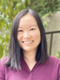 Shao Sichen合作论文数National Institute of Child Health and Human Development, National Institutes of Health20
 Robert J. Keenan合作论文数17Vishwanath R. Lingappa合作论文数Prosetta Bioconformat16
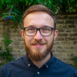 Szymon Juszkiewicz合作论文数MRC Laboratory of Molecular Biology, Francis Crick Avenue, Cambridge CB2 0QH, UK14Robert W Robey合作论文数12Yvona Ward合作论文数12Akina Tamaki合作论文数11Caterina Ierano合作论文数10Susan E. Bates合作论文数10Ajay Sharma合作论文数9Basseville Agnes合作论文数9Neena S. Rane合作论文数9Shana Trostel合作论文数8
 Snapp Erik L合作论文数Dept Anat & Struct Biol, Yeshiva Univ8Zavodszky Eszter合作论文数Cambridge Institute for Medical Research, University of Cambridge8
 Jack Taunton合作论文数School of Medicine, University of California, San Francisco;Helen Diller Family Comprehensive Cancer Center, University of California, San Francisco8
 Oishee Chakrabarti合作论文数National Institute of Child Health and Human Development, National Institutes of Health8Sew-Yeu Peak-Chew合作论文数7Aaron Lewis合作论文数7
 Aarthi Ashok合作论文数Department of Biological Sciences, University of Toronto Scarborough7
 Malaiyalam Mariappan合作论文数Biochemistry, Quantitative Biology, Biophysics, and Structural Biology, Biological & Biomedical Sciences, Yale University7
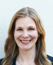 Jennifer Garrison合作论文数School of Medicine, University of California, San Francisco;Buck Institute for Research on Aging6
 D. Thomas Rutkowski合作论文数Howard Hughes Medical Institute, University of Michigan Medical Center6Soo Jung Kim合作论文数5
 Zhewang Lin合作论文数Inst Bioengn & Nanotechnol, Singapore 138669, Singapore5
 Jennifer Lippincott-Schwartz合作论文数Howard Hughes Medical Institute5Agnieszka Mateja合作论文数5
 Luka Smalinskaite合作论文数4Eric J. Kunkel合作论文数4
 Alina Guna合作论文数Whitehead Institute for Biomedical Research, Massachusetts Institute of Technology4
 Alan Brown合作论文数Laboratory of Molecular Biology, Medical Research Council (MRC)4
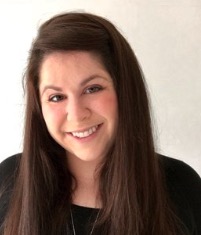 Rebecca Voorhees合作论文数Division of Biology and Biological Engineering, California Institute of Technology4Eisuke Itakura合作论文数4
 Venkatraman Ramakrishnan合作论文数MRC Laboratory of Molecular Biology;Trinity College, University of Cambridge4Viswanathan Chandrasekaran合作论文数4Ivana Gasic合作论文数3Sabine Voigt合作论文数3Robert Kurzbauer合作论文数3
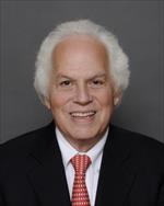 Stanley B. Prusiner合作论文数Institute for Neurodegenerative Diseases, University of California, San Francisco;Department of Neurology, University of California, San Francisco3
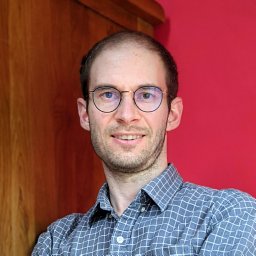 Markus Hopfler合作论文数Max F. Perutz Laboratories, Medical University of Vienna3Chitwood Patrick J合作论文数Department of Molecular, Cellular, and Developmental Biology, University of Colorado3
 Stephen De Armond合作论文数Department of Pathology, School of Medicine, University of California, San Francisco;UCSF Health-UCSF Medical Center3
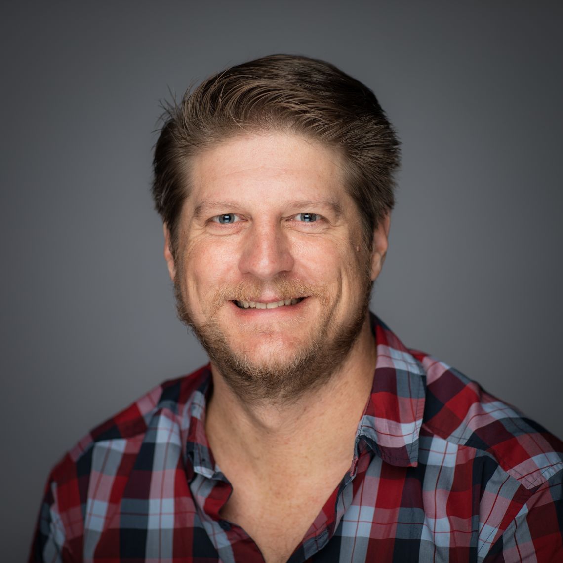 John Christianson合作论文数Nuffield Department of Orthopaedics, Rheumatology and Musculoskeletal Sciences, University of Oxford3
 Norbert Volkmar合作论文数3Yagita Yuichi合作论文数3Sang-Wook Kang合作论文数3
 Tom Rapoport合作论文数Department of Cell Biology, Harvard Medical School, Harvard University;Howard Hughes Medical Institute3
 Tim Clausen合作论文数Research Institute for Molecular Pathology3Devarati Mitra合作论文数Laboratory of Cellular Oncology, National Cancer Institute3




