Dual-mobility designs aim to reduce dislocation with a dual head, creating a larger effective head size. However, concerns exist regarding corrosion from dissimilar metal interfaces. An 80-year-old female patient exhibited elevated cobalt (2.6 μg/L) and titanium (7.8 μg/L) but normal chromium (1.2 μg/L) levels after 5 years. She had staged bilateral total hip arthroplasty with identical modular liners, a 22 mm cobalt-chrome and a 28 mm ceramic head in the left and right hips, respectively. Magnetic resonance imaging showed left-sided ALTR. At revision, the trunnion and head showed gross corrosion. Biomechanical analysis confirmed trunnionosis of the 22-mm cobalt-chrome head without liner corrosion, suggesting dual mobility may increase the risk for trunnionosis at this size. This case highlights cobalt-chrome head risks and identifies trunnion corrosion as a cause of metallosis.
OBJECTIVE:The synovial tissue pathotype may determine the treatment response in rheumatoid arthritis (RA); however, biopsies are not widely available. Synovial fluid is a promising tissue surrogate. Our purpose was to compare RA synovial fluid cell counts with histopathology and use synovial fluid to predict tissue inflammation. METHODS:Synovial fluid and tissue were collected during knee arthroplasty. Patients were stratified based on their medication treatment history. Synovial lymphocytic inflammation (SLI) was graded from low to high. Synovial fluid white blood cell (WBC) count and differentials were performed in the clinical laboratory. Descriptive statistics, correlations, receiver operating characteristic curve analysis, and multivariable regression were performed to determine the associations with tissue SLI. RESULTS:Sixty-four patients with RA had paired synovial tissue and synovial fluid data available. The mean Clinical Disease Activity Index (CDAI) score was 17.9. High tissue SLI was observed in 29 patients, and low SLI was observed in 35 patients, with roughly equal distribution among treatment groups. The mean synovial fluid WBC count was 5,661 cells/μL and was not correlated with CDAI but correlated positively with SLI and percentage polymorphonuclear cells (PMN%). Synovial fluid WBC count ≥1,400 cells/μL was sensitive (0.86) and specific (0.91) for high SLI (area under the curve 0.91). In a multivariable regression, PMN% was associated with high SLI (odds ratio [OR] 1.46 [95% confidence interval (CI) 1.14-1.85]). Synovial fluid monocyte percentage was negatively associated with high SLI (OR 0.44 [95% CI 0.27-0.73]). CONCLUSION:Synovial fluid WBC count is sensitive and specific for differentiating high and low lymphocytic synovial inflammation. Further analysis of the synovial fluid as it relates to the adjacent tissue in different cohorts is needed.
OBJECTIVE:Patients with psoriatic arthritis (PsA) experience higher postoperative complication rates after total hip or total knee arthroplasty (THA and TKA, respectively) than patients with osteoarthritis, but contributing factors to this risk are poorly defined. We examined clinical factors predicting postoperative complications to determine whether PsA-specific disease activity is associated with adverse events (AEs). METHODS:We conducted a prospective study of adults with PsA undergoing THA or TKA. Baseline assessments included demographics, BMI, Charlson Comorbidity Index (CCI), PsA disease activity measures, patient global assessment, medications, and patient-reported outcomes. Operative synovial tissue was assessed for inflammation. Postoperative AEs collected within 1 year were graded for severity (1-4) using the Clavien-Dindo (CD) classification. Logistic regression, adjusting for age and sex, assessed predictors of AEs and AE severity. RESULTS:In total, 57 patients were included (24 THA, 33 TKA). At surgery, patients had moderate PsA activity (mean Disease Activity Index for Psoriatic Arthritis [DAPSA] score of 15.8) with minimal skin disease or enthesitis. CD grade 3-4 AEs occurred in 10 patients (18%). BMI and CCI were higher in THA patients with any AEs and all patients with grade 3-4 AEs. In the adjusted analysis, higher CCI was associated with grade 2-4 and 3-4 AEs, and BMI with grade 3-4 AEs. No PsA-specific measures, including DAPSA or synovial histologic inflammation, were associated with AEs. CONCLUSION:In patients with PsA undergoing THA/TKA, AEs were driven by comorbidity burden and obesity rather than PsA-specific disease activity or joint inflammation. This finding highlights the importance of perioperative optimization of comorbidities and weight.
Objectives:To identify synovial transcriptional clusters in human knee osteoarthritis (OA) and determine how these relate to synovial histologic features, cell-type-associated gene expression, and cartilage degeneration severity. Methods:Bulk RNA sequencing (RNA-seq) of synovial tissue from n = 135 patients with knee OA was analyzed using consensus clustering. Clusters were compared by clinical and histologic features, including cartilage degeneration severity (OARSI score). Single-cell RNA-seq (n = 18) and spatial transcriptomics were used to relate cartilage degeneration-associated gene expression patterns to synovial cell populations. Results:Four synovial transcriptional clusters that differed in synovial histologic features and cartilage degeneration severity were identified. Greater cartilage degeneration was associated with enrichment of lining fibroblast- and inflammatory myeloid-associated gene expression, whereas lesser cartilage degeneration was associated with enrichment of sublining fibroblast, endothelial, mural cell, and adipocyte-associated gene expression. Conclusions:Human knee OA synovium segregates into transcriptional clusters associated with cartilage degeneration severity. Synovial transcriptional heterogeneity corresponds to cell-type-associated gene expression.
In the rheumatoid arthritis (RA) synovium, resident fibroblast-like synoviocytes (FLS) express MHC class II molecules (HLA-D) but lack the costimulatory signals typically required for T cell activation. Here, we demonstrate that antigen presentation by FLS induces a distinct T cell activation state characterized by high CD69 yet reduced CD25 and HLA-DR expression, suppressed proliferation, and decreased effector cytokine production compared with professional antigen-presenting cells (APCs), such as macrophages. FLS were also capable of suppressing macrophage-induced T cell activation, underscoring their dominant immunomodulatory role in the synovial microenvironment. Mechanistically, we identify indoleamine 2,3-dioxygenase-mediated (IDO1-mediated) tryptophan depletion as the primary driver of FLS-induced T cell hyporesponsiveness. Spatial transcriptomics revealed colocalization of IDO1 and CD69 within ectopic lymphoid structures in RA synovium, further supporting the in vivo relevance of this pathway. These findings provide the groundwork for positioning FLS as critical T cell regulators in RA and highlight the importance of preserving their immunosuppressive properties when therapeutically targeting pathogenic FLS functions.
Single-cell RNA sequencing studies have revealed the heterogeneity of cell states present in the rheumatoid arthritis (RA) synovium. However, it remains unclear how these cell types interact with one another in situ and how synovial microenvironments shape observed cell states. Here, we use spatial transcriptomics (ST) to define stable microenvironments across eight synovial tissue samples from six RA patients and characterize the cellular composition of ectopic lymphoid structures (ELS). To identify disease-relevant cellular communities, we developed DeepTopics, a scalable reference-free deconvolution method based on a Dirichlet variational autoencoder architecture. DeepTopics identified 22 topics across tissue samples that were defined by specific cell types, activation states, and/or biological processes. Some topics were defined by multiple colocalizing cell types, such as CD34+ fibroblasts and LYVE1+ macrophages, suggesting functional interactions. Within ELS, we discovered two divergent cellular patterns that were stable across ELS in each patient and typified by the presence or absence of a "germinal-center-like" topic. DeepTopics is a versatile and computationally efficient method for identifying disease-relevant microenvironments from ST data, and our results highlight divergent cellular architectures in histologically similar RA synovial samples that have implications for disease pathogenesis.
Objective We sought to develop computer vision methods to quantify aggregates of cells in synovial tissue and compare these with clinical and gene expression parameters. Methods We assembled a computer vision pipeline to quantify five features encompassing synovial cell density and aggregates and compared these with pathologist scores, disease classification, autoantibody status, and RNA expression in a cohort of 156 patients with rheumatoid arthritis (RA) and 149 patients with osteoarthritis (OA). Results All five features were associated with pathologist scores of synovial lymphocytic inflammation ( P < 0.0001). Three features that related to the cells per unit of tissue were significantly increased in patients with both seronegative and seropositive RA compared with those with OA; on the other hand, aggregate features (number and diameter) were significantly increased in seropositive, but not seronegative, RA compared with OA. Aggregate diameter was associated with the gene expression of immunoglobulin heavy‐chain genes in the synovial tissue. Compared with blood, synovial immunoglobulin isotypes were skewed from IGHM and IGHD to IGHG3 and IGHG1 . Further, patients with RA with high levels of lymphocytic infiltrates in the synovium demonstrated parallel skewing in their blood with a relative decrease in IGHGM ( P < 0.002) and IGHD ( P < 0.03) and an increase in class‐switched immunoglobulin genes IGHG3 ( P < 0.03) and IGHG1 ( P < 0.002). Conclusion High‐resolution automated identification and quantification of synovial immune cell aggregates uncovered skewing in the synovium from naïve IGHD and IGHM to memory IGHG3 and IGHG1 and revealed that this process is reflected in the blood of patients with high inflammatory synovium. image
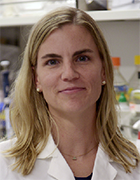 Laura Donlin合作论文数HSS Research Institute;Hospital for Special Surgery, Weill Cornell Medicine7
 Melanie Smith合作论文数7
 Susan M. Goodman合作论文数Department of Rheumatology, Hospital for Special Surgery;Weill Cornell Medical College7
 Edward F. Dicarlo合作论文数Medical College, New York Hospital-Cornell University7
 Bella Mehta合作论文数6
 Dana E Orange合作论文数5Robert L Jacobs合作论文数5
 Charles P. Andrews合作论文数Lung Diagnostics, Biogenics Research Institute5Orange Dana E合作论文数Laboratory of Molecular Neuro-oncology, The Rockefeller University4Otero Miguel合作论文数4
 Anne-Marie Malfait合作论文数Dept. of Rheumatology, Ghent University Hospital4Caryn Hale合作论文数Department of Biochemistry and Molecular Biology;Department of Genetics;University of Georgia;Laboratoire d'Enzymologie et Biochimie;Centre National de la Recherche Scientifique4N. Harper合作论文数3Yiyuan Wu合作论文数3
 Tristan Maerz合作论文数Department of Biomedical Engineering, College of Engineering, University of Michigan-Ann Arbor3Ahuja Sunil K合作论文数3W He合作论文数3Rachel E. Miller合作论文数2Amit Lakhanpal合作论文数2R EVANS合作论文数2Edoardo Spolaore合作论文数2
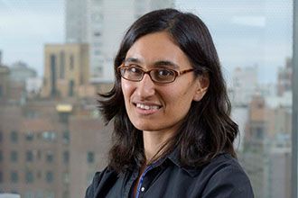 Christina Leslie合作论文数Computational Biology, Weill Cornell Graduate School of Medical Sciences, Cornell University2A. Helena Jonsson合作论文数2
 Martin Lotz合作论文数Department of Molecular and Cellular Biology, The Scripps Research Institute;Department of Molelcular Medicine, The Scripps Research Institute2
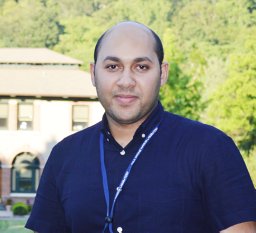 Shady Younis合作论文数Science for Life Laboratory Uppsala, Uppsala University;Department of Animal Production, Ain Shams University;Uppsala University, Ain Shams University2
 William H. Robinson合作论文数Division of Immunology and Rheumatology, Stanford University2
 Meghana Kshirsagar合作论文数Microsoft2Thomas Carroll合作论文数1Damon Frezza合作论文数1Preethi K. Periyakoil合作论文数1Sami L. Bahna合作论文数1Alia M. Obeidat合作论文数1Aimee C Colbath合作论文数1Spencer Fullam合作论文数1Saimun Habib合作论文数1WA NISH合作论文数1Y Liu (刘义)合作论文数Sch Astronaut, Beihang Univ1
 Wei Wang合作论文数Department of Chemistry and Biochemistry, University of California, San Diego;Department of Cellular and Molecular Medicine, University of California, San Diego1DW GOETZ合作论文数1David Slater合作论文数The MITRE Corporation1
 Kristine Carandang合作论文数Child and Adolescent Services Research Center, San Diego, California.1
 Weiqiang Chen (陈伟强)合作论文数School of Biomedical Engineering, Nanjing University1
 Alexander Rudensky合作论文数Graduate School of Medical Sciences, Weill Cornell Medicine;Weill Cornell Medical College;Memorial Sloan‐Kettering Cancer Center1
 Puraskar Ingale合作论文数Department of Biology, University of Texas at San Antonio1Frank Mayu O合作论文数1Sculco Peter K合作论文数Hospital for Special Surgery – NYC1Thompson James R合作论文数Special Education, Illinois State University1Li Jun合作论文数1
 Merissa Olmer合作论文数1




