Prostate cancer is the most common solid malignancy in men and requires a biopsy for diagnosis. This manuscript describes a freehand micro-ultrasound guided transperineal technique performed under local anesthesia, which maintains accuracy, keeps patients comfortable, has low adverse events, and minimizes the need for disposables. Prior micro-ultrasound-guided transperineal techniques required general or spinal anesthesia. The key steps described in the protocol include (1) the placement of the local anesthesia, (2) micro-ultrasound imaging, (3) and the visualization of the anesthetic/biopsy needle while uncoupled from the insonation plane. A retrospective review of 100 patients undergoing this technique demonstrated a 68% clinically significant cancer detection rate. Pain scores were prospectively collected in a subset of patients (N = 20) and showed a median procedural pain score of 2 out of 10. The 30 day Grade III adverse event rate was 3%; one of these events was probably related to the prostate biopsy. Overall, we present a simple, accurate, and safe technique for performing a micro-ultrasound-guided transperineal prostate biopsy.
Prostate cancer is the most common internal malignancy among males. Micro-Ultrasound is a promising imaging modality for cancer identification and computer-assisted visualization. Identifying the prostate capsule area is essential in active surveillance monitoring and treatment planning. In this paper, we present a pilot study that assesses prostate capsule segmentation using the U-Net deep neural network framework. To the best of our knowledge, this is the first study on prostate capsule segmentation in Micro-Ultrasound images. For our study, we collected multi-frame volumes of Micro-Ultrasound images, and then expert prostate cancer surgeons annotated the capsule border manually. The lack of clear boundaries and variation of shapes between patients make the task challenging, especially for novice Micro-Ultrasound operators. In total 2099 images were collected from 8 subjects, 1296 of which were manually annotated and were split into a training set (1008), a validation set (112), and a test set from a different subject (176). The performance of the model was evaluated by calculating the Intersection over Union (IoU) between the manually annotated area of the capsule and the segmentation mask computed from the trained deep neural network. The results demonstrate high IoU values for the training set (95.05%), the validation set (93.18%) and the test set from a separate subject (85.14%). In 10-fold cross-validation, IoU was 94.25%, and accuracy was 99%, validating the robustness of the model. Our pilot study demonstrates that deep neural networks can produce reliable segmentation of the prostate capsule in Micro-Ultrasound images and pave the road for the segmentation of other anatomical structures within the capsule, which will be the subject of our future studies.
Creatinine (Cr) is often used as a standalone gold standard marker of kidney function. Cystatin C (Cys C) is a less physiologically labile marker of renal function, particularly in certain subgroups. Herein, we analyze trends in cystatin C as compared to creatinine in men on testosterone replacement therapy with varying body mass indices and percent body fat (PBF). This retrospective analysis observes 227 men with testosterone-induced muscle hypertrophy who visited a men's health tertiary care clinic. All participants were characterized as competitive or recreational athletes. In patients with a normal body mass index (BMI), there was no clinically significant correlation between Cr and Cys C. Slight correlation was seen with overweight (R-2 = .27) patients (p < .0001) and obese (R-2 = .29) patients (p < .0001). Patients with PBF of 0%-10% (n = 22) exhibited minimal (R-2 = .23) positive correlation between Cys C and Cr (p = .03). Positive correlation between Cys C and Cr in patients with PBF of 10%-20% was clinically negligible (R-2 = .17, n = 87), modest (R-2 = .49) in patients with PBF of 20%-30% (n = 42), and evident (R-2 = 1.00) in patients >30% (n = 3) (p < .0001, respectively). Cystatin C measurements display less variance compared with creatinine at differing BMI distinctions. At the upper limit of BMI or PBF in our patient population, cystatin C exhibits minimal to moderate variability compared with creatinine.
Over the past several decades, carbon monoxide (CO) has been studied as a potential cytoprotective treatment for neurological diseases. This chapter mainly discusses the mechanisms by which CO is neuroprotective after acute brain injury through Nrf2 and heme oxygenase 1 (HO-1) upregulation. It provides a synopsis of preclinical studies using CO gas or CO-releasing molecules (CORMs) to treat acute brain injuries, including ischemic stroke, neonatal hypoxic-ischemic encephalopathy, hemorrhagic stroke, and traumatic brain injury (TBI). CO gas and CORMs are being tested in clinical trials as putative therapeutic agents for various conditions, including chronic headaches, acute respiratory distress syndrome, vascular disorders, pulmonary fibrosis, chronic obstructive pulmonary disease, pulmonary hypertension, and traumatic brain injury. Finally, clinical trials that use CO gas and CORMs to treat disease in humans are discussed, as will the risks and benefits of beginning clinical trials using CO gas or CORMs to treat acute brain injuries in humans.
Circulating hemopexin is the primary protein responsible for the clearance of heme; therefore, it is a systemic combatant against deleterious inflammation and oxidative stress induced by the presence of free heme. This role of hemopexin is critical in hemolytic pathophysiology. In this review, we outline the current research regarding how the dynamic activity of hemopexin is implicated in sickle cell disease, which is characterized by a pathological aggregation of red blood cells and excessive hemolysis. This pathophysiology leads to symptoms such as acute kidney injury, vaso-occlusion, ischemic stroke, pain crises, and pulmonary hypertension exacerbated by the presence of free heme and hemoglobin. This review includes in vivo studies in mouse, rat, and guinea pig models of sickle cell disease, as well as studies in human samples. In summary, the current research indicates that hemopexin is likely protective against these symptoms and that rectifying depleted hemopexin in patients with sickle cell disease could improve or prevent the symptoms. The data compiled in this review suggest that further preclinical and clinical research should be conducted to uncover pathways of hemopexin in pathological states to evaluate its potential clinical function as both a biomarker and therapy for sickle cell disease and related hemoglobinopathies.
Vitamin D supplementation has been shown to improve outcomes for patients suffering from a variety of illnesses such as stroke and cancer. Vitamin D deficiencies have been associated with longer hospital stays, greater severity of symptoms, and death in some complex cases. Due to vitamin D’s burgeoning role in improving patient outcomes, a new sector of research is focusing on the lesser-known implications of vitamin D on health. Traumatic brain injury (TBI) affects approximately 69 million people worldwide per year. Here, we summarize the current scientific understanding of vitamin D dynamics with TBI to elucidate a potential way to lessen the cascade of secondary damage after an initial insult, with the goal of improving overall patient outcomes. Because vitamin D supplementation has been correlated with better outcomes in other pathologies involving immune and inflammatory molecules, it is important to study the potential effect of vitamin D deficiency (VDD) and supplementation on TBI outcomes. Research on vitamin D supplementation in TBI remains in the preliminary stages. There is still much to learn about vitamin D deficiency, dosage, variants of supplementary forms, mechanisms, and its role in TBI.
Vitamin D deficiency, if left untreated, is associated with bone disorders, cardiovascular damage, and an increased risk of ischemic stroke. While there are various nutritional options for the natural intake of vitamin D, we hope to elucidate the potential mechanisms dietary vitamin D may play in hemorrhagic stroke pathology. This scoping review outlines findings from studies relevant to the biochemical activity of vitamin D, the impact of vitamin D deficiency on hemorrhagic stroke outcomes, and the potential benefit of nutritional vitamin D on hemorrhagic stroke outcomes. Here, we analyze the relevant factors that can lead to vitamin D deficiency, and subsequently, a higher risk of hemorrhagic stroke incidence with worsened subsequent outcomes. The neuroprotective mechanisms through which vitamin D works to attenuate hemorrhagic stroke onset and post-stroke outcomes have not yet been thoroughly examined. However, researchers have proposed several potential protective mechanisms, including reduction of blood brain barrier disturbance by inhibiting the production of reactive oxygen species, mitigation of inflammation through a reduction of levels of proinflammatory cytokines, and prevention of cerebral vasospasm and delayed cerebral ischemia following subarachnoid hemorrhage and intracerebral hemorrhage. While more research is needed and there are limitations to vitamin D supplementation, vitamin D as a whole may play a significant role in the dynamics of hemorrhagic stroke. Further research should focus on expanding our understanding of the neuroprotective capacity and mechanisms of vitamin D, as well as how vitamin D supplementation could serve as an effective course of treatment of hemorrhagic strokes.
Soluble receptors are widely understood to be freestanding moieties formed via cleavage from their membrane-bound counterparts. They have unique structures, are found among various receptor families, and have intriguing mechanisms of generation and release. Soluble receptors' ability to exhibit pleiotropic action by receptor modulation or by exhibiting a dual role in cytoprotection and neuroinflammation is concentration dependent and has continually mystified researchers. Here, we have compiled findings from preclinical and clinical studies to provide insights into the role of soluble/decoy receptors, focusing on the soluble cluster of differentiation 36, the soluble cluster of differentiation 163, and soluble lipoprotein-related protein 1 (sCD36, sCD163, and sLRP1, respectively) and the functions they could likely serve in the management of stroke, as they would notably regulate the bioavailability of the hemoglobin and heme after red blood cell lysis. The key roles that these soluble receptors play in inflammation, oxidative stress, and the related pharmacotherapeutic potential in improving stroke outcomes are described. The precise pleiotropic physiological functions of soluble receptors remain unclear, and further scientific investigation/validation is required to establish their respective role in diagnosis and therapy.
Intracerebral hemorrhage (ICH) is the second most prevalent type of stroke, after ischemic stroke, and has exceptionally high morbidity and mortality rates. After spontaneous ICH, one primary goal is to restrict hematoma expansion, and the second is to limit brain edema and secondary injury. Various types of transfusion therapies have been studied as treatment options to alleviate the adverse effects of ICH etiopathology. The objective of this work is to review transfusions with platelets, fresh frozen plasma (FFP), prothrombin complex concentrate (PCC), and red blood cells (RBCs) in patients with ICH. Furthermore, tranexamic acid infusion studies have been included due to its connection to ICH and hematoma expansion. As stated, the first line of therapy is limiting bleeding in the brain and hematoma expansion. Platelet transfusion is used to promote recovery and mitigate brain damage, notably in patients with severe thrombocytopenia. Additionally, tranexamic acid infusion, FFP, and PCC transfusion have been shown to affect hematoma expansion rate and volume. Although there is limited available research, RBC transfusions have been shown to cause higher tissue oxygenation and lower mortality, notably after brain edema, increases in intracranial pressure, and hypoxia. However, these types of transfusion have varied results depending on the patient, hemostasis status/blood thinner, hemolysis, anemia, and complications, among other variables. Inconsistencies in published results on various transfusion therapies led us to review the data and discuss issues that need to be considered when establishing future guidelines for patients with ICH.
Acute ischemic stroke treatment has progressed significantly in recent decades, and in most industrialized countries, it ranks among the deadliest diseases. Stroke is essentially caused by a blockage of a cerebral artery. Currently, there are only two modes of interventions approved: injection of a thrombolytic agent or thrombectomy. Both protocols must be performed by extremely limited World Stroke Centers that have unique brain-imaging facilities and highly skilled neurosurgeons, neurologists, and intensive care teams. Moreover, success in these interventions is dependent on a very narrow window of time after the onset of stroke, and while several neuroprotective therapies have been tested, they often fail due to the time they are administered. When considering many other neurological disorders, the issue of reaching a target by needing to cross the blood-brain barrier (BBB) presents a significant problem. Thus, by understanding the etiopathology of stroke over time, after the acute insult, one can mitigate given neuropathological events by providing specific treatment. Though similar to other effective treatments for complex diseases (e.g., breast tumor, HIV), targeting a single pathway is not sufficient. Restoration of the loss of brain homeostasis after stroke should be a priority. Therefore, targeting a transcription factor that can restore this overall imbalance is a logical step in acute treatment, and it can also build brain resistance against further attack. Nrf2 is now recognized as one of the foremost transcription factors with the required pleiotropic actions [1]. Monomodal “neuroprotective„ drugs with a single mechanism of action have failed to support long-term results. Activation of a multipronged approach by small Nrf2 activators is likely what is necessary to add to current interventions to limit acute and secondary ischemic damage and prevent further brain attack, overall enhancing the brain’s resilience to stroke to improve patient-level health outcomes.
Intracerebral hemorrhage (ICH) is the second most prevalent type of stroke, after ischemic stroke, and has exceptionally high morbidity and mortality rates. After spontaneous ICH, one primary goal is to restrict hematoma expansion, and the second is to limit brain edema and secondary injury. Various types of transfusion therapies have been studied as treatment options to alleviate the adverse effects of ICH etiopathology. The objective of this work is to review transfusions with platelets, fresh frozen plasma (FFP), prothrombin complex concentrate (PCC), and red blood cells (RBCs) in patients with ICH. Furthermore, tranexamic acid infusion studies have been included due to its connection to ICH and hematoma expansion. As stated, the first line of therapy is limiting bleeding in the brain and hematoma expansion. Platelet transfusion is used to promote recovery and mitigate brain damage, notably in patients with severe thrombocytopenia. Additionally, tranexamic acid infusion, FFP, and PCC transfusion have been shown to affect hematoma expansion rate and volume. Although there is limited available research, RBC transfusions have been shown to cause higher tissue oxygenation and lower mortality, notably after brain edema, increases in intracranial pressure, and hypoxia. However, these types of transfusion have varied results depending on the patient, hemostasis status/blood thinner, hemolysis, anemia, and complications, among other variables. Inconsistencies in published results on various transfusion therapies led us to review the data and discuss issues that need to be considered when establishing future guidelines for patients with ICH.
Introduction: Clinical and experimental studies have linked the presence of oxidative damage and neuroinflammation from free hemoglobin (Hb) to the innate immune response. CD36 has been identified as a macrophage/microglia surface receptor as part of a scavenging system, notably by RBC phagocytosis. CD163 is another surface receptor binding to Haptoglobin-Hb complexes with high affinity and Hb with lower affinity. These receptors participate in the clearance of extravasated erythrocytes and Hb, and also prevent triggering a pro-inflammatory cascade. Hypothesis: We hypothesize CD36 and CD163 knockout would play a role in oxidative stress and inflammation as indicated by neurological function, hemispheric, cortical volumes, and iron staining. Methods: Wildtype and CD36 -/- , CD163 -/- and double CD36 -/- xCD163 -/- mice were injected with 40μL of cerebrospinal fluid (vehicle) or stroma-free Hb. Behavioral rotarod, latency of fall and ambulatory distance tests were utilized to score neurological dysfunction. Brain sections were stained with Cresyl violet and Perls’ Prussian blue. Scanned images were analyzed by an observer blinded to genotypes using algorithm positive pixel counts. Results: At 24h, the CD36 -/- xCD163 -/- mice resulted in an average neurological deficit score of 4.75 (n=9, p<.001), CD36 -/- of 6.34 (n=8, p<.05), CD163 -/- of 6.50 (n=10, p<.05) and WT of 8.23 (n=13). The CD36 -/- ipsilateral and contralateral hemispheric volumes increased respectively 12.4±1.6% (n=11, p=.0005) and 19.4±2.7% that of WT mice (n=6, p=.0001). Double knockouts showed 4.3±1.1% and 6.8±1.9% decreases in ipsilateral and contralateral cortical volumes (n=6, p=.0422, p=.0396). The CD36, CD36 -/- xCD163 -/- and CD163 -/- showed 68.5±13.7% (n=10, p=.003), 41.2±10.3% (n=10, p=.0317), and 30.0±9.8% (n=7, p=.0216) increase in iron staining than WT mice, respectively. Conclusions: Together, the behavioral data, the variations in brain volume, and the significant changes in deposited iron suggest the CD36 and CD163 receptors do play major roles in brain damage and neurological deficits following intracranial bleeding and could be targeted as a potential therapeutic intervention.
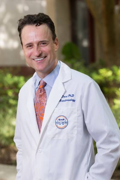 Sylvain Doré合作论文数Department of Anesthesiology, College of Medicine, University of Florida9
 Madison Fangman合作论文数7Brianna Nguyen合作论文数2
 Wayne Brisbane合作论文数Deparment of Urology, University of Florida2
 Ali Solhpour合作论文数Imam Khomeini Hospital, Tehran University of Medical Sciences2Oscar Velazquez合作论文数1Jeremy Archer合作论文数1Michael Maidaa合作论文数1Kiana Saadatmand合作论文数1Raymond Hautamaki合作论文数1Alicia Burris合作论文数1Abdullah Ahmad合作论文数1
 Wenbin Guo合作论文数Digital Worlds Institute, University of Florida1Josh Lua合作论文数1
 Sara M Falzarano合作论文数1
 Joseph R Grajo合作论文数1
 Pradip Kumar Kamat合作论文数1Miriam O Ezenwa合作论文数1
 Garrett Timothy J合作论文数1
 Paul L Crispen合作论文数Department of Urology, University of Florida Health1
 Larry I Lipshultz合作论文数Dan L Duncan Comprehensive Cancer Center, Baylor College of Medicine1A. Bhattacharya合作论文数1
 Kevin Campbell合作论文数Department of Molecular Physiology and Biophysics, Carver College of Medicine, University of Iowa;Wellstone Muscular Dystrophy Specialized Research Center, Carver College of Medicine, University of Iowa;Howard Hughes Medical Institute1Li-Ming Su合作论文数1
 Yahya Ayhan Acar合作论文数1
 Padraic O'Malley合作论文数New York University Langone Medical Center1David Lizdas合作论文数1
 Angelos Barmpoutis合作论文数CISE Department, University of Florida1
 Samsun Lampotang合作论文数Department of Anesthesiology, College of Medicine, University of Florida;Department of Biomedical Engineering, College of Engineering, University of Florida;UF Health Shands Experiential Learning Center1




