Précis: This study of inter-test comparability of a novel visual field application installed on an augmented-reality portable headset and Humphrey field analyzer Swedish interactive thresholding algorithm (SITA) Standard visual field test demonstrates the excellent correlation of mean deviation (MD) and mean sensitivity (MS). Purpose: To determine the correlation between visual field testing with novel software on a wearable headset versus standard automated perimetry. Patients and Methods: Patients with and without visual field defects attributable to glaucoma had visual field testing in one eye of each patient with 2 methods: re:Imagine Strategy (Heru, Inc.) and the Humphrey field analyzer (Carl Zeiss Meditec, Inc.) SITA Standard 24-2 program. Main outcome measures included MS and MD, which were evaluated by linear regression, intraclass correlation coefficient (ICC), and Bland Altman analysis for assessment of the mean difference and limits of agreement. Results: Measurements from 89 eyes of 89 patients (18 normal and 71 glaucomas) were compared with both instruments. Linear regression analysis demonstrated an excellent Pearson correlation coefficient of r = 0.94 for MS and r = 0.95 for MD. ICC analysis demonstrated high levels of concordance (ICC = 0.95, P < 0.001 for MS and ICC = 0.94, P < 0.001 for MD). Bland-Altman analysis determined a small mean difference between the two devices (Heru minus Humphrey) of 1.15 dB for MS and 1.06 dB for MD. Conclusions: The Heru visual field test correlated well with SITA Standard in a population of normal eyes and eyes with glaucoma.
Purpose To evaluate see-through Augmented Reality Digital spectacles (AR DSpecs) for improving the mobility of patients with peripheral visual field (VF) losses when tested on a walking track. Design Prospective Case Series. Participants 21 patients with peripheral VF defects in both eyes, with the physical ability to walk without assistance. Methods We developed the AR DSpecs as a wearable VF aid with an augmented reality platform. Image remapping algorithms produced personalized visual augmentation in real time based on the measured binocular VF with the AR DSpecs calibration mode. We tested the device on a walking track to determine if patients could more accurately identify peripheral objects. Main outcome measures We analyzed walking track scores (number of recognized/avoided objects) and eye tracking data (six gaze parameters) to measure changes in the kinematic and eye scanning behaviors while walking, and assessed a possible placebo effect by deactivating the AR DSpecs remapping algorithms in random trials. Results Performance, judged by the object detection scores, improved with the AR DSpecs (P<0.001, Wilcoxon rank sum test) with an average improvement rate of 18.81%. Two gaze parameters improved with the activated algorithm (P<0.01, paired t-test), indicating a more directed gaze on the central path with less eye scanning. Determination of the binocular integrated VF with the DSpecs correlated with the integrated standard automated perimetry (R = 0.86, P<0.001), mean sensitivity difference 0.8 ± 2.25 dB (Bland-Altman). Conclusions AR DSpecs may improve walking maneuverability of patients with peripheral VF defects by enhancing detection of objects in a testing environment.
PURPOSE:To examine an image remapping method for peripheral visual field (VF) expansion with novel virtual reality digital spectacles (DSpecs) to improve visual awareness in glaucoma patients. DESIGN:Prospective case series. METHODS:Monocular peripheral VF defects were measured and defined with a head-mounted display diagnostic algorithm. The monocular VF was used to calculate remapping parameters with a customized algorithm to relocate and resize unseen peripheral targets within the remaining VF. The sequence of monocular VF was tested and customized image remapping was carried out in 23 patients with typical glaucomatous defects. Test images demonstrating roads and cars were used to determine increased awareness of peripheral hazards while wearing the DSpecs. Patients' scores in identifying and counting peripheral objects with the remapped images were the main outcome measurements. RESULTS:The diagnostic monocular VF testing algorithm was comparable to standard automated perimetric determination of threshold sensitivity based on point-by-point assessment. Eighteen of 23 patients (78%) could identify safety hazards with the DSpecs that they could not previously. The ability to identify peripheral objects improved with the use of the DSpecs (P = 0.024, chi-square test). Quantification of the number of peripheral objects improved with the DSpecs (P = 0.0026, Wilcoxon rank sum test). CONCLUSIONS:These novel spectacles may enhance peripheral objects awareness by enlarging the functional field of view in glaucoma patients.
 Mohamed Abou Shousha合作论文数The Artificial Intelligence and Computer Augmented Vision Laboratory, Bascom Palmer Eye Institute, University of Miami14
 Ahmed Sayed合作论文数8
 Richard K. Parrish合作论文数8Georgeana Mijares合作论文数7John McSoley合作论文数7
 Michael H. Chen合作论文数4Valeria Lopez合作论文数4
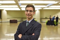 Gustavo Rosa Gameiro合作论文数Dept Ophthalmol & Visual Sci, Fed Univ Sao Paulo EPM UNIFESP4
 Mostafa Abdel-Mottaleb合作论文数Dept Elect & Comp Engn, Univ Miami4
 Collins Opoku-Baah合作论文数4654
 Mary K. Durbin合作论文数Carl Zeiss Meditec, Inc., Carl Zeiss Meditec Inc.4Nadine Rady合作论文数3Christian Duque合作论文数3Abdulla R. Shaheen合作论文数3Catherine Johnson合作论文数3
 Vatookarn Roongpoovapatr合作论文数Bascom Palmer Eye Inst, Univ Miami Hlth Syst3
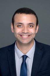 Taher K Eleiwa合作论文数Dept Ophthalmol, Benha Univ3
 Esdras A Arrieta合作论文数3Alexandra Nicklin合作论文数2Feras Mohder合作论文数2Sina Bonyadi合作论文数2Meghan Sharma合作论文数2Alex Hirschfeld Goldbach合作论文数2
 Amr Elsawy合作论文数Electrical and Computer Engineering, University of Miami2
 Mohamed Abdel Mottaleb合作论文数Department of Electrical and Computer Engineering, University of Miami2
 David Taylor Gonzalez合作论文数1Jane C Cook合作论文数1Susan Su合作论文数1
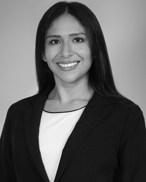 Olga Jumbo合作论文数1
 Md Baharul Islam合作论文数1
 Ozcan Eyup合作论文数1
 William Feuer合作论文数Miller School of Medicine, University of Miami1Richard K. Parrish Ii合作论文数1
 Mohamed A. Tolba合作论文数1




