varices (66.7% vs. 36.6%,p = 0.05) and related bleeding (44.4% vs. 19.5%,p = 0.06).Mortality was not statistically significantly different (22.2% vs. 39.0%,p = 0.18) over the study period.Those with PVTs on average had higher serum albumin levels (3.14 vs. 2.59, p = 0.01), were less likely to have CVC present (0.0% vs. 26.8%,p = 0.04), be smokers, have a prior history of NSVTE, and have an ICU admission during diagnosis of the NSVTE, but were more likely to have a history of cancer and worse thrombocytopenia.There was no significant difference in BMI or baseline MELD.Conclusions: This study demonstrated increased incidence of esophageal varices and variceal bleeding in cirrhotic patients with a history of both PVT and NSVTE.Our results suggest that patients diagnosed with NSVTE and PVT may have a higher incidence of esophageal varices and variceal hemorrhage.Although this study failed to detect an impact on survival, early diagnosis of PVT in patients with NSVTE may help guide therapy and prevent further complications in this patient population.A Doppler ultrasound may be beneficial in this group of patients at the time of HCC screening.Larger, multi-center or prospective studies are needed to confirm these findings.
Background: Gallstone disease is characterized by an impaired gallbladder (GB) emptying and a reduced GB turnover of bile, being the latter assessed by a mathematical analysis of UltraSonographic (US) data (1).In these patients administration of ursodeoxycholic (UDCA) acid seems to be associated to a reduced risk of bifiary pain and complications.The effect of UDCA on GB function is still matter of debate.Aim: To assess by means of statistical analysis of the US motility data, and GB bile flow with computer fluid dynamics the effect of UDCA therapy.Patients and Methods: in 3 GS patients, postprandial US assessment of GB volumes, for a study period of 90 minutes, with a time sampling of 1 minute, has been performed before and after 30 days of UDCA therapy (lOmg/Kg/die).The measured time series were successively analyzed with statistical tools and Fourier analysis.Results: The average volume of the gallbladder of patients is increased upon UDCA treatment (10.6%, 21.1%, 23.9%, respectively).The increase is statistically significant for the three patients.Indeed, a hypothesis test (a T-test) on the means of two distributions led to the conclusions that, with 95% significance, the null hypothesis of equal mean volumes pre and posttreatment must be rejected for the three patients.On the other hand, the organ motility is only slightly affected by UDCA administration, so that the characteristic GB-dimension oscillations remain essentially unaltered.In particular, the time series analysis shows that the volume oscillations have the same amplitude before and after UDCA treatment, and a Fourier analysis shows that also the characteristic frequencies (pre and post treatment) are basically unchanged.A simple geometrical reasoning proves that UDCA treatment improves the intra-GB bile turnover.Conclusions: Short-time treatment with UDCA induces an improvement of postprandial GB bile exchanges in GS patients.Application of standard statistical tests and time series analysis in the frequency domain to US GB motility data could provide additional insights into the assessment of intra-GB bile stasis, a key-event in the pathogenesis of GS disease. 1
Oral tissue samples were studied using mid-IR fiber-optic attenuated total reflectance spectroscopy and other spectral techniques. The 1745 cm(-1) band, which is assigned to the ester group (C==O) vibration of triglycerides, is a reliable marker that is present in normal tissues but absent or a weak band in malignant oral tissues. Other bands such as C--H stretching bands and the amide bands are also helpful in distinguishing malignant tissues from normal tissues. Subtraction spectra confirmed the above conclusion. In addition, Raman spectroscopic measurements were in agreement with the results observed from FTIR spectra.
nism of LPC-induced mucus glycoprotein secretion in the gallbladder (GB) epithelial cells (EC).METHODS: Mucus glycoprotein secretion was studied using cultured dog GBEC (Lab Invest 64:682,1991).PG E2 and 6-ketoPG Fl oin media was assayed by ELISA.Intracellular calcium ([Ca2+]i) was assayed by fluorecent dye method using Fra-2.Effects of LPC (50p,M) on PG synthesis and mucus glycoprotein secretion with or without various inhibitors of arachidonic acid cascades or calcium chelator (EDTA) were examined.Effects of calcium ionophore (ionomycin) or PKC-activator (PMA) on the mucin secretion was also studied.RESULTS: LPC significantly promoted mucus glycoprotein secretion in GBEC.This was associated with increases in PG E2_ and 6keto-PG FI_aproduction as well as in [Caz+]i.Treatment with cyclooxygenase inhibitor (indomethacin) or phospholipase A2 inhibitors (DEPA, OBAA) partially inhibited the LPC-induced mucin secretion.Treatment with Ca z+ chelator (EGTA) also partially inhibited the LPC-induced mucin secretion.Calcium ionophore (ionomycin) and calcium-dependent PKC-activator (PMA) both induced the mucus glycoprotein secretion.CONCLUSIONS: Gallbadder mucus glycoproten secretion is mediated by both arachidonic acid cascade and calcium-dependent pathway.LPC promotes mucus glycoprotein secretion by activating both pathways.
nism of LPC-induced mucus glycoprotein secretion in the gallbladder (GB) epithelial cells (EC).METHODS: Mucus glycoprotein secretion was studied using cultured dog GBEC (Lab Invest 64:682,1991).PG E2 and 6-ketoPG Fl oin media was assayed by ELISA.Intracellular calcium ([Ca2+]i) was assayed by fluorecent dye method using Fra-2.Effects of LPC (50p,M) on PG synthesis and mucus glycoprotein secretion with or without various inhibitors of arachidonic acid cascades or calcium chelator (EDTA) were examined.Effects of calcium ionophore (ionomycin) or PKC-activator (PMA) on the mucin secretion was also studied.RESULTS: LPC significantly promoted mucus glycoprotein secretion in GBEC.This was associated with increases in PG E2_ and 6keto-PG FI_aproduction as well as in [Caz+]i.Treatment with cyclooxygenase inhibitor (indomethacin) or phospholipase A2 inhibitors (DEPA, OBAA) partially inhibited the LPC-induced mucin secretion.Treatment with Ca z+ chelator (EGTA) also partially inhibited the LPC-induced mucin secretion.Calcium ionophore (ionomycin) and calcium-dependent PKC-activator (PMA) both induced the mucus glycoprotein secretion.CONCLUSIONS: Gallbadder mucus glycoproten secretion is mediated by both arachidonic acid cascade and calcium-dependent pathway.LPC promotes mucus glycoprotein secretion by activating both pathways.
Background: Vascular endothelial growth factor (YEGF) is an angiogenic factor that increases vascular permeability .YEGF stimulates capillary formation and has mitogenic effects on vascular endothelial cells.The development of malignant ascites causes significant morbidity .We hypothesized that increased levels of VEGF plays a role in development of malignant ascites in patients with pancreatic cance r.Methods: Athymic mice underwent orthotopic implantation of human pancreatic ductal adenocarcinoma tumors into the body of the pancreas.Tumors were allowed to grow over 3 months until development of ascites was observed in a subset of mice.Paracentesis was performed on mice with noticeable ascites.Saline lavage was performed in mice with equivalent pancreatic tumor sizes without ascites.Both ascites and tumor masses were harvested for YEGF protein quantitation by immunosorbent assay.Results: VEGF protein levels were elevated in malignant ascites by 18 fold as compared to control mice with equivalent tumors (N= 6, P
Corticosteroids (CS) are the main drugs for the treatment of the patients with autoimmune diseases.Dyslipidemia and blood cholesterol (Ch) increase due to prednisone (PS) were observed in these patients.The main Ch catabolism is its transformation to bile acids (BA).Ratio Ch to BA has the significant role in the normal process of bile formation and bile secretion.The aims of the present study were two-fold: first, to evaluate blood lipid profile in patients with systemic lupus erythematosus (SLE), that had constantly used prednisone for a long time and second, to determine the frequency of gallstones in SLE patients depending on prednisone dose and duration of its application.Material and methods.60 patients with SLE were divided in two groups: 28 patients didn t received PS (Group A) and 32 patients with SLE that had received PS for a different time duration (Group B).Gallbladder Ultrasonic examination and blood lipid profile were investigated in all patients.The structure and chemical composition of gallstones were examined with colour cathodoluminescence scanning electron microscopy (CCL SEM).Results .The patients of Group A hadn t gallstones.The gallstones were diagnosed in 22 (61%) patients of Group B. The presence of cholelithiasis in patients with SLE was correlated with the treatment duration and doses of prednisone.All patients, that were treated with prednisone more than 5 years had cholelithiasis.The gallstones received during the cholecystectomy were examined by CCL SEM.Cholesterol and protein component deposits were revealed on the all surface of the scanned gallstone.Bilirubine was demonstrated as impregnation on the gallstone s periphery.Conclusion .• There was correlation of cholelithiasis with prednosone doses and its treatment duration in patients with SLE • Preferably the protein-cholesterol structure of gallstones was determined by the CCL SEM method in patients with SLE and cholelithiasis.
nism of LPC-induced mucus glycoprotein secretion in the gallbladder (GB) epithelial cells (EC).METHODS: Mucus glycoprotein secretion was studied using cultured dog GBEC (Lab Invest 64:682,1991).PG E2 and 6-ketoPG Fl oin media was assayed by ELISA.Intracellular calcium ([Ca2+]i) was assayed by fluorecent dye method using Fra-2.Effects of LPC (50p,M) on PG synthesis and mucus glycoprotein secretion with or without various inhibitors of arachidonic acid cascades or calcium chelator (EDTA) were examined.Effects of calcium ionophore (ionomycin) or PKC-activator (PMA) on the mucin secretion was also studied.RESULTS: LPC significantly promoted mucus glycoprotein secretion in GBEC.This was associated with increases in PG E2_ and 6keto-PG FI_aproduction as well as in [Caz+]i.Treatment with cyclooxygenase inhibitor (indomethacin) or phospholipase A2 inhibitors (DEPA, OBAA) partially inhibited the LPC-induced mucin secretion.Treatment with Ca z+ chelator (EGTA) also partially inhibited the LPC-induced mucin secretion.Calcium ionophore (ionomycin) and calcium-dependent PKC-activator (PMA) both induced the mucus glycoprotein secretion.CONCLUSIONS: Gallbadder mucus glycoproten secretion is mediated by both arachidonic acid cascade and calcium-dependent pathway.LPC promotes mucus glycoprotein secretion by activating both pathways.
Introduction: LAN a proton pump inhibitor, effects healing of ERO through potent acid inhibition, and CIS a prokinetic agent, improves oesophageal peristalsis, lower oesophageal sphincter tone and gastro-duodenal emptying.Aim: To compare the efficacy and safety of LAN vs CIS in the acute treatment of ERO in an open, randomized, parallel, multicentre study.Patients and methods: Outpatients (aged 18 to 75 years) with symptomatic ERO (SavarylMiller grade I to III) were randomized to receive LAN 30mg once daily (n= 132), or CIS 20mg bd (n= 130) for 8 weeks.Efficacy was assessed as endoscopic healing of the ERO (primary variable) and absence/improvement of reflux symptoms (secondary variable) at cessation of therapy.Routine laboratory tests were done at entry.Results: Endoscopic healing rates were 96%(118/123) vs 47%(38/81) after 8 weeks' treatment for LAN and CIS respectively (ITT, p < 0.0001).The mean change in reflux symptom score(Johnson tables)from baseline at 8 weeks was statistically in favour of LAN-treated patients (-15.78 vs -12.27).Most frequent adverse events (LAN and CIS respectively) were headaches (2.3 vs 3.1%), diarrhoea (1.5 vs 3.1%),dizziness (1.5 vs 0.8%), abdominal pain and nausea (0 vs 2.3%).Conclusion: LAN is significantly more efficacious in healing and symptom relief of ERO compared to CIS.
UCP-2 and J3-actin primer s were carried out.UCP-2 expression was normalized to J3-actin expres sion .UCP-2 e xpression of HCC and nontumorou s liver tissue will be compared using paired t-test, Results: The ratio of UCP-2 to J3-act in exp ressio n in HCC and in non-tumorou s tissue is 3.00 :': 0. 19 (SO) and 2.30 :': 0.17 (SD)respectivel y.The ratio is higher in HCC than non-tumoro us tissue in 35 patients (76 %).Using paired t-test, the observed differen ce in UCP-2 expressio n between tumor and nontum orous tissue is highly significant (p = 0.00 16).Conclusion: UCP-2 expression in HCC is higher than non-tumorous cirrhotic liver tissue.The progress from cirrhotic liver to HCC is associated with an up-re gulation of UCP-2 gene expression.However, the biological significance of such observation is to be further defined .
modification of cytosine to uracil.B converts all cytosines to uracil, except methylated (5-methycytosine) which are resistant and remain as cytosine.Primers were designed to discriminate between methylated and unmethylated alleles following B treatment.Our results demonstrated PI6 amplification with the methylated primer set in naive ECL and ECLoma cells.Unmethylated amplification was however only evident in ECLomas.Ecadherin exhibited no evidence for methylation in the MSP assay but MSP analysis revealed the presence of unmethylated alleles in both naive mastomy and ECLoma.These data indicate alterations in both PI6 and E Cadherin methylation patterns during transformation and suggest that DNA methylation is an important issue in ECL cell hypergastrinemic transformation.The further delineation of this pattern of methylation heterogeneity present in ECLoma may be helpful in defining the mechanistic nature of the transformation process.Since E-cadherin, is often silenced by aberrant methylation of the 5'promoter in endocrine driven neoplasia including breast and prostate, the expression of E-cadherin may represent a mechanism involved in the mastomys genomic aberration.
modification of cytosine to uracil.B converts all cytosines to uracil, except methylated (5-methycytosine) which are resistant and remain as cytosine.Primers were designed to discriminate between methylated and unmethylated alleles following B treatment.Our results demonstrated PI6 amplification with the methylated primer set in naive ECL and ECLoma cells.Unmethylated amplification was however only evident in ECLomas.Ecadherin exhibited no evidence for methylation in the MSP assay but MSP analysis revealed the presence of unmethylated alleles in both naive mastomy and ECLoma.These data indicate alterations in both PI6 and E Cadherin methylation patterns during transformation and suggest that DNA methylation is an important issue in ECL cell hypergastrinemic transformation.The further delineation of this pattern of methylation heterogeneity present in ECLoma may be helpful in defining the mechanistic nature of the transformation process.Since E-cadherin, is often silenced by aberrant methylation of the 5'promoter in endocrine driven neoplasia including breast and prostate, the expression of E-cadherin may represent a mechanism involved in the mastomys genomic aberration.
 Jg Wu合作论文数9
 Yizhuang Xu (徐怡庄)合作论文数College of Chemistry and Molecular Engineering, Peking University7S.-F. Weng合作论文数5LM Yang合作论文数4X.-F. Lin合作论文数4George Xiangguo Xu合作论文数4Xs Zhou合作论文数3Z-L Yang合作论文数3
 Shifu Weng (翁诗甫)合作论文数Analytical Instrumentation Center, Peking University3YL Su合作论文数2Js Wang合作论文数2Shu-Ping Lan合作论文数2YF Zhang合作论文数2W Tian合作论文数2JS Shi合作论文数2X. Li合作论文数2
 John Lachin合作论文数Department of Biostatistics and Bioinformatics, Milken Institute School of Public Health, George Washington University2
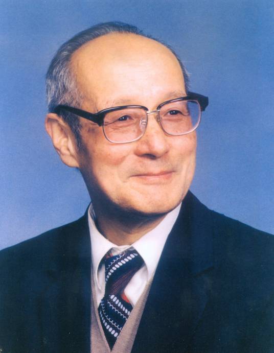 Guangxian Xu (徐光宪)合作论文数Peking University2
 Duanfu Xu (徐端夫)合作论文数Institute of Chemistry, Chinese Academy of Sciences2
 Yong Sun合作论文数School of Pharmacy, Qingdao University2D.-F. Xu合作论文数2
 Datao Xie (谢大弢)合作论文数Institute of Heavy lon Physics, School of Physics, Peking University2
 William F. Balistreri合作论文数Cincinnati Children's Hospital Medical Center2Archie H. Baggenstoss合作论文数2H-Z Jia合作论文数2Marcelle J. Shapiro合作论文数2RA BAUM合作论文数1S M Grundy合作论文数1N C Hightower合作论文数1T Hersh合作论文数1randall j mack合作论文数1Wayne N. Crow合作论文数1R F Hanson合作论文数1Bijun Yang合作论文数1Mg Geall合作论文数1Yy Song合作论文数1Fanxiu Guo合作论文数1SP HELMRICH合作论文数1PD STOLLEY合作论文数1Jl Thistle合作论文数1Fb Thomas合作论文数1Jw Marks合作论文数1LJ SCHOENFIELD合作论文数1Lj Schoenfield合作论文数1Xf Ling合作论文数1XJ Sun合作论文数1Bl Stubbs合作论文数1Js Schwartz合作论文数1AG Zheng合作论文数1




