Plants possess cell-surface recognition receptors that detect molecular patterns from microbial invaders and initiate an immune response. Understanding the conservation of pattern-triggered immunity within different plant organs and across species is crucial to its sustainable and effective use in plant disease management but is currently unclear.We examined the activation and immune response patterns of three pattern recognition receptors (PRRs: SlFLS2, SlFLS3, and SlCORE) in different developmental regions of roots and in leaves of multiple accessions of domesticated and wild tomato (Solanum lycopersicum and S. pimpinellifolium) using biochemical and genetic assays.Roots from different tomato accessions differed in the amplitude and dynamics of their immune response, but all exhibited developmental-specific PTI responses in which the root early differentiation zone was the most sensitive to molecular patterns. PRR signaling pathways also showed distinct but occasionally overlapping responses downstream of each immune receptor in tomato roots.These results reveal that each PRR initiates a unique PTI pathway and suggest that the specificity and complexity of tomato root immunity are tightly linked to the developmental stage, emphasizing the importance of spatial and temporal regulation in PTI.
Plant disease limits crop production, and host genetic resistance is a major means of control. Plant pathogenic Ralstonia causes bacterial wilt disease and is best controlled with resistant varieties. Tomato wilt resistance is multigenic, yet the mechanisms of resistance remain largely unknown. We combined metaRNAseq analysis and functional experiments to identify core Ralstonia-responsive genes and the corresponding biological mechanisms in wilt-resistant and wilt-susceptible tomatoes. While trade-offs between growth and defence are common in plants, wilt-resistant plants activated both defence responses and growth processes. Measurements of innate immunity and growth, including reactive oxygen species production and root system growth, respectively, validated that resistant plants executed defence-related processes at the same time they increased root growth. In contrast, in wilt-susceptible plants roots senesced and root surface area declined following Ralstonia inoculation. Wilt-resistant plants repressed genes predicted to negatively regulate water stress tolerance, while susceptible plants repressed genes predicted to promote water stress tolerance. Our results suggest that wilt-resistant plants can simultaneously promote growth and defence by investing in resources that act in both processes. Infected susceptible plants activate defences, but fail to grow and so succumb to Ralstonia, likely because they cannot tolerate the water stress induced by vascular wilt.
This paper reports molecular diffusion behavior in two bolaamphiphile-based organic nanotubes having inner carboxyl groups with different inner dimeters (10 and 20 nm) and wall structures, COOH-ONT10nm and COOH-ONT20nm, using imaging fluorescence correlation spectroscopy (imaging FCS). The results were compared to those previously obtained in a similar nanotube with inner amine groups (NH2-ONT10nm). COOH-ONT10nm, as with NH2-ONT10nm, were formed from a rolled bolaamphiphile layer incorporating triglycine moieties, whereas COOH-ONT20nm consisted of four stacks of triglycine-free bolaamphiphile layers. Imaging FCS measurements were carried out for anionic sulforhodamine B (SRB), zwitterionic/cationic rhodamine B (RB), and cationic rhodamine-123 (R123) diffusing within ONTs (1-9 mu m long) at different pH (3.4-8.4) and ionic strengths (1.6-500 mM). Diffusion coefficients (D) of these dyes in the ONTs were very small (0.01-0.1 mu m(2)/s), reflecting the significant contributions of molecule-nanotube interactions to diffusion. The D of SRB was larger at higher pH and ionic strength, indicating the essential role of electrostatic repulsion that was enhanced by the deprotonation of the inner carboxyl groups. Importantly, the D of SRB was virtually independent of nanotube inner diameter and wall structure, indicating the diffusion of the hydrophilic molecule was controlled by short time scale adsorption/desorption processes onto the inner surface. In contrast, pH effects on D were less clear for relatively hydrophobic R123 and RB, suggesting the significant contributions of non-Coulombic interactions. Interestingly, the diffusion of these molecules in COOH-ONT20nm was slower than in COOH-ONT10nm. Slower diffusion in COOH-ONT20nm was attributable to relatively efficient partitioning of the hydrophobic dyes into the bolaamphiphile layers, which was reduced in COON-ONT10nm due to the stabilization of its layer by polyglycine-II-type hydrogen bonding networks. These results show that, by tuning the bolaamphiphile structures and their intermolecular interactions, unique environments can be created within the nanospaces for enhanced molecular separations and reactions.
Bart Bartlett, a professor of chemistry at the University of Michigan, was an ACS Scholar from 1998 to 2000, during his junior and senior years of college at Washington University in St. Louis. In this interview, Bartlett talks about what inspired him to go into chemistry and the importance of having an alumni network. This interview was edited for length and clarity. What motivated you to go into chemistry? I got into chemistry largely because I had a good experience with chemistry in high school. I had a really good high school chemistry teacher, and I liked the subject. The summer after my junior year, I started doing research at the Washington University School of Medicine with the Department of Genetics. There's a program called the Young Scientist Program. It was instrumental in getting me to see how a lab functioned. Unlike in a high school lab class where you
Viruses are a prolific force able to infect any form of life on the planet, including plants. Around the world, plant viruses cause considerable yield loss in many agriculture crops every year. Antiviral immunity in plants is mediated by RNA silencing and RNA decay mechanisms, which proceed as follows: once a virus inserts its genetic material into a cell, dicer‐like ribonucleases (DCLs) detect the viral RNA and fragment it into small interfering RNAs (siRNAs). These RNA fragments are then bound by Argonaute proteins and form an RNA‐induced silencing complex (RISC). The RISC binds to complementary sequences of the viral RNA and remains bound, thus deactivating or degrading the fragment by enzymatically cleaving the RNA. siRNA antiviral RNA silencing is utilized for acute or cellular antiviral defense, but it also plays a role in systemic antiviral defense. In plants the systemic silencing mechanism is mediated by enzymes called RNA‐dependent RNA polymerases (RDR) that synthesize double stranded RNA (dsRNA) from single strand RNA. These dsRNAs are then processed through the same siRNA‐silencing pathway, producing new siRNAs that can silence viral genes. The supplementary siRNAs can then be transported between cells through the plants’ vasculature and the plasmodesmata. This leads to systemic treatment and defense in the plant. Though the core genetic components of antiviral RNA silencing have been determined, additional components remain to be identified and characterized. For example, the A. thaliana genome encodes six RDR genes. It is known that RDR1 and RDR6 have major roles in antiviral RNA silencing and that RDR2 mediates biogenesis of cellular siRNAs. However, the role of the RDR3, RDR4, and RDR5 genes is not known. Though single mutants of RDR3, RDR4, and RDR5, have been constructed, double or triple mutants cannot be obtained by tDNA mutation because all three genes are linked together on the second chromosome. Our hypothesis was that RDR3 and RDR4 can be inactivated by site‐specific genome editing using the CRISPR/Cas9 technique to develop mutants lacking these genes. Further experiments show the expression of the Cas9 protein in the second generation, confirming transformation of A. thaliana proteins and RNA. We expect that creating this sextuple mutant, in which all six RDR genes are deactivated, allows the continued exploration of the role of RDRs in antiviral silencing. The RDR3 and RDR4 inactive mutants will also facilitate the necessary positive and negative control mutants needed for future analytical studies.Support or Funding InformationUndergraduate Creative Activities and Research Experience Project, funded by the PepsiQuasi Endowment and Union Bank & Trust. National Institute of Health (R01GM120108). Institute of Agriculture and Natural Resources, Agricultural Research Division, Undergraduate Student Research ProgramCRISPR single guide RNAs.A) Target Sites and sgRNAs in RDR3a an dRDR3b found using CCTOP (Stemmer et al. 2015). Small guide RNAs are the target sites minus the PAM colored in red. B) Single guide RNAs targeting RDR3a and RDR3b.Figure 1
 Anjali S Iyer-Pascuzzi合作论文数Biology Department, Duke University2
 Caitilyn Allen合作论文数Department of Plant Pathology, College of Agricultural & Life Sciences, University of Wisconsin-Madison1
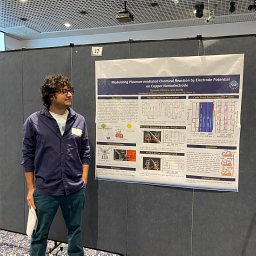 Govinda Ghimire合作论文数Physics Department, Florida International University1
 Rachel Hiles合作论文数Department of Botany and Plant Pathology, Purdue University1
 Valerian Meline合作论文数Dept Bot & Plant Pathol, Purdue Univ1
 Denise Lynn Caldwell合作论文数Department of Botany and Plant Pathology and Center for Plant Biology, Purdue University1
 Tri Tran合作论文数1Danielle M Stevens合作论文数1Connor G. Hendrich合作论文数1
 Ito Takashi合作论文数Dept Chem, Kansas State Univ1
 Alicia N Truchon合作论文数Department of Plant Pathology, University of Wisconsin-Madison1
 Roberts Robyn合作论文数Department of Plant Pathology, University of Wisconsin-Madison1Daniel A. Higgins合作论文数Department of Chemistry;University of WisconsirrMadison;Department of Chemistry, University of WisconsirrMadison1
 Mitsutoshi Masuda合作论文数Nanoarchitectonics Research Center (NARC),, National Institute of Advanced Industrial Science and Technology (AIST),1
 Gitta Coaker合作论文数Department of Plant Pathology, University of California, Davis1Raka M. Mitra合作论文数Department of Plant Biology;Plant Genomics Institute;University of Minnesota;Plant Genomics Institute, University of Minnesota1Shinobu Nagasaka合作论文数1
 Naohiro Kameta合作论文数Faculty of Science, Ibaraki University1




