Despite advances in the treatment of B-cell non-Hodgkin lymphomas (B-NHL) and B-cell acute lymphoblastic leukemia (B-ALL), disease resistance, relapse, and treatment-related toxicities remain significant challenges. Antibody-drug conjugates (ADCs) have transformed the treatment of many cancers by enabling the selective delivery of cytotoxic payloads to malignant cells, potentially reducing systemic toxicities and improving patient outcomes. Here we describe AZD4512, a novel, first-in-class ADC targeting CD22, a surface antigen with expression restricted to the B-cell lineage and B-cell malignancies. The consistent expression of CD22 across different B-NHL and B-ALL subtypes and its rapid internalization upon antibody binding make it an ideal target for ADC therapies. AZD4512 consists of an anti-CD22 human monoclonal antibody conjugated to a topoisomerase 1 inhibitor payload via a novel cleavable linker. The primary mechanism of action for AZD4512 involves the intracellular delivery of the topoisomerase 1 inhibitor payload to CD22- expressing tumor cells via target-mediated internalization, leading to drug-release, DNA damage and apoptotic cell death. In vitro, AZD4512 exhibited potent and specific cytotoxicity against CD22-expressing tumor cells, with IC50 values in the picomolar to sub-nanomolar range. Importantly, AZD4512 did not exhibit cross-resistance in B-NHL cell lines resistant to an ADC containing the microtubule inhibitor MMAE, a payload deployed in multiple ADCs used for the treatment of B-NHL. Notably, AZD4512 demonstrated a substantially greater selectivity in primary cell settings by specifically targeting and killing CD22-positive B cells while not killing CD22-negative T cells, in contrast to a benchmark CD22-calicheamicin ADC, which showed a much narrower selective window. In vivo, AZD4512 exhibited robust antitumor activity and provided significant survival benefits across diverse models of B-NHL and B-ALL. In a study of 11 human B-NHL patient-derived xenografts (PDXs) encompassing diffuse large B-cell lymphoma (DLBCL), mantle cell lymphoma (MCL), and follicular lymphoma (FL), a single intravenous dose of 1.6 mg/kg achieved a 100% overall response rate, defined as at least a 30% reduction in tumor volume from baseline, with a 36% response rate observed at 0.8 mg/kg. In a study of four B-ALL PDX models, including both Ph-positive, Ph-negative, and KMT2A-rearranged subtypes, treatment with AZD4512 at both 1.0 mg/kg and 2.4 mg/kg resulted in a significant reduction of disease burden in peripheral blood in three of the four models across most assessed time points over a five-week post-treatment period. Subsequently, AZD4512 provided a substantial survival benefit in these responsive models: median survival ranged from 35 to 41 days at 1.0 mg/kg and from 45 to 73 days at 2.4 mg/kg, compared to median survival of 27 to 32 days in untreated groups. Toxicology studies showed that AZD4512 was well-tolerated, with no unexpected side effects. Collectively, these findings highlight AZD4512 as a promising therapeutic candidate for the treatment of patients with B-ALL and B-NHL.
KRAS mutations frequently co-occur with alterations in STK11/LKB1 and/or KEAP1, defining an aggressive subset of lung cancers resistant to immuno- and chemotherapy. While LKB1 loss is associated with vulnerability to DNA damage response-based therapies, the impact of KEAP1 alterations remains unknown. We demonstrate that KEAP1-NRF2 pathway drives a compensatory modulation of ATR-CHK1 signaling, enhancing vulnerability to ATR inhibitors (ATRi), particularly in the setting of increased replication stress associated with LKB1 loss. ATRi shows enhanced anti-tumor activity in LKB1 and/or KEAP1-deficient non-small cell lung cancer (NSCLC) models and synergizes with gemcitabine. ATRi also enhances antitumor immunity and mitigates the immunosuppressed phenotype of LKB1/KEAP1-deficient tumors. In the HUDSON trial, LKB1/KEAP1-deficient NSCLC patients demonstrate enhanced benefits to the ATRi ceralasertib plus durvalumab. These findings suggest that alterations in the KEAP1-NRF2 pathway and/or LKB1 are associated with enhanced sensitivity to ATRi and could serve as biomarkers for predicting response to ATRi combination regimens.
Despite the recent advances in our understanding of the role of lipids, metabolites and related enzymes in mediating kidney injury, there is limited integrated multi-omics data identifying potential metabolic pathways driving impaired kidney function. The limited availability of kidney biopsies from living donors with acute kidney injury has remained a major constraint. Here, we validated the use of deceased transplant donor kidneys as a good model to study acute kidney injury in humans and characterized these kidneys using imaging and multi-omics approaches. We noted consistent changes in kidney injury and inflammatory markers in donors with reduced kidney function. Neighborhood and correlation analyses of imaging mass cytometry data showed that subsets of kidney cells (proximal tubular cells and fibroblasts) are associated with the expression profile of kidney immune cells, potentially linking these cells to kidney inflammation. Integrated transcriptomic and metabolomic analysis of human kidneys showed that kidney arachidonic acid metabolism and seven other metabolic pathways were upregulated following diminished kidney function. To validate the arachidonic acid pathway in impaired kidney function we demonstrated increased levels of cytosolic phospholipase A2 protein and related lipid mediators (prostaglandin E2) in the injured kidneys. Further, inhibition of cytosolic phospholipase A2 reduced injury and inflammation in human kidney proximal tubular epithelial cells in vitro. Thus, our study identified cell types and metabolic pathways that may be critical for controlling inflammation associated with impaired kidney function in humans.
Trastuzumab deruxtecan (T-DXd; DS-8201; ENHERTU®) is a human epithelial growth factor receptor 2 (HER2)-directed antibody drug conjugate (ADC) with demonstrated antitumor activity against a range of tumor types. Aiming to understand the relationship between antigen expression and downstream efficacy outcomes, T-DXd was administered in tumor-bearing mice carrying NCI-N87, Capan-1, JIMT-1, and MDA-MB-468 xenografts, characterized by varying HER2 levels. Plasma pharmacokinetics (PK) of total antibody, T-DXd, and released DXd and tumor concentrations of released DXd were evaluated, in addition to monitoring γΗ2AX and pRAD50 pharmacodynamic (PD) response. A positive relationship was observed between released DXd concentrations in tumor and HER2 expression, with NCI-N87 xenografts characterized by the highest exposures compared to the remaining cell lines. γΗ2AX and pRAD50 demonstrated a sustained increase over several days occurring with a time delay relative to tumoral-released DXd concentrations. In vitro investigations of cell-based DXd disposition facilitated the characterization of DXd kinetics across tumor cells. These outputs were incorporated into a mechanistic mathematical model, utilized to describe PK/PD trends. The model captured plasma PK across dosing arms as well as tumor PK in NCI-N87, Capan-1, and MDA-MB-468 models; tumor concentrations in JIMT-1 xenografts required additional parameter adjustments reflective of complex receptor dynamics. γΗ2AX longitudinal trends were well characterized via a unified PD model implemented across xenografts demonstrating the robustness of measured PD trends. This work supports the application of a mechanistic model as a quantitative tool, reliably projecting tumor payload concentrations upon T-DXd administration, as the first step towards preclinical-to-clinical translation.
Ataxia-telangiectasia mutated gene (ATM) is a key component of the DNA damage response (DDR) and double-strand break repair pathway. The functional loss of ATM (ATM deficiency) is hypothesised to enhance sensitivity to DDR inhibitors (DDRi). Whole-exome sequencing (WES), immunohistochemistry (IHC), and Western blotting (WB) were used to characterise the baseline ATM status across a panel of ATM mutated patient-derived xenograft (PDX) models from a range of tumour types. Antitumour efficacy was assessed with poly(ADP-ribose)polymerase (PARP, olaparib), ataxia- telangiectasia and rad3-related protein (ATR, AZD6738), and DNA-dependent protein kinase (DNA-PK, AZD7648) inhibitors as a monotherapy or in combination to associate responses with ATM status. Biallelic truncation/frameshift ATM mutations were linked to ATM protein loss while monoallelic or missense mutations, including the clinically relevant recurrent R3008H mutation, did not confer ATM protein loss by IHC. DDRi agents showed a mixed response across the PDX's but with a general trend toward greater activity, particularly in combination in models with biallelic ATM mutation and protein loss. A PDX with an ATM splice-site mutation, 2127T > C, with a high relative baseline ATM expression and KAP1 phosphorylation responded to all DDRi treatments. These data highlight the heterogeneity and complexity in describing targetable ATM-deficiencies and the fact that current patient selection biomarker methods remain imperfect; although, complete ATM loss was best able to enrich for DDRi sensitivity.
Fliegende Elefanten und empfindliche Fracht. Die Elektrospray-Ionisierung von Proteinen wurde damit verglichen, molekulare Elefanten fliegen zu lassen. Helen J. Cooper und Mitarbeiter (e202202075) demonstrieren den massenspektrometrischen Nachweis und die Bildgebung eines in vivo erzeugten nicht-kovalenten Komplexes, der aus einem endogenen Protein und einem exogen verabreichten Wirkstoffmolekül besteht, direkt in Lebergewebe. Die Methode sollte bessere Einblicke in Protein-Wirkstoff-Komplexe zur Unterstützung der Wirkstoffentwicklung ermöglichen.
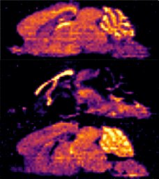
: Here, we demonstrate detection by mass spectrometry of an intact protein–drug complex directly from liver tissue from rats that had been orally dosed with the drug. The protein–drug complex comprised fatty acid binding protein 1, FABP1, non-covalently bound to the small molecule therapeutic bezafibrate. Moreover, we demonstrate spatial mapping of the [FABP1 + bezafibrate] complex across a thin section of liver by targeted mass spectrometry imaging. This work is the first demonstration of in situ mass spectrometry analysis of a non-covalent protein–drug complex formed in vivo and has implications for early stage drug discovery by providing a route to target-drug characterization directly from the physiological environment.
Introduction: Trastuzumab deruxtecan (T-DXd) is an antibody-drug conjugate comprised of an anti-HER2 antibody, a cleavable tetrapeptide-based linker, and a cytotoxic topoisomerase I inhibitor approved for the treatment of HER2 positive metastatic breast and gastric cancer. T-DXd has demonstrated antitumor activity in both HER2+ and HER2-low patient populations. Methods: To establish exposure profiles of T-DXd and link with tumor biomarker changes, we administered a single IV dose of T-DXd at 10 mg/kg in human tumor xenograft models representing HER2-positive (NCI-N87; nude) and Her2-low (Capan-1; NOD-SCID) and collected tumor and plasma from 6 h to 336 h post dose. We measured tumor volume in addition to total ADC, total antibody, and free payload in the plasma and assessed biomarkers related to DNA damage in the tumor by western blot (WB) and immunohistochemistry (IHC). Results: In HER2+ NCI-N87 tumor-bearing mice, T-DXd plasma AUC was 342.6ug/ml*day and T1/2 was 3.5 days, while in the HER2-low Capan-1 tumor-bearing mice, T-DXd plasma AUC was 297.2ug/ml*day and T1/2 was 1.4 days. Plasma exposures of free payload (DXd) were less than 1 ng/mL. Both models responded to T-DXd, demonstrating regression over the 14 day study (T/C =-6.08%, NCI-N87 and -96.1%, Capan-1). In NCI-N87, we observed rapid and sustained increases in gamma H2AX (gH2AX), with a 3.5-fold increase in % positive staining by IHC (H-score p<0.001) with gH2AX foci as early as 24h post treatment, and sustained out to 96h. Western blot analysis and quantification of gH2AX revealed a 4.3-fold increase at 48h (p<0.0001), which was sustained out to 96h. Further, we observed significant increases in pRAD50 at 24h (3.6-fold increase by IHC, H-score p<0.001) which was sustained out to 96h. In Capan-1, we observed more diffuse staining of gH2AX and non-significant 1.4-fold increase in gH2AX at 24h. pRAD50 increases were delayed in the Capan-1 model with a 2.8-fold increase observed at 48h and sustained out to 168h post treatment. Conclusions: Plasma exposure of T-DXd in NCI-N87 tumor bearing mice was prolonged compared to Capan-1, possibly due to mouse strain differences. The increased systemic exposure resulted in more rapid and sustained DNA damage as measured by gH2AX and pRAD50 in the NCI-N87 tumor compared to Capan-1. This profile suggests exploration of combinations with DNA damage response inhibitors to inform design of dose and schedule of combination therapy may be warranted. Citation Format: Theresa Proia, Jelena Urosevic, Christina Vasalou, Rebecca Sargeant, Matthew Griffin, Jiaqi Yuan, Anton I. Rosenbaum, Jerome Mettetal. Pharmacokinetic and pharmacodynamic evaluation of human tumor xenograft models treated upon administration of trastuzumab deruxtecan [abstract]. In: Proceedings of the American Association for Cancer Research Annual Meeting 2022; 2022 Apr 8-13. Philadelphia (PA): AACR; Cancer Res 2022;82(12_Suppl):Abstract nr 1141.
Here, we demonstrate detection by mass spectrometry of an intact protein-drug complex directly from liver tissue from rats that had been orally dosed with the drug. The protein-drug complex comprised fatty acid binding protein 1, FABP1, non-covalently bound to the small molecule therapeutic bezafibrate. Moreover, we demonstrate spatial mapping of the [FABP1+bezafibrate] complex across a thin section of liver by targeted mass spectrometry imaging. This work is the first demonstration of in situ mass spectrometry analysis of a non-covalent protein-drug complex formed in vivo and has implications for early stage drug discovery by providing a route to target-drug characterization directly from the physiological environment.
Current clinical poly (ADP-ribose) polymerase (PARP) inhibitors target both PARP1 and PARP2 and they all cause clinical cytopenias with varying severity. Understanding the mechanism underlying the hematological toxicity of these agents is key for the rational design of a best-in-class molecule with greater therapeutic potential, both as monotherapy and in combination with chemotherapy. We validated the rat as a physiologically competent translational model to investigate PARP inhibitor-driven hematological toxicity. Here we demonstrate that in comparison with a representative PARP1/2 inhibitor, olaparib, the novel highly potent PARP1-selective inhibitor and trapper, AZD5305, does not cause hematological toxicity as a monotherapy in pre-clinical rat models at predicted clinically efficacious exposures. Thus, monotherapy toxicity of PARP1/2 inhibitors likely depends on PARP2 inhibition. Next, we proceeded to investigate whether PARP1-selective inhibition would be better tolerated in chemotherapy combinations than PARP1/2 inhibition. We performed a rat in vivo study comparing daily olaparib or daily AZD5305 at matched exposures in combination with one cycle of intravenous carboplatin for 14 days. We show that olaparib and PARP1-selective AZD5305 cause comparable exacerbation of carboplatin-induced peripheral blood effects implicating PARP1 inhibition in combination-driven hematological toxicity. Importantly however, AZD5305+carboplatin showed improved hematological tolerability over olaparib+carboplatin because peripheral reticulocytes and bone marrow erythroid precursor cells recover in the presence of continuous AZD5305 but not in the presence of continuous olaparib. Importantly, this differentiation was maintained in a subsequent rat in vivo study, where daily olaparib or daily AZD5305 were combined with two three-weekly cycles of a higher dose of carboplatin to more closely mimic clinical protocol. AZD5305+carboplatin was associated with a more rapid recovery of reticulocytes, red blood cells and hemoglobin following both cycles of carboplatin. In contrast, olaparib+carboplatin was associated with a slower recovery resulting in a more sustained reduction in red cells and hemoglobin during both the first and second cycle of carboplatin. Thus, in rodents the novel potent PARP1-selective inhibitor AZD5305 has improved hematological tolerability over dual PARP1/2 inhibitors, both as a monotherapy and in carboplatin combinations. Citation Format: Sonja J. Gill, Ruth Macdonald, Carmen Pin, Rob Collins, Emilyanne Leonard, Gareth Maglennon, Andy Pike, Peter Cotton, Glen Hawthorne, Jordan Pugh, Rebecca Sargeant, Daniel Sutton, James Atkinson, Stewart Jones, Sarah Chinery, Mark Anderton. The novel PARP1-selective inhibitor AZD5305 has reduced hematological toxicity when compared to PARP1/2 inhibitors in pre-clinical models [abstract]. In: Proceedings of the American Association for Cancer Research Annual Meeting 2021; 2021 Apr 10-15 and May 17-21. Philadelphia (PA): AACR; Cancer Res 2021;81(13_Suppl):Abstract nr 1374.
Cardiomyocytes (CMs) derived from induced pluripotent stem cells (iPSCs) provide an in vitro model of the human myocardium. Complex 3D scaffolded culture methods improve the phenotypical maturity of iPSC-CMs, although typically at the expense of throughput. We have developed a novel, scalable approach that enables the use of iPSC-CM 3D spheroid models in a label-free readout system in a standard 96-well plate-based format. Spheroids were accurately positioned onto recording electrodes using a magnetic gold–iron oxide nanoparticle approach. Remarkably, both contractility (impedance) and extracellular field potentials (EFPs) could be detected from the actively beating spheroids over long durations and after automated dosing with pharmacological agents. The effects on these parameters of factors, such as co-culture (including human primary cardiac fibroblasts), extracellular buffer composition, and electrical pacing, were investigated. Beat amplitudes were increased greater than 15-fold by co-culture with fibroblasts. Optimization of extracellular Ca2+ fluxes and electrical pacing promoted the proper physiological response to positive inotropic agonists of increased beat amplitude (force) rather than the increased beat rate often observed in iPSC-CM studies. Mechanistically divergent repolarizations in different spheroid models were indicated by their responses to BaCl2 compared with E-4031. These studies demonstrate a new method that enables the pharmacological responses of 3D iPSC-CM spheroids to be determined in a label-free, standardized, 96-well plate-based system. This approach could have discovery applications across cardiovascular efficacy and safety, where parameters typically sought as readouts of iPSC-CM maturity or physiological relevance have the potential to improve assay predictivity.
A B S T R A C T Background: Extracellular microRNAs enter kidney cells and modify gene expression. We used a Dicer-hepa- tocyte-speci fi c microRNA conditional-knock-out (Dicer-CKO) mouse to investigate microRNA transfer from liver to kidney. Methods: Dicer fl ox/ fl ox mice were treated with a Cre recombinase-expressing adenovirus (AAV8) to selectively inhibit hepatocyte microRNA production (Dicer-CKO). Organ microRNA expression was measured in health and following paracetamol toxicity. The functional consequence of hepatic microRNA transfer was determined by measuring the expression and activity of cytochrome P450 2E1 (target of the hepatocellular miR- 122), and by measuring the effect of serum extracellular vesicles (ECVs) on proximal tubular cell injury. In humans with liver injury we measured microRNA expression in urinary ECVs. A murine model of myocardial infarction was used as a non-hepatic model of microRNA release. Findings: Dicer-CKO mice demonstrated a decrease in kidney miR-122 in the absence of other microRNA changes. During hepatotoxicity, miR-122 increased in kidney tubular cells; this was abolished in Dicer-CKO mice. Depletion of hepatocyte microRNA increased kidney cytochrome P450 2E1 expression and activity. Serum ECVs from mice with hepatotoxicity increased proximal tubular cell miR-122 and prevented cisplatin toxicity. miR-122 increased in urinary ECVs during human hepatotoxicity. Transfer of microRNA was not restricted to liver injury (cid:2) miR-499 was released following cardiac injury and correlated with an increase in the kidney. Interpretation: Physiological transfer of functional microRNA to the kidney is increased by liver injury and this signalling represents a new paradigm for understanding the relationship between liver injury and renal function.
Extracellular microRNAs have been demonstrated to have the ability to enter kidney tubular cells and modify gene expression. We have used a Dicer-hepatocyte-specific microRNA conditional knock-out (Dicer-CKO) mouse to investigate functional microRNA transfer from liver to kidney under physiological conditions and in the context of drug toxicity. Dicer-CKO mice demonstrated a time-dependent decrease in the hepatocyte-derived microRNA, miR-122, in the kidney in the absence of other microRNA changes. During hepatotoxicity, miR-122 increased in kidney tubular cells; this was abolished in Dicer-CKO mice. Depletion of hepatocyte microRNAs increased expression and activity of the miR-122 target - cytochrome (CYP) P450 2E1 - in the kidney. Serum extracellular vesicles (ECVs) from mice with hepatotoxicity increased proximal tubular cell miR-122 and prevented cisplatin proximal tubular cell toxicity. miR-122 also increased in urinary ECVs during hepatotoxicity in humans. Transfer of microRNA was not restricted to liver injury – we detected miR-499 release with murine cardiac injury, and this correlated with an increase in the kidney. In summary, a physiological transfer of microRNA to the kidney exists, which is increased by liver injury. Regulation of renal drug response due to signalling by microRNA of hepatic origin represents a new paradigm for understanding and preventing nephrotoxicity.
Significant advances in immunotherapies have resulted in the increasing need of predictive preclinical models to improve immunotherapeutic drug development, treatment combination, and to prevent or minimize toxicity in clinical trials. Immunodeficient mice reconstituted with human immune system (HIS), termed humanized mice or HIS mice, permit detailed analysis of human immune biology, development, and function. Although this model constitutes a great translational model, some aspects need to be improved as the incomplete engraftment of immune cells, graft versus host disease and the lack of human cytokines and growth factors. In this review, we discuss current HIS platforms, their pathology, and recent advances in their development to improve the quality of human immune cell reconstitution. We also highlight new technologies that can be used to better understand these models and how improved characterization is needed for their application in immuno-oncology safety, efficacy, and new modalities therapy development.
Multicellular tumor spheroids (MTS) are a well-established model system for drug development and are a valuable in vitro research tool for use prior to employing animal models. These 3D-cell cultures are thought to display chemical gradients of oxygen and nutrients throughout their structure, giving rise to distinct microenvironments in radial layers, thus, mimicking the pathophysiological environment of a tumor. Little is known about the localized distributions of metabolites within these microenvironments. To address this, here we utilize high spectral resolution Fourier-transform ion cyclotron resonance (FT-ICR), MALDI mass spectrometry imaging (MSI) to image the distribution of endogenous metabolites in breast cancer MCF-7 spheroids. We show that known specific metabolite markers (adenosine phosphates and glutathione) indicate that the central region of these cell culture models experiences increased hypoxic and oxidative stress. By using discriminatory analysis, we have identified which m/z values localize toward the outer proliferative or central hypoxic regions of an MTS. Elemental formulae were assigned with sub-ppm mass accuracy, allowing metabolite assignment. Using this information, we have mapped these metabolites back to distinct pathways to improve our understanding of the molecular environment and biochemistry of these tumor models.
Pharmaceutical agents despite their efficacy to treat disease can cause additional unwanted cardiovascular side effects. Cardiotoxicity is characterized by changes in either the function and/or structure of the myocardium. Over recent years, functional cardiotoxicity has received much attention, however morphological damage to the myocardium and/or loss of viability still requires improved detection and mechanistic insights. A human 3D cardiac microtissue containing human induced pluripotent stem cell-derived cardiomyocytes (hiPS-CMs), cardiac endothelial cells and cardiac fibroblasts was used to assess their suitability to detect drug induced changes in cardiac structure. Histology and clinical pathology confirmed these cardiac microtissues were morphologically intact, lacked a necrotic/apoptotic core and contained all relevant cell constituents. High-throughput methods to assess mitochondrial membrane potential, endoplasmic reticulum integrity and cellular viability were developed and 15 FDA approved structural cardiotoxins and 14 FDA approved non-structural cardiotoxins were evaluated. We report that cardiac microtissues provide a high-throughput experimental model that is both able to detect changes in cardiac structure at clinically relevant concentrations and provide insights into the phenotypic mechanisms of this liability.
 Goodwin Richard J A合作论文数Department of Pharmaceutical Biosciences, Uppsala University5James W. Hughes合作论文数4
 Andreas Dannhorn合作论文数4Stewart Jones合作论文数4
 Oliver J Hale合作论文数Sch Biosci, Univ Birmingham4
 Helen J. Cooper合作论文数School of Biosciences, University of Birmingham4
 Strittmatter Nicole合作论文数4Christina Vasalou合作论文数3Eva Illes-Toth合作论文数3Olivia Matthews合作论文数2
 Jelena Urosevic合作论文数2Jayati Basak合作论文数2John D Tranter合作论文数2Matthew Griffin合作论文数2
 Kylie P Matchett合作论文数Centre for Inflammation Research, The Queen's Medical Research Institute, Edinburgh BioQuarter, University of Edinburgh, Edinburgh EH16 4TJ, UK.2Theresa Proia合作论文数AstraZeneca, Waltham, MA USA2
 Abhishek Srivastava合作论文数2
 Jiaqi Yuan合作论文数AstraZeneca, BioPharmaceut R&D2Pointon Amy合作论文数Innovative Medicines and Early Development, AstraZeneca R&amp;D, Macclesfield SK10 4TG, UK Amy.Pointon@astrazeneca.com.2Williams Dominic P合作论文数2
 Jonathan A. Rose合作论文数2Pilling James E合作论文数2
 Adina Hughes合作论文数2Rollison Helen E合作论文数2Archer Caroline R合作论文数2
 Kevin Park合作论文数Department of Pharmacology and Therapeutics, University of Liverpool2Jerome Mettetal合作论文数2
 Anton I. Rosenbaum合作论文数2
 Kourosh Saeb-Parsy合作论文数Saeb-Parsy Lab, Department of Surgery, University of Cambridge;Revalia Bio;Excelect Ltd;Avatrial;Oxford Immune Algorithmics Ltd;Cambridge University Hospitals NHS Trust;Fitzwilliam College2
 Alan F Lau合作论文数2Simon T. Barry合作论文数1Barbara Musial合作论文数1Sara Talbot合作论文数1Maelle Mairesse合作论文数1
 Jen Barnes合作论文数1John Meekin合作论文数1Harini Shandilya合作论文数1Anna Przybyła合作论文数1Alissa Poteete合作论文数1Rachel Harvey合作论文数1Daniel Jachimowicz合作论文数1Kavya Ramkumar合作论文数1Susan E. Critchlow合作论文数1
 Daniel Sutton合作论文数1
 Alba Matas合作论文数1Sarah Chinery合作论文数1Lulu H Tucker合作论文数1Emanuela M. Cuomo合作论文数1Fynn N Krause合作论文数1




