Transcription factors (TFs) regulate gene expression through chromatin where nucleosomes restrict DNA access. To study how TFs bind nucleosome-occupied motifs, we focused on the reprogramming factors OCT4 and SOX2 in mouse embryonic stem cells. We determined TF engagement throughout a nucleosome at base-pair resolution in vitro, enabling structure determination by cryo-electron microscopy at two preferred positions. Depending on motif location, OCT4 and SOX2 differentially distort nucleosomal DNA. At one position, OCT4-SOX2 removes DNA from histone H2A and histone H3; however, at an inverted motif, the TFs only induce local DNA distortions. OCT4 uses one of its two DNA-binding domains to engage DNA in both structures, reading out a partial motif. These findings explain site-specific nucleosome engagement by the pluripotency factors OCT4 and SOX2, and they reveal how TFs distort nucleosomes to access chromatinized motifs.
Molecular glue compounds induce protein–protein interactions that, in the context of a ubiquitin ligase, lead to protein degradation1. Unlike traditional enzyme inhibitors, these molecular glue degraders act substoichiometrically to catalyse the rapid depletion of previously inaccessible targets2. They are clinically effective and highly sought-after, but have thus far only been discovered serendipitously. Here, through systematically mining databases for correlations between the cytotoxicity of 4,518 clinical and preclinical small molecules and the expression levels of E3 ligase components across hundreds of human cancer cell lines3–5, we identify CR8—a cyclin-dependent kinase (CDK) inhibitor6—as a compound that acts as a molecular glue degrader. The CDK-bound form of CR8 has a solvent-exposed pyridyl moiety that induces the formation of a complex between CDK12–cyclin K and the CUL4 adaptor protein DDB1, bypassing the requirement for a substrate receptor and presenting cyclin K for ubiquitination and degradation. Our studies demonstrate that chemical alteration of surface-exposed moieties can confer gain-of-function glue properties to an inhibitor, and we propose this as a broader strategy through which target-binding molecules could be converted into molecular glues. The cyclin-dependent kinase inhibitor CR8 acts as a molecular glue compound by inducing the formation of a complex between CDK12–cyclin K and DDB1, which results in the ubiquitination and degradation of cyclin K.
In this Article, in Fig. 1a, the 5′ and 3′ labels were reversed in the DNA sequence, and Fig. 4 was missing panel labels a–e. These errors have been corrected online.
Access to DNA packaged in nucleosomes is critical for gene regulation, DNA replication and DNA repair. In humans, the UV-damaged DNA-binding protein (UV-DDB) complex detects UV-light-induced pyrimidine dimers throughout the genome; however, it remains unknown how these lesions are recognized in chromatin, in which nucleosomes restrict access to DNA. Here we report cryo-electron microscopy structures of UV-DDB bound to nucleosomes bearing a 6-4 pyrimidine-pyrimidone dimer or a DNA-damage mimic in various positions. We find that UV-DDB binds UV-damaged nucleosomes at lesions located in the solvent-facing minor groove without affecting the overall nucleosome architecture. In the case of buried lesions that face the histone core, UV-DDB changes the predominant translational register of the nucleosome and selectively binds the lesion in an accessible, exposed position. Our findings explain how UV-DDB detects occluded lesions in strongly positioned nucleosomes, and identify slide-assisted site exposure as a mechanism by which high-affinity DNA-binding proteins can access otherwise occluded sites in nucleosomal DNA.
4-hydroxy-2-oxoglutarate aldolase (HOGA1) is a mitochondrial enzyme that plays a gatekeeper role in hydroxyproline metabolism. Its loss of function in humans causes primary hyperoxaluria type 3 (PH3), a rare condition characterised by excessive production of oxalate. In this study, we investigated the significance of the associated oxaloacetate decarboxylase activity which is also catalysed by HOGA1. Kinetic studies using the recombinant human enzyme (hHOGA1) and active site mutants showed both these dual activities utilise the same catalytic machinery with micromolar substrate affinities suggesting that both are operative in vivo. Biophysical and structural studies showed that pyruvate was a competitive inhibitor with an inhibition constant in the micromolar range. By comparison α-ketoglutarate was a weak inhibitor with an inhibition constant in the millimolar range and could only be isolated as an adduct with the active site Lys196 in the presence of sodium borohydride. These studies suggest that pyruvate inhibits HOGA1 activity during gluconeogenesis. We also propose that loss of HOGA1 function could increase oxalate production in PH3 by decreasing pyruvate availability and metabolic flux through the Krebs cycle.
The small molecule drugs thalidomide, lenalidomide, and pomalidomide induce the ubiquitination and proteasomal degradation of Ikaros (IKZF1) and Aiolos (IKZF3) by mediating their interaction with Cereblon (CRBN), the substrate receptor of the CRL4CRBN ubiquitin ligase. Here we screened the human Cys2-His2 (C2H2) zinc finger (ZF) proteome for degradation by CRL4CRBN in the presence of thalidomide analogues, identifying 11 ZF targets. Structural and functional characterization of the C2H2 zinc finger degron demonstrates how diverse ZF domains bind the drug-CRBN interface. Computational ZF docking, in conjunction with biochemical analysis, predicts that at least 50 zinc-fingers bind the drug-CRBN complex in vitro, a larger number than previously anticipated. These results provide strategies to degrade other zinc finger transcription factors. Citation Format: Quinlan L. Sievers, Georg Petzold, Richard D. Bunker, Aline Renneville, Brian Liddicoat, Wassim Abdulrahman, Tarjei Mikkelsen, Benjamin L. Ebert, Nicolas H. Thoma. The zinc-finger degrome [abstract]. In: Proceedings of the AACR-NCI-EORTC International Conference on Molecular Targets and Cancer Therapeutics; 2019 Oct 26-30; Boston, MA. Philadelphia (PA): AACR; Mol Cancer Ther 2019;18(12 Suppl):Abstract nr PL02-03. doi:10.1158/1535-7163.TARG-19-PL02-03
In mammals, ∼100 deubiquitinases act on ∼20,000 intracellular ubiquitination sites. Deubiquitinases are commonly regarded as constitutively active, with limited regulatory and targeting capacity. The BRCA1-A and BRISC complexes serve in DNA double-strand break repair and immune signaling and contain the lysine-63 linkage-specific BRCC36 subunit that is functionalized by scaffold subunits ABRAXAS and ABRO1, respectively. The molecular basis underlying BRCA1-A and BRISC function is currently unknown. Here we show that in the BRCA1-A complex structure, ABRAXAS integrates the DNA repair protein RAP80 and provides a high-affinity binding site that sequesters the tumor suppressor BRCA1 away from the break site. In the BRISC structure, ABRO1 binds SHMT2α, a metabolic enzyme enabling cancer growth in hypoxic environments, which we find prevents BRCC36 from binding and cleaving ubiquitin chains. Our work explains modularity in the BRCC36 DUB family, with different adaptor subunits conferring diversified targeting and regulatory functions.
The COP9 signalosome (CSN) is a large multi-protein complex in eukaryotes that is an essential regulator of intracellular protein degradation. Structures of human CSN were determined by X-ray crystallography from weakly diffracting crystals variably affected by twinning and rotational pseudo-symmetry. Strategies were developed to overcome further challenges of crystal instability and non-isomorphism that were strongly influenced by handling, radiation damage, and difficulty obtaining heavy atom derivatives. Many subunits of the same fold class were distinguished at low-resolution aided by combinatorial selenomethionine labelling. Here, a detailed account of the crystallographic structure determination of CSN is given as it was approached using low-resolution cluster compound MIRAS phasing, MR-SAD phasing with electron density models, and cross-crystal averaging and co-refinement of multiple structures, exploiting non-isomorphism across unit-cell variants of the same crystal form.
The small molecules thalidomide, lenalidomide, and pomalidomide induce the ubiquitination and proteasomal degradation of the transcription factors Ikaros (IKZF1) and Aiolos (IKZF3) by recruiting a Cys(2)-His(2) (C2H2) zinc finger domain to Cereblon (CRBN), the substrate receptor of the CRL4(CRBN) E3 ubiquitin ligase. We screened the human C2H2 zinc finger proteome for degradation in the presence of thalidomide analogs, identifying 11 zinc finger degrons. Structural and functional characterization of the C2H2 zinc finger degrons demonstrates how diverse zinc finger domains bind the permissive drug-CRBN interface. Computational zinc finger docking and biochemical analysis predict that more than 150 zinc fingers bind the drug-CRBN complex in vitro, and we show that selective zinc finger degradation can be achieved through compound modifications. Our results provide a rationale for therapeutically targeting transcription factors that were previously considered undruggable.
Significance The room temperature structure of natively formed protein nanocrystals consisting of 9,000 unit cells has been solved to 2 Å resolution using an unattenuated X-ray free-electron laser (XFEL) beam, representing, by far, the smallest protein crystals used for protein structure determination by X-ray crystallography to date. Radiation damage limits structure determination from protein crystals using synchrotron techniques, whereas femtosecond X-ray pulses from free-electron lasers enable much higher tolerable doses, extracting more signal per molecule, allowing the study of submicrometer crystals. Radiation-sensitive features, such as disulfide bonds, are well resolved in the XFEL structure despite the extremely high dose (1.3 GGy) used. Analysis of signal levels obtained in this experiment indicates that structure determination from even smaller protein crystals could be possible.
In yeast, Rif1 is part of the telosome, where it inhibits telomerase and checkpoint signaling at chromosome ends. In mammalian cells, Rif1 is not telomeric, but it suppresses DNA end resection at chromosomal breaks, promoting repair by nonhomologous end joining (NHEJ). Here, we describe crystal structures for the uncharacterized and conserved ∼125-kDa N-terminal domain of Rif1 from Saccharomyces cerevisiae (Rif1-NTD), revealing an α-helical fold shaped like a shepherd's crook. We identify a high-affinity DNA-binding site in the Rif1-NTD that fully encases DNA as a head-to-tail dimer. Engagement of the Rif1-NTD with telomeres proved essential for checkpoint control and telomere length regulation. Unexpectedly, Rif1-NTD also promoted NHEJ at DNA breaks in yeast, revealing a conserved role of Rif1 in DNA repair. We propose that tight associations between the Rif1-NTD and DNA gate access of processing factors to DNA ends, enabling Rif1 to mediate diverse telomere maintenance and DNA repair functions.
The cullin–RING ubiquitin E3 ligase (CRL) family comprises over 200 members in humans. The COP9 signalosome complex (CSN) regulates CRLs by removing their ubiquitin-like activator NEDD8. The CUL4A–RBX1–DDB1–DDB2 complex (CRL4A DDB2 ) monitors the genome for ultraviolet-light-induced DNA damage. CRL4A DBB2 is inactive in the absence of damaged DNA and requires CSN to regulate the repair process. The structural basis of CSN binding to CRL4A DDB2 and the principles of CSN activation are poorly understood. Here we present cryo-electron microscopy structures for CSN in complex with neddylated CRL4A ligases to 6.4 Å resolution. The CSN conformers defined by cryo-electron microscopy and a novel apo-CSN crystal structure indicate an induced-fit mechanism that drives CSN activation by neddylated CRLs. We find that CSN and a substrate cannot bind simultaneously to CRL4A, favouring a deneddylated, inactive state for substrate-free CRL4 complexes. These architectural and regulatory principles appear conserved across CRL families, allowing global regulation by CSN.
The COP9 signalosome (CSN) is an essential multi-protein complex in eukaryotes. CSN is a master regulator of intracellular protein degradation, controlling the vast family of cullin-RING ubiquitin (E3) ligases (CRLs). Important in many cellular processes, CSN has prominent roles in DNA repair, cell-cycle control and differentiation. The recent crystal structure of human CSN provides insight into its exquisite regulation and functionality [Lingaraju et al. (2014), Nature (London), 512, 161-165]. Structure determination was complicated by low-resolution diffraction from crystals affected by twinning and rotational pseudo-symmetry. Crystal instability and non-isomorphism strongly influenced by flash-cooling, radiation damage and difficulty in obtaining heavyatom derivatives, were overcome. Many different subunits of the same fold class were distinguished at low resolution aided by combinatorial selenomethionine labelling. As an example of how challenging projects can be approached, the structure determination of CSN is described as it unfolded using clustercompound MIRAS phasing, MR-SAD with electron-density models and crosscrystal averaging exploiting non-isomorphism among unit-cell variants of the same crystal form.
Protein 3D structure can be a powerful predictor of function, but it often faces a critical roadblock at the crystallization step. Rv1738, a protein from Mycobacterium tuberculosis that is strongly implicated in the onset of nonreplicating persistence, and thereby latent tuberculosis, resisted extensive attempts at crystallization. Chemical synthesis of the L- and D-enantiomeric forms of Rv1738 enabled facile crystallization of the D/L-racemic mixture. The structure was solved by an ab initio approach that took advantage of the quantized phases characteristic of diffraction by centrosymmetric crystals. The structure, containing L- and D-dimers in a centrosymmetric space group, revealed unexpected homology with bacterial hibernation-promoting factors that bind to ribosomes and suppress translation. This suggests that the functional role of Rv1738 is to contribute to the shutdown of ribosomal protein synthesis during the onset of nonreplicating persistence of M. tuberculosis.
The great benefits that chemical pesticides have brought to agriculture are partly offset by widespread environmental damage to nontarget species and threats to human health. Microbial bioinsecticides are considered safe and highly specific alternatives but generally lack potency. Spindles produced by insect poxviruses are crystals of the fusolin protein that considerably boost not only the virulence of these viruses but also, in cofeeding experiments, the insecticidal activity of unrelated pathogens. However, the mechanisms by which spindles assemble into ultra-stable crystals and enhance virulence are unknown. Here we describe the structure of viral spindles determined by X-ray microcrystallography from in vivo crystals purified from infected insects. We found that a C-terminal molecular arm of fusolin mediates the assembly of a globular domain, which has the hallmarks of lytic polysaccharide monooxy-genases of chitinovorous bacteria. Explaining their unique stability, a 3D network of disulfide bonds between fusolin dimers covalently crosslinks the entire crystalline matrix of spindles. However, upon ingestion by a new host, removal of the molecular arm abolishes this stabilizing network leading to the dissolution of spindles. The released monooxygenase domain is then free to disrupt the chitin-rich peritrophic matrix that protects insects against oral infections. The mode of action revealed here may guide the design of potent spindles as synergetic additives to bioinsecticides.
The COP9 signalosome (CSN) is a multifunctional ~350 kDa eight-protein complex with highly-specific isopeptidase activity, which regulates the vast cullin-RING family of ubiquitin ligases (CRLs). Ubiquitin ligases direct target-specific ubiquitination and are involved in a multitude of regulatory processes, ultimately linked to protein turnover by the proteosome. Humans have a repertoire of approximately 240 CRLs, more than any other ubiquitin ligase family. The entire human CSN has been structurally characterised de novo using low-resolution (4-Å) X-ray crystallography. The crystallographic analysis was complicated by rotational pseudo-symmetry and twinning, non-isomorphism as well as the absence of experimental phase information beyond ~8 Å resolution. Details of the structure determination procedures, which combined a cluster compound-based phasing strategy, analysis of seven differentially substituted selenomethionyl derivatives, multi-crystal averaging among non-isomorphous data sets with a multi-crystal co-refinement approach, will be described. Some implications of the structure and insight into the regulation of CSN will also be presented.
The 'dissolvasome', composed of TopIIIα, BLM and RMI proteins, coordinates DNA-helicase and DNA-topoisomerase activities to resolve double Holliday junctions (dHJs) generated during DNA recombination and repair. The first crystal structure of a human TopIIIα–Rmi1 subcomplex provides insights into how topoisomerase is stimulated to promote dHJ decatenation. Repair of DNA double-strand breaks via homologous recombination can produce double Holliday junctions (dHJs) that require enzymatic separation. Topoisomerase IIIα (TopIIIα) together with RMI1 disentangles the final hemicatenane intermediate obtained once dHJs have converged. How binding of RMI1 to TopIIIα influences it to behave as a hemicatenane dissolvase, rather than as an enzyme that relaxes DNA topology, is unknown. Here, we present the crystal structure of human TopIIIα complexed to the first oligonucleotide-binding domain (OB fold) of RMI1. TopIII assumes a toroidal type 1A topoisomerase fold. RMI1 attaches to the edge of the gate in TopIIIα through which DNA passes. RMI1 projects a 23-residue loop into the TopIIIα gate, thereby influencing the dynamics of its opening and closing. Our results provide a mechanistic rationale for how RMI1 stabilizes TopIIIα-gate opening to enable dissolution and illustrate how binding partners modulate topoisomerase function.
Yeast telomeres comprise irregular TG₁₋₃ DNA repeats bound by the general transcription factor Rap1. Rif1 and Rif2, along with Rap1, form the telosome, a protective cap that inhibits telomerase, counteracts SIR-mediated transcriptional silencing, and prevents inadvertent recognition of telomeres as DNA double-strand breaks. We provide a molecular, biochemical, and functional dissection of the protein backbone at the core of the yeast telosome. The X-ray structures of Rif1 and Rif2 bound to the Rap1 C-terminal domain and that of the Rif1 C terminus are presented. Both Rif1 and Rif2 have separable and independent Rap1-binding epitopes, allowing Rap1 binding over large distances (42-110 Å). We identify tetramerization (Rif1) and polymerization (Rif2) modules that, in conjunction with the long-range binding, give rise to a higher-order architecture that interlinks Rap1 units. This molecular Velcro relies on Rif1 and Rif2 to recruit and stabilize Rap1 on telomeric arrays and is required for telomere homeostasis in vivo.
In Mycobacterium tuberculosis, the protein MbtN (Rv1346) catalyzes the formation of a double bond in the fatty-acyl moiety of the siderophore mycobactin, which is used by this organism to acquire essential iron. MbtN is homologous to acyl-CoA dehydrogenases, whose general role is to catalyze the α,β-dehydrogenation of fatty-acyl-CoA conjugates. Mycobactins, however, contain a long unsaturated fatty-acid chain with an unusual cis double bond conjugated to the carbonyl group of the mycobactin core. To characterize the role of MbtN in the dehydrogenation of this fatty-acyl moiety, the enzyme has been expressed, purified and crystallized. The crystals diffracted to 2.3 Å resolution at a synchrotron source and were found to belong to the hexagonal space group H32, with unit-cell parameters a = b = 139.10, c = 253.09 Å, α = β = 90, γ = 120°.
In mammals, the enzyme D-xylulokinase (XK; EC 2.7.1.17) catalyses the last step of the glucuronate-xylulose pathway, in which the ketopentose sugar D-xylulose is phosphorylated to yield D-xylulose 5-phosphate (Xu5P). Xu5P is also a metabolite of the pentose phosphate pathway and acts as a signalling molecule that regulates lipogenesis and glycolysis in the liver. To date, no eukaryotic XK has been structurally characterized. A putative human XK was expressed in Escherichia coli aided by molecular chaperones, purified and crystallized. A seeding procedure involving repeated rounds of seeding was developed and proved to be essential for obtaining diffraction-quality crystals. Preliminary X-ray diffraction analysis was performed using synchrotron radiation. This resulted in the collection of a complete diffraction data set to 2.7 Å resolution from a crystal belonging to the trigonal space group P3(1) or P3(2) with unit-cell parameters a = b = 101.87, c = 158.85 Å.
 Nicolas H. Thomä合作论文数Friedrich Miescher Institute for Biomedical Research, Basel, Switzerland13
 Edward Baker合作论文数Faculty of Science, University of Auckland;School of Biological Sciences, University of Auckland11
 Simone Cavadini合作论文数8
 Wassim Abdulrahman合作论文数5
 Kerry Loomes合作论文数School of Biological Sciences, Faculty of Science, The University of Auckland5Mahamadou Faty合作论文数5Matsumoto Syota合作论文数4Georg Petzold合作论文数4Andreas D. Schenk合作论文数4Alessandro Potenza合作论文数3J. M. Dickson合作论文数3
 Hassiepen Ulrich合作论文数3
 Sievers Quinlan L合作论文数Broad Inst Harvard & MIT3Julius Rabl合作论文数Institute of Molecular Biology and Biophysics, ETH Zürich3
 Kaoru Sugasawa (菅澤薫)合作论文数Department of Biology, Graduate School of Science, Kobe University;Biosignal Research Center, Kobe University3
 Grand Ralph S合作论文数University of Auckland, Massey University3
 Dirk Schübeler合作论文数Friedrich Miescher Institute for Biomedical Research;University of Basel3
 Eric Sebastian Fischer合作论文数Department of Biological Chemistry and Molecular Pharmacology, Blavatnik Institute, Harvard Medical School;Department of Cancer Biology, Dana Farber Cancer Institute3Gondichatnahalli M. Lingaraju合作论文数3T. T. Caradoc-Davies合作论文数3
 Benjamin Levine Ebert合作论文数Harvard Medical School;Dana-Farber Cancer Institute;Harvard Stem Cell Institute;Broad Institute3Stefano Mattarocci合作论文数2Tianlai Shi合作论文数2Kenneth N Goldie合作论文数2
 Zuzanna Kozicka合作论文数Friedrich, Miescher Institute for Biomedical Research2
 Brian Liddicoat合作论文数2Hitoshi Kurumizaka合作论文数Lab Struct Biol, Waseda Univ2Renneville Aline合作论文数2Yamamoto Junpei合作论文数2
 David A. Shore合作论文数Department of Molecular Biology, University of Geneva2
 Daniel Hess合作论文数Laboratoire de Chimie Biologique;Universitedes Sciences et Technologies;Laboratoire de Chimie Biologique, Universitedes Sciences et Technologies2
 Ulrich Rass合作论文数2
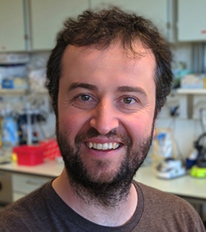 Mikolaj Slabicki合作论文数Benjamin Ebert’s laboratory, Broad Institute of MIT and Harvard;Dana-Farber Cancer Institute2Elaine Chiu合作论文数2
 Thomas Proft合作论文数Department of Molecular Medicine and Pathology, School of Medical Sciences, Faculty of Medical and Health Sciences, The University of Auckland2
 Christian Linke-Winnebeck合作论文数Max Planck Institute for Molecular Genetics, Berlin, Germany2Martin Renatus合作论文数Dana Farber Cancer Institute and Brigham and Women’s Hospital, Harvard Medical School2
 Peter Metcalf合作论文数European Molecular Biology Laboratory, Heidelberg, D-6900, Germany2
 Tarjei S. Mikkelsen合作论文数2Heinz Gut合作论文数Biochemisches Institut der Universität Zürich, Winterthurerstrasse 190, CH-8057 Zürich, Switzerland2
 Shaun Lott合作论文数School of Biological Sciences, Faculty of Science, The University of Auckland2Shigenori Iwai合作论文数2Seth M. Rubin合作论文数Structural Biology Program and Howard Hughes Medical Institute, Memorial Sloan-Kettering Cancer Center2
 Martin J Middleditch合作论文数School of Biological Sciences and Centre for Molecular Biodiscovery;University of Auckland2
 Tewis Bouwmeester合作论文数2Yen Der Li合作论文数1Mark E. Gill合作论文数1
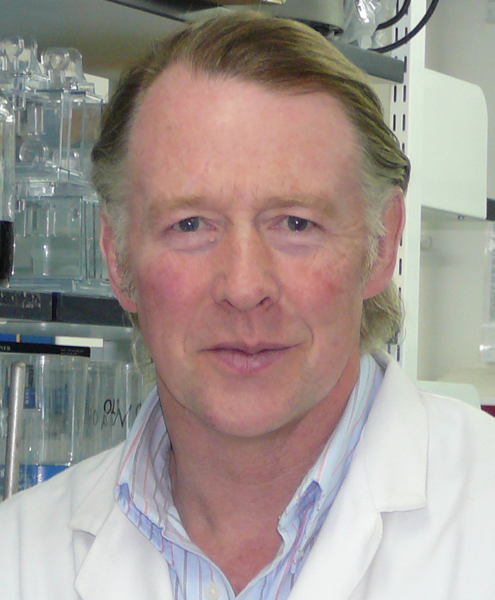 Ronald Hay合作论文数The Division of Molecular, Cell and Developmental Biology, School of Life Sciences, University of Dundee;College of Life Sciences, University of Dundee1Stefan Fröhling合作论文数1




