PURPOSE:Multiuse eye drops must maintain sterility and typically accomplish this by added preservatives. However, preservatives often cause harmful side effects. A gauze barrier dressing ("BIOGUARD®") recently cleared by the FDA has an immobilized poly diallyldimethylammonium chloride (p-DADMAC) coating and is an effective antimicrobial with minimal compound release into solution. To implement use of this dressing as a replacement for preservatives in multidose eye drop bottles, its ability to maintain sterility without interacting with the active ingredient (AI) of the ophthalmic medication was tested.METHODS:To determine immobilized p-DADMAC's microbicidal efficacy, it was added to eye drop bottles, then contaminated with Staphylococcus aureus (SA113) bacteria. To assess interference with AI in eye drops, high performance liquid chromatography was used to determine whether the AIs timolol and dorzolamide were affected after exposure to p-DADMAC. To further investigate effects on AI, the microbicidal activity of Vigamox® (moxifoxacin) was assessed after p-DADMAC gauze exposure.RESULTS:S. aureus bacteria were eliminated by p-DADMAC-treated gauze for all samples. The concentrations of both timolol and dorzolamide increased after exposure to p-DADMAC-treated gauze, but spectrometric analysis showed that this did not occur when the p-DADMAC-coated material was presoaked in deionized water. The microbicidial activity of moxifloxacin was unaffected by exposure to p-DADMAC-treated gauze.CONCLUSIONS:Due to its lack of effect on eye drop AI and its microbicidal efficacy, p-DADMAC treatment would make an excellent candidate for replacing preservatives in eye drops.
IMPORTANCE:The Cornea Donor Study (CDS) showed that donor age is not a factor in survival of most penetrating keratoplasties for endothelial disease. Secondary analyses confirm the importance of surgical indication and presence of glaucoma in outcomes at 10 years.OBJECTIVE:To assess the relationship between donor and recipient factors and corneal graft survival in the CDS.DESIGN, SETTING, AND PARTICIPANTS:Multicenter prospective, double-masked, controlled clinical trial conducted at 80 clinical sites. One hundred five surgeons enrolled 1090 participants undergoing corneal transplant for a moderate-risk condition, principally Fuchs dystrophy or pseudophakic or aphakic corneal edema (PACE). Forty-three eye banks provided corneas.INTERVENTIONS:Corneas from donors younger than 66 years and donors 66 years or older were assigned, masked to donor age. Surgery and postoperative care were performed according to the surgeons' usual routines. Participants were followed up for as long as 12 years.MAIN OUTCOMES AND MEASURES:Graft failure, defined as a regrafting procedure or a cloudy cornea for 3 consecutive months.RESULTS:The 10-year cumulative probability of graft failure was higher in participants with PACE than in those with Fuchs dystrophy (37% vs 20%; hazard ratio [HR], 2.1 [99% CI, 1.4-3.0]; P < .001) and in participants with a history of glaucoma before penetrating keratoplasty, particularly with prior glaucoma surgery (58% with prior glaucoma surgery and use of medications to lower intraocular pressure at the time of surgery vs 22% with no history of glaucoma surgery or medication use; HR, 4.1 [99% CI, 2.2-7.5]; P < .001). We found trends toward increased graft failure in recipients who were 70 years or older compared with those younger than 60 years (29% vs 19%; HR, 1.2 [99% CI, 0.7-2.1]; P = .04) or were African American (HR, 1.5; P = .11) or who had a history of smoking (35% vs 24%; HR, 1.6 [99% CI, 0.9-2.8]; P = .02). Lower endothelial cell density (ECD) and higher corneal thickness (CT) at 6 months (6% vs 41% for ECD ≥2700 vs <1700 cells/mm2 [P < .001]; 14% vs 36% for CT <500 vs ≥600 μm [P = .001]), 1 year (4% vs 39% for ECD ≥2700 vs <1700 cells/mm2 [P < .001]; 18% vs 28% for CT <500 vs ≥600 μm [P = .04]), and 5 years (2% vs 29% for ECD ≥1500 vs <500 cells/mm2 [P < .001]; 7% vs 34% for CT <550 vs ≥650 μm [P < .001]) were associated with subsequent graft failure.CONCLUSIONS AND RELEVANCE:Most penetrating corneal grafts for Fuchs dystrophy or PACE remain clear at 10 years. The risk for failure is greater for graft recipients with PACE and those with a history of glaucoma. Measurements of ECD and CT during the course of postkeratoplasty follow-up are associated with a risk for failure. However, even with very low ECD and high CT at 5 years, most corneas remain clear at 10 years.
Purpose To assess the efficacy and safety of lifitegrast ophthalmic solution 5.0% compared with placebo in subjects with dry eye disease. Design Prospective, randomized, double-masked, placebo-controlled, parallel arm, multicenter clinical trial. Participants A total of 588 adult subjects with dry eye disease. Methods Eligible subjects were randomized 1:1 to receive topically administered lifitegrast (5.0%) or placebo (vehicle) twice daily for 84 days after a 14-day open-label placebo run-in period. After enrollment (day 0), subjects were evaluated at days 14, 42, and 84. Key objective (fluorescein and lissamine staining scores [Ora scales]) and subjective (Ocular Surface Disease Index [OSDI], 7-item visual analog scale, and ocular discomfort score [Ora scale]) measures were assessed at all visits. Main Outcome Measures The primary objective efficacy measure (sign) was mean change from baseline inferior corneal staining score (ICSS) at day 84. The co-primary subjective efficacy measure (symptom) was the mean change from baseline in the visual-related function subscale score of the Ocular Surface Disease Index (VR-OSDI). Supportive measures included corneal fluorescein scores (superior, central, total region) and conjunctival lissamine scores (nasal, temporal, total region) and symptom scores at day 84. Results The study met the primary objective efficacy ICSS end point in demonstrating superiority of lifitegrast compared with placebo (P = 0.0007). Lifitegrast significantly reduced corneal fluorescein staining (superior, P = 0.0392; total cornea, P = 0.0148) and conjunctival lissamine staining (nasal, P = 0.0039; total conjunctiva, P = 0.0086) at day 84 versus placebo. Significant (P < 0.05) improvements in nasal and total lissamine scores were observed at day 14 and maintained through day 84. The study did not meet the co-primary subjective VR-OSDI measure (P = 0.7894). However, significant improvements were observed at day 84 in ocular discomfort (P = 0.0273) and eye dryness (P = 0.0291), the most common and severe symptoms reported at baseline in both groups. There were no unanticipated or serious ocular adverse events (AEs). The most frequent reported ocular AEs were transient intermittent instillation site symptoms (irritation, discomfort) primarily on the initial lifitegrast dose at day 0. Conclusions Lifitegrast ophthalmic solution 5.0% significantly reduced corneal fluorescein and conjunctival lissamine staining and improved symptoms of ocular discomfort and eye dryness compared with placebo when administered twice daily over 84 days.
Purpose: To examine the confocal microscopic findings in a case of late- onset central toxic keratopathy.Methods: A 46-year-old man with central toxic keratopathy 2 months after IntraLASIK underwent confocal microscopy to examine the cellular findings within the flap interface.Results: The confocal microscopy in central toxic keratopathy reveals activated keratocytes without aggregation of inflammatory cells.Conclusions: The lack of inflammatory cells in the interface is consistent with the noninflammatory nature of central toxic keratopathy. To our knowledge, this is the first report of confocal microscopy in presumed central toxic keratopathy.
Surgical options for patients with ectasic disorders continue to evolve. Collagen cross-linking and INTACS hold promise for delaying or avoiding transplantation. Laser applications to previously manual procedures bring new precision to tissue modifications and visual rehabilitation Here, we discuss new technologies that may strengthen weak corneas to address ectasia and advanced vision correction procedures to aid those with keratoconus and keratoectasia.
To report four cases of ocular infection caused by Gemella species.
A 34-year-old white woman sought medical care for a 2-month history of redness and foreign body sensation in both eyes. The patient's medical history was significant for 10 to 15 years of primary hyperparathyroidism, which had been treated with parathyroidectomy.
 dale devore合作论文数10william b telfair合作论文数4
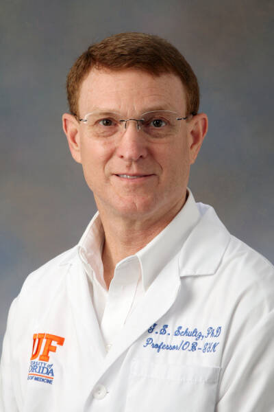 Gregory Schultz合作论文数Department of Obstetrics & Gynecology, College of Medicine, University of Florida;Institute for Wound Research, University of Florida;Center for Vision Research, University of Florida4bruce dewoolfson合作论文数3
 Pedram Hamrah合作论文数2Larry E. Magargal合作论文数2eugene b mclaurin合作论文数2Kennedy Kathryn S合作论文数2Lonsdale John D合作论文数2Torkildsen Gail L合作论文数2Yoder P R合作论文数2
 Alan Sugar合作论文数Cornea & Refractive Surgery Clinic, Kellogg Eye Center, University of Michigan2
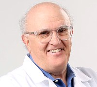 Jeffrey P. Callen合作论文数Division of Dermatology, Department of Medicine, School of Medicine, University of Louisville2Robert E. Nordquist合作论文数2Henry H. Zenzie合作论文数2
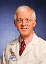 David C. Musch合作论文数Department of Ophthalmology and Visual Sciences, Kellogg Eye Center, University of Michigan;Department of Epidemiology, School of Public Health, University of Michigan2Carsten Bekker合作论文数2Chris Batich合作论文数Department of Materials Science and Engineering, University of Florida2Edwin U. Keates合作论文数2H. Kaz Soong合作论文数2Charles P. Semba合作论文数2
 Larry A Donoso合作论文数2Hanna J. Hoffman合作论文数2Ronald S. Fishman合作论文数1Malcolm Luxenberg合作论文数1Whitney Lomazow合作论文数1W M Pierce合作论文数1Ivey L. Thornton合作论文数1Peter Macdowell合作论文数1Deborah Harrison合作论文数1R F Meyer合作论文数1Charles Wm. Stewart合作论文数1Qais A. Farjo合作论文数1Ayad A Farjo合作论文数1Jeffrey G Penta合作论文数1Garvin H Davis合作论文数1Jf Gordon合作论文数1H. J. Hoffman合作论文数1
 Bernd Liesenfeld合作论文数1Jonathan B Rubenstein合作论文数1Thomas Boland合作论文数1
 Jessica Knowlton合作论文数1Dontchev Mariya合作论文数1Joseph M. Thomas合作论文数1Bernie Iliakis合作论文数1Michael W. Belin合作论文数1Montoya Monty合作论文数1Kenneth R. Kenyon合作论文数1Jh Lass合作论文数1




