It should be noted that "buckling" in this sense refers to the arrangement of the phospholipid molecules rather than macroscopic folding of the membrane, although this may also occur.
Background Device-assisted enteroscopy (DAE) has become a well-established diagnostic and therapeutic tool for the management of small-bowel pathology. We aimed to evaluate the performance measures for DAE across the UK against the quality benchmarks proposed by the European Society of Gastrointestinal Endoscopy (ESGE). Methods We retrospectively collected data on patient demographics and DAE performance measures from electronic endoscopy records of consecutive patients who underwent DAE for diagnostic and therapeutic purposes across 12 enteroscopy centers in the UK between January 2017 and December 2022. Results A total of 2005 DAE procedures were performed in 1663 patients (median age 60 years; 53% men). Almost all procedures (98.1%) were performed for appropriate indications. Double-balloon enteroscopy was used for most procedures (82.0%), followed by single-balloon enteroscopy (17.2%) and spiral enteroscopy (0.7%). The estimated depth of insertion was documented in 73.4% of procedures. The overall diagnostic yield was 70.0%. Therapeutic interventions were performed in 42.6% of procedures, with a success rate of 96.6%. Overall, 78.0% of detected lesions were marked with a tattoo. Patient comfort was significantly better with the use of deep sedation compared with conscious sedation (99.7% vs. 68.5%; P<0.001). Major adverse events occurred in only 0.6% of procedures. Conclusions Performance measures for DAE in the UK meet the ESGE quality benchmarks, with high diagnostic and therapeutic yields, and a low incidence of major adverse events. However, there is room for improvement in optimizing sedation practices, standardizing the depth of insertion documentation, and adopting marking techniques to aid in the follow-up of detected lesions.
Global incidence of non-alcoholic fatty liver disease: A systematic review and meta-analysis of 63 studies and 1,201,807 personsJournal of HepatologyVol. 79Issue 2PreviewThe prevalence of non-alcoholic fatty liver disease (NAFLD) is increasing. We aimed to estimate the pooled global NAFLD incidence. Full-Text PDF We read with interest the systematic review and meta-analysis by Le et al. which demonstrates the rapidly increasing worldwide incidence of non-alcoholic fatty liver disease (NAFLD) likely associated with the obesity epidemic.[1]Le MH Le DM Baez TC Wu Y Ito T Lee EY Global incidence of non-alcoholic fatty liver disease: a systematic review and meta-analysis of 63 studies and 1,201,807 persons.J Hepatol. 2023; 79: 287-295Abstract Full Text Full Text PDF PubMed Scopus (20) Google Scholar Recently, we have demonstrated that NAFLD is the second most common cause of liver disease in patients admitted to hospital with decompensated cirrhosis in the UK (14.4% of admissions).[2]The Trainee Collaborative for Research and Audit in Hepatology UKRegional variation in characteristics of patients with decompensated cirrhosis admitted to hospitals in the UK.Lancet Gastroenterol Hepatol. 2023; 8: 604-606Abstract Full Text Full Text PDF Scopus (2) Google Scholar We strongly agree that targeted public health interventions to reduce the incidence and prevalence of NAFLD combined with optimal outpatient management strategies are needed to mitigate against NAFLD-related complications.[3]McPherson S Armstrong MJ Cobbold JF Corless L Anstee QM Aspinall RJ et al.Quality standards for the management of non-alcoholic fatty liver disease (NAFLD): consensus recommendations from the British Association for the Study of the Liver and British Society of Gastroenterology NAFLD Special Interest Group.Lancet Gastroenterol Hepatol. 2022; 7: 755-769Abstract Full Text Full Text PDF PubMed Scopus (28) Google Scholar However, efforts to better characterise and stratify this cohort are similarly required. Utilising more robust phenotypic data should allow for the development of NAFLD-specific risk prediction models and may encourage implementation of precision approaches to improve overall patient outcomes. We analysed data from a UK multicentre, retrospective observational cohort study, including patients admitted to hospitals with decompensated cirrhosis in November 2019[4]The Trainee Collaborative for Research and Audit in Hepatology UKAdmission care bundles for decompensated cirrhosis are poorly utilised across the UK: results from a multi-centre retrospective study.Clin Med (Lond). 2023; 23: 193-200Crossref PubMed Scopus (4) Google Scholar (Table S1 provides regional submission data). We compared admissions for patients with NAFLD to the rest of the predominately alcohol-related liver disease (ARLD) cohort. Details of methods and statistical analyses are presented in the supplementary materials. The NAFLD cohort were significantly older (69.0 (IQR 62.3-77.0) vs. 55.5 (IQR 47.0-65.0), p <0.0001∗) and less likely to be male (50.6% vs. 63.5%, p = 0.001∗) (Table 1A). Whilst we note Le et al. demonstrated a higher incidence of NAFLD amongst male patients, this may reflect previous findings that mortality is comparable across male and female patients with NAFLD, reflecting a similar prevalence of advanced disease.[5]Simon T.G. Roelstraete B. Khalili H. Hagström H. Ludvigsson J.F. Mortality in biopsy-confirmed nonalcoholic fatty liver disease: results from a nationwide cohort.Gut. 2021; 70: 1375-1382Crossref PubMed Scopus (248) Google Scholar No differences were demonstrated in the proportion of admissions with a previous history of decompensation or known liver disease, or a history of hepatocellular carcinoma (HCC) between cohorts. Patients admitted with NAFLD were significantly less likely to regularly consume alcohol (18.0% vs. 61.8%, p <0.0001∗) than the rest of the predominant ARLD cohort (Table 1A). However, alcohol consumption has been shown to be underreported in previous cohorts of NAFLD and markers of alcohol use may have highlighted individual’s regularly consuming alcohol above recommended limits.[6]Staufer K Huber-Shonauer U Strebinger G Pimingstorfer P Suesse S Scherzer TM et al.Ethyl glucuronide in hair detects a high rate of harmful alcohol consumption in presumed non-alcoholic fatty liver disease.J Hepatol. 2022; 77: 918-930Abstract Full Text Full Text PDF PubMed Scopus (43) Google Scholar Admissions for patients with NAFLD were predominantly related to the management of ascites (40.9% vs. 32.1%, p = 0.02∗) or encephalopathy (25.6% vs. 15.8%, p = 0.002∗), and less likely to be for jaundice (2.8% vs. 17.2%, p <0.0001∗) (Table 1A). Ascites is associated with the highest risk of readmission for patients with NAFLD.[7]Paik JM Eberly KE Kabbara K Harring M Younossi Y Henry L et al.Non-alcoholic fatty liver disease is associated with greater risk of 30-day hospital readmission in the United States (U.S.).Ann Hepatol. 2023; 28: 101108Crossref PubMed Scopus (2) Google Scholar Patient-centred elective outpatient paracentesis provision is therefore a requisite component of modern hepatology services. Recently, data has implicated the premature onset of encephalopathy in NAFLD, while the association of hyperammonaemia with deleterious outcomes is also well-described.[8]Thomsen KL Eriksen PL Kerbert AJC De Chiara F Jalan R Vilstrup H Role of ammonia in NAFLD: an unusual suspect.JHEP Reports. 2023; 5: 100780Abstract Full Text Full Text PDF PubMed Scopus (2) Google Scholar A high index of suspicion for encephalopathy is required in this cohort, in addition to a low threshold in initiating therapies for hyperammonaemia. The reduction in the proportion of patients admitted with jaundice likely reflects the cohort of patients from the predominately ARLD cohort presenting with alcohol-related hepatitis.Table 1Characterising UK NAFLD admissions.A) VariableNNAFLD, n = 176NAlternate aetiology of liver disease, n = 1,048p valueAge17669.0 (62.3-77.0)1,04855.5 (47.0-65.0)<0.0001∗Male sex17689 (50.6%)1,048665 (63.5%)0.001∗Current alcohol use12222 (18.0%)813502 (61.8%)<0.0001∗Previously known liver disease176151 (85.8%)1,048890 (84.9%)0.82Previous known decompensation176116 (65.9%)1,048706 (67.4%)0.73Known HCC17610 (5.7%)1,04854 (5.2%)0.72Reason for admission Ascites17672 (40.9%)1,048336 (32.1%)0.02∗ Encephalopathy17645 (25.6%)1,048166 (15.8%)0.002∗ Gastrointestinal bleeding17619 (10.8%)1,048161 (15.4%)0.13 Jaundice1765 (2.8%)1,048180 (17.2%)<0.0001∗ Sepsis17610 (5.7%)1,04869 (6.6%)0.74Prognostic scores MELD score14713.0 (11.0-18.0)95517.0 (12.0-21.0)<0.0001∗ UKELD score14753.0 (51.0-60.0)95557.0 (52.0-62.0)<0.0001∗ Child-Pugh score1398.0 (7.0-10.0)9329.0 (8.0-11.0)0.0005∗Post 24-hour care Managed by a specialist176125 (71.0%)1,048776 (74.1%)0.41 Predominately managed on a specialist ward17693 (52.8%)1,043597 (57.2%)0.29 Critical care admission during stay1769 (5.1%)1,048123 (11.7%)0.008∗ Transfer to another centre1765 (2.8%)1,04819 (1.8%)0.37 Admission mortality17127 (15.8%)1,029160 (15.6%)0.91 Length of stay1427.0 (4.0-13.0)8597.00 (3.0-13.0)0.60B) VariableNSurvivors, n = 144NNon-survivors, n = 27p valueAge14469.0 (62.0-76.0)2773.0 (65.0-78.0)0.17Male sex14472 (50.0%)2715 (55.6%)0.68Current alcohol use10220 (19.6%)172 (11.8%)0.74Previously known liver disease144125 (86.8%)2722 (81.5%)0.54Previous known decompensation14496 (66.7%)2717 (63.0%)0.83Known HCC1448 (5.6%)272 (7.4%)0.66Reason for admission Ascites14459 (41.0%)2711 (40.7%)>0.9999 Encephalopathy14438 (26.4%)275 (18.5%)0.47 Gastrointestinal bleeding14416 (11.1%)272 (7.4%)0.74 Jaundice1444 (2.8%)271 (3.7%)0.58 Sepsis14410 (6.9%)270 (0.0%)0.37Prognostic scores MELD score11913.0 (10.0-17.0)2321.0 (15.0-27.0)<0.0001∗ UKELD score11953.0 (50.0-57.0)2359.0 (54.0-61.0)0.0001∗ Child-Pugh score1138.0 (7.0-9.0)2210.0 (8.8-11.0)0.0002∗Post 24-hour care Managed by a specialist144100 (69.4%)2721 (77.8%)0.49 Predominately managed on a specialist ward14478 (54.2%)2712 (44.4%)0.40 Critical care admission during stay1445 (3.5%)274 (14.8%)0.04A) Comparison of admissions for patients with NAFLD compared to patients with alternate aetiologies of liver disease. B) Comparison of NAFLD admissions resulting in patient survival with those that did not. Non-normally continuous data were analysed using Mann-Whitney U tests and presented as median (IQR). Categorical data were analysed using Fisher’s exact tests and presented as number (%). ∗Statistical significance set as per Benjamini-Hochberg procedure with a false discovery rate of 0.05.HCC, hepatocellular carcinoma; MELD, model for end-stage liver disease; NAFLD, non-alcoholic fatty liver disease; UKELD, UK end-stage liver disease. Open table in a new tab A) Comparison of admissions for patients with NAFLD compared to patients with alternate aetiologies of liver disease. B) Comparison of NAFLD admissions resulting in patient survival with those that did not. Non-normally continuous data were analysed using Mann-Whitney U tests and presented as median (IQR). Categorical data were analysed using Fisher’s exact tests and presented as number (%). ∗Statistical significance set as per Benjamini-Hochberg procedure with a false discovery rate of 0.05. HCC, hepatocellular carcinoma; MELD, model for end-stage liver disease; NAFLD, non-alcoholic fatty liver disease; UKELD, UK end-stage liver disease. Following admission, no differences were noted between cohorts in the proportion of patients managed by a specialist Gastroenterologist/Hepatologist on specialist wards, or in patients transferred to specialist centres. No differences were demonstrated between patient cohorts for mortality during admission (15.8% vs. 15.6%, p = 0.91) despite admissions with NAFLD having significantly lower prognostic scores and being less likely to access critical care (5.1% vs. 11.7%, p = 0.008∗) (Table 1A). Whilst this may reflect the older age of this cohort and concomitant comorbidity, previous concerns have been raised regarding limited access to critical care for patients with ARLD with stigma amongst healthcare professionals suggested as a potential barrier for this cohort.[9]Mitchison H. Saksena S. Hudson M. NCEPOD and alcohol-related liver disease, what are the views of those who deliver the service? A survey of consultants and trainees in North Eastern England.J R Coll Physicians Edinb. 2018; 48: 293-298Crossref PubMed Google Scholar Understanding potential barriers to patients with NAFLD accessing critical care is required to optimise management. Admissions for patients with NAFLD were not more likely to result in mortality after adjustment for age, critical care admission or MELD score (adjusted odds ratio 1.16; 95% CI 0.65-2.00; Fig. S1). Whilst comparisons between non-survivors and survivors are likely underpowered, conventional prognostic models discriminated admission survival (Table 1B). However, no prognostic scores significantly outperformed other models (p = 0.41), with no model achieving an AUC of greater than 0.8 (Fig. S2). After exclusion of admissions resulting in mortality or critical care admission, length of stay was no different to the rest of the cohort (Table 1A). Limitations of these analyses are discussed in the supplementary materials and include the retrospective design, coverage of only a single month, incomplete coverage of the UK with potential selection bias, lack of data regarding comorbidities and the subjective nature of aetiology assignment. Whilst accepting these limitations, this study is representative of a large, real-world cohort. With the likely increased incidence of patients being admitted to hospital with decompensated NAFLD, further work to understand how to optimally manage this cohort are necessary. This cohort is older, often more co-morbid and will likely require a tailored approach to their care. Understanding barriers to providing best care, including access to critical care, is essential when developing the hepatology services of the future. We are grateful for the funding support provided by Guts-UK. We are grateful for the support and endorsement from the British Society of Gastroenterology, British Association for the Study of the Liver, Scottish Society of Gastroenterology and the Welsh Association for Gastroenterology and Endoscopy. The authors declare no conflicts of interest that pertain to this work. Please refer to the accompanying ICMJE disclosure forms for further details. The following are the supplementary data to this article. Download .pdf (.35 MB) Help with pdf files Multimedia component 1 Download .pdf (.31 MB) Help with pdf files Multimedia component 2 Download .docx (.03 MB) Help with docx files Multimedia component 3
Major cardiovascular diseases (CVDs) are associated with (regional) dysfunction of the left ventricle. Despite the 3-D nature of the heart and its dynamics, the assessment of myocardial function is still largely based on 2-D ultrasound imaging, thereby making diagnosis heavily susceptible to the operator's expertise. Unfortunately, to date, 3-D echocardiography cannot provide adequate spatiotemporal resolution in real-time. Hence, tri-plane imaging has been introduced as a compromise between 2-D and true volumetric ultrasound imaging. However, tri-plane imaging typically requires high-end ultrasound systems equipped with fully populated matrix array probes embedded with expensive and little flexible electronics for two-stage beamforming. This article presents an advanced ultrasound system for real-time, high frame rate (HFR), and tri-plane echocardiography based on low element count sparse arrays, i.e., the so-called spiral arrays. The system was simulated, experimentally validated, and implemented for real-time operation on the ULA-OP 256 system. Five different array configurations were tested together with four different scan sequences, including multi-line and planar diverging wave transmission. In particular, the former can be exploited to achieve, in tri-plane imaging, the same temporal resolution currently used in clinical 2-D echocardiography, at the expenses of contrast (-3.5 dB) and signal-to-noise ratio (SNR) (-8.7 dB). On the other hand, the transmission of planar diverging waves boosts the frame rate up to 250 Hz, but further compromises contrast (-10.5 dB), SNR (-9.7 dB), and lateral resolution (+46%). In conclusion, despite an unavoidable loss in image quality and sensitivity due to the limited number of elements, HFR tri-plane imaging with spiral arrays is shown to be feasible in real-time and may enable real-time functional analysis of all left ventricular segments of the heart.
Transducers with a larger aperture size are desirable in ultrasound imaging to improve resolution and image quality. A coherent multi-transducer ultrasound imaging system (CoMTUS) enables an extended effective aperture through the coherent combination of multiple transducers. In this study, the discontinuous extended aperture created by CoMTUS and its performance for deep imaging and through layered media are investigated by both simulations and experiments. Typical image quality metrics-resolution, contrast and contrast-to-noise ratio-are evaluated and compared with a standard single probe imaging system. Results suggest that the image performance of CoMTUS depends on the relative spatial location of the arrays. The resulting effective aperture significantly improves resolution, while the separation between the arrays may degrade contrast. For a limited gap in the effective aperture (less than a few centimetres), CoMTUS provides benefits to image quality compared to the standard single probe imaging system. Overall, CoMTUS shows higher sensitivity and reduced loss of resolution with imaging depth. In general, CoMTUS imaging performance was unaffected when imaging through a layered medium with different speed of sound values and resolution improved up to 80% at large imaging depths.
Molecular targeted nanodroplets that can extravasate beyond the vascular space have great potential to improve tumor detection and characterisation. High-frame-rate ultrasound, on the other hand, is an emerging tool for imaging at a frame rate one to two orders of magnitude higher than those of existing ultrasound systems. In this study, we used high-frame-rate ultrasound combined with optics to study the acoustic response and size distribution of folate receptor (FR)-targeted versus non-targeted (NT)-nanodroplets in vitro with MDA-MB-231 breast cancer cells immediately after ultrasound activation. A flow velocity mapping technique, Stokes’ theory and optical microscopy were used to estimate the size of both floating and attached vaporised nanodroplets immediately after activation. The floating vaporised nanodroplets were on average more than seven times larger than vaporised nanodroplets attached to the cells. The results also indicated that the acoustic signal of vaporised FR-targeted-nanodroplets persisted after activation, with 70% of the acoustic signals still present 1 s after activation, compared with the vaporised NT-nanodroplets, for which only 40% of the acoustic signal remained. The optical microscopic images revealed on average six times more vaporised FR-targeted-nanodroplets generated with a wider range of diameters (from 4 to 68 µm) that were still attached to the cells, compared with vaporised NT-nanodroplets (from 1 to 7 µm) with non-specific binding after activation. The mean size of attached vaporised FR-targeted-nanodroplets was on average about threefold larger than that of attached vaporised NT-nanodroplets. Taking advantage of high-frame-rate contrast-enhanced ultrasound and optical microscopy, this study offers an improved understanding of the vaporisation of the targeted nanodroplets in terms of their size and acoustic response in comparison with NT-nanodroplets. Such understanding would help in the design of optimised methodology for imaging and therapeutic applications.
High-frame-rate (HFR) ultrasound (US) imaging and contrast-enhanced US (CEUS) are often implemented using multipulse transmissions, to enhance image quality. Multipulse approaches, however, suffer from degradation in the presence of motion, especially when coherent compounding and CEUS are combined. In this paper, we investigate this effect on the intensity of HFR CEUS in deep tissue imaging using simulations and in vivo contrast echocardiography (CE). The simulation results show that the motion artifact is much higher when the flow is in an axial direction than a lateral direction. Using a pulse repetition frequency suitable for cardiac imaging, a motion of 35 cm/s can cause as much as 28.5 dB decrease in image intensity, where compounding can contribute up to 18.7 dB of intensity decrease (11 angles). These motion effects are also demonstrated for in vivo cardiac HFR CE, where the large velocities of both the myocardium and the blood are present. Intensity reductions of 10.4 dB are readily visible in the chamber. Finally, we demonstrate how performing motion-correction before pulse inversion compounding greatly reduces such motion artifact and improve image signal-to-noise ratio and contrast.
In order to improve resolution, transducers with larger aperture size are desirable in ultrasound imaging. However, the practical aperture size is limited. Inhomogeneities and aberrating layers cause phase errors restricting the improvements provided by large arrays. Recently, we have shown the feasibility of a fully coherent multitransducer ultrasound imaging system (CMTUS), formed by several ultrasound transducers that are synchronized and freely located in space. The transducer locations along with the average speed of sound in the medium are deduced by maximizing the coherence between backscattered echoes from targeted point-like scattereres in the common field of view of the transducers. An improved image is obtained through coherent combination of the multiple transducers acting as a single larger effective aperture. In this study, the behavior of CMTUS in the presence of aberration is further investigated with simulations. A parametric study is presented, in which the geometry of the system, defined by two linear arrays, and the presence of acoustic clutter are investigated. In this framework, typical image quality metrics - resolution, contrast and contrast-to-noise ratio - are evaluated. Results suggest that the imaging enhancement made by the CMTUS is limited by the location of the transducers in space. Based on image metrics, an optimal spatial location is proposed for a CMTUS formed by two linear arrays. In addition, results show that, in the presence of clutter, image quality diminishes at larger aperture sizes. Nevertheless, CMTUS successfully corrects the aberration effects, without degrading the gains made by the large effective aperture.
An algorithm was developed for the correction of ring artifacts in phase-insensitive ultrasound computed tomography attenuation images. Differences in the measurement sensitivity between the ultrasound transducer array elements cause discontinuities in the sinogram which manifest as rings and arcs in the reconstructed image. The magnitudes of the discontinuities are potentially time-varying and dependent on the attenuation being measured. The algorithm dynamically determines the measurement sensitivity of each transducer in the array during the scan by comparison with both the elements to its left and the elements to its right. Elements at either end of the array are corrected, assuming a zero-attenuation path. The two estimates of sensitivity are combined using a weighted mean similar to a Kalman filter. The algorithm was tested on simulated and experimentally acquired data. It was demonstrated to reduce the root-mean-square error (RMSE) of simulated images against ground-truth images by up to a factor of 50 compared with uncorrected images and to visibly reduce artifacts on images reconstructed from the experimentally acquired data.
Motion during image acquisition can cause image degradation in all medical imaging modalities. This is particularly relevant in 2-D ultrasound imaging, since out-of-plane motion can only be compensated for movements smaller than elevational beamwidth of the transducer. Localization based super-resolution imaging creates even a more challenging motion correction task due to the requirement of a high number of acquisitions to form a single super-resolved frame. In this study, an extension of two-stage motion correction method is proposed for 3-D motion correction. Motion estimation was performed on high volumetric rate ultrasound acquisitions with a handheld probe. The capability of the proposed method was demonstrated with a 3-D microvascular flow simulation to compensate for handheld probe motion. Results showed that two-stage motion correction method reduced the average localization error from 136 to 18 μm.
A coherent multi-transducer ultrasound imaging system (CoMTUS) enables an extended effective aperture through coherent combination of multiple transducers. The resulting larger effective aperture improves the ultrasound imaging performance. The optimal beamforming parameters, which include the transducer locations and the average speed of sound in the medium, are deduced by maximizing the coherence of the received radio frequency data by cross-correlation. In this technique, the detection of multiple isolated point-like targets in the overlap of the insonated regions is mandatory to determine the relative probe-to-probe position. This study proposes the use of microbubbles to generate the point-like targets that the CoMTUS approach requires. The first phantom images produced using CoMTUS and microbubbles are presented here.
Background: Variations in lymph node (LN) microcirculation can be indicative of metastasis. The identification and quantification of metastatic LNs remains essential for prognosis and treatment planning, but a reliable noninvasive imaging technique is lacking. Three-dimensional super-resolution (SR) US has shown potential to noninvasively visualize microvascular networks in vivo. Purpose: To study the feasibility of three-dimensional SR US imaging of rabbit LN microvascular structure and blood flow by using microbubbles. Materials and Methods: In vivo studies were carried out to image popliteal LNs of two healthy male New Zealand white rabbits aged 6-8 weeks. Three-dimensional, high-frame-rate, contrast material-enhanced US was achieved by mechanically scanning with a linear imaging probe. Individual microbubbles were identified, localized, and tracked to form three-dimensional SR images and super-resolved velocity maps. Acoustic subaperture processing was used to improve image contrast and to generate enhanced power Doppler and color Doppler images. Vessel size and blood flow velocity distributions were evaluated and assessed by using Student paired t test. Results: SR images revealed microvessels in the rabbit LN, with branches clearly resolved when separated by 30 mu m, which is less than half of the acoustic wavelength and not resolvable by using power or color Doppler. The apparent size distribution of most vessels in the SR images was below 80 mu m and agrees with micro-CT data, whereas most of those detected with Doppler techniques were larger than 80 mu m in the images. The blood flow velocity distribution indicated that most of the blood flow in rabbit popliteal LN was at velocities lower than 5 mm/sec. Conclusion: Three-dimensional super-resolution US imaging using microbubbles allows noninvasive nonionizing visualization and quantification of lymph node microvascular structures and blood flow dynamics with resolution below the wave diffraction limit. This technology has potential for studying the physiologic functions of the lymph system and for clinical detection of lymph node metastasis.
Current localization-based super-resolution ultrasound imaging requires a low concentration of flowing microbubbles to visualize microvasculature beyond the diffraction limit and acquisition is slow. Nanodroplets offer a promising solution as they can be sparsely activated and deactivated on-demand. In this study, acoustic wave sparsely-activated localization microscopy (AWSALM) using activation and deactivation of nanodroplets, an acoustic counterpart of photo-activated localization microscopy (PALM) which is less dependent on agent concentration and the presence of flow, is demonstrated for super-resolution imaging in deep tissues in vivo. An in vivo super-resolution image of a rabbit kidney is obtained in 1.1 seconds using AWSALM, where micro-vessels with apparent sizes far below the half-wavelength of 220 μm were visualized. This preliminary result demonstrates the feasibility of applying AWSALM for in vivo super-resolution imaging.
One of the crucial challenges in the application of ultrasound super-resolution imaging using microbubble contrast agents to the clinic is the long acquisition time. Recently acoustic wave sparsely activated localization microscopy (AWSALM) was developed to activate, image, and destroy nanodroplets to achieve ultrasound super-resolution imaging. The activation and destruction of nanodroplets using focused pulses in AWSALM can generate new localization signals without relying on flow and shows the potential to achieve faster ultrasound super-resolution. However, the technique requires optimization to achieve good activation efficiency with the minimum amount of activation time. This work investigates how the different activation strategies affect the activation time and efficiency. A range of activation sequences with different F-numbers and spacing between transmit foci are used and their activation efficiency and the data acquisition time are quantified. The results show that sweeping the focus of the activation pulse with an F-number of 0.4 and a step size of 2.5 wavelengths can generate the highest droplet activation. By using a larger or smaller spacing between consecutive focused activation beams generated lower contrast, which suggests that densely spaced activation pulses may destroy the activated nanodroplets generated by the previous activation pulse. In summary, this study demonstrates that the rate of activation of nanodroplets can be increased by optimizing the Fnumber and the sweep step size. This finding can inform the development of droplet-based super-resolution imaging to further minimize the data acquisition time.
Transducers with larger aperture size are desirable in ultrasound imaging to improve resolution and image quality. However, in practice inhomogeneities and aberrating layers cause phase errors that limit the benefits of increased aperture size. A coherent multi-transducer ultrasound imaging system (CoMTUS) enables an extended effective aperture through coherent combination of multiple transducers. The optimal beamforming parameters, which include the transducer locations and the average speed of sound in the medium, are deduced by maximizing the coherence of the received radio frequency data by crosscorrelation. In this work, the robustness of the CoMTUS approach in the presence of acoustic clutter and aberration is investigated by both simulated and in-vitro results. Results show that CoMTUS improves ultrasound imaging quality in terms of resolution, and also suppresses phase aberration effects along with clutter, providing benefits to image quality compared with a single probe imaging system. Overall resolution was unaffected by aberration and improved up to 70% in the CoMTUS.
The measurement of cardiac and aortic pressures enables diagnostic insight into cardiac contractility and stiffness. However, these pressures are currently assessed invasively using pressure catheters. It may be possible to estimate these pressures less invasively by applying microbubble ultrasound contrast agents as pressure sensors. The aim of this study was to investigate the subharmonic response of the microbubble ultrasound contrast agent SonoVue (Bracco Spa, Milan, Italy) at physiological pressures using a static pressure phantom. A commercially available cell culture cassette with Luer connections was used as a static pressure chamber. SonoVue was added to the phantom, and radio frequency data were recorded on the ULtrasound Advanced Open Platform (ULA-OP). The mean subharmonic amplitude over a 40% bandwidth was extracted at 0-200-mmHg hydrostatic pressures, across 1.7-7.0-MHz transmit frequencies and 3.5%-100% maximum scanner acoustic output. The Rayleigh-Plesset equation for single-bubble oscillations and additional hysteresis experiments were used to provide insight into the mechanisms underlying the subharmonic pressure response of SonoVue. The subharmonic amplitude of SonoVue increased with hydrostatic pressure up to 50 mmHg across all transmit frequencies and decreased thereafter. A decreasing microbubble surface tension may drive the initial increase in the subharmonic amplitude of SonoVue with hydrostatic pressure, while shell buckling and microbubble destruction may contribute to the subsequent decrease above 125-mmHg pressure. In conclusion, a practical operating regime that may be applied to estimate cardiac and aortic blood pressures from the subharmonic signal of SonoVue has been identified.
This paper describes the application of a pulse pileup correction algorithm based on maximum likelihood estimation to simulated and experimentally acquired signals from a pyroelectric ultrasound sensor. The sensor forms part of a prototype phase-insensitive ultrasound computed tomography (piUCT) system for breast cancer detection that generates quantitative maps of acoustic attenuation. The effectiveness of the piUCT technique has been previously demonstrated through imaging of cylindrical, polyurethane phantoms. For the technique to be applied clinically, the system must be capable of measuring high acoustic attenuations (>30 dB) and completing a scan in a clinically acceptable time frame of approximately 5 min. High attenuations present a challenge because the sensor responds directly to acoustic power and therefore, an attenuation of 30 dB causes a drop in signal level of three orders of magnitude, resulting in a large dynamic range in the received signal set. Completion of a scan in 5 min requires a period between measurements shorter than the response time of the sensor, causing pyroelectric pulse pileup. The maximum likelihood estimation method recovers the amplitudes of closely spaced superimposed pyroelectric signals while improving the coefficient of variation of the measurement by a factor of two compared to direct pulse amplitude measurement, simultaneously increasing the maximum measurable attenuation and measurement rate of the system.
 Meng-Xing Tang合作论文数Department of Bioengineering, Imperial College London113
 DO Cosgrove合作论文数37Kirsten Christensen-Jeffries合作论文数School of Biomedical Engineering & Imaging Sciences, Faculty of Life Sciences & Medicine, King’s College London34
 Christopher Dunsby合作论文数Department of Physics, Faculty of Natural Sciences, Imperial College London32
 Sevan Harput合作论文数Acoustic Group;Sabanci University;Faculty of Engineering and Natural Sciences Orhanli-Tuzla;Faculty of Engineering and Natural Sciences Orhanli-Tuzla, Sabanci University28
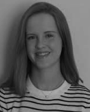 Jemma Brown合作论文数Biomedical Engineering Department, King’s College London27
 Eleanor P. J. Stride合作论文数Institute of Biomedical Engineering, University of Oxford;Department of Engineering Science, University of Oxford;St Catherine's College, University of Oxford26N Patel合作论文数21
 Charles A Sennoga合作论文数20Mj Blomley合作论文数19MJ Blomley合作论文数19
 Ge Zhang (张桂扬)合作论文数19Thomas Albrecht合作论文数18V Jayaram合作论文数17
 Helen Mulvana合作论文数Biomed Engn, Univ Strathclyde16Chris J Harvey合作论文数16
 Jo Hajnal合作论文数Department of Biomedical Computing, School of Biomedical Engineering & Imaging Sciences, Faculty of Life Sciences & Medicine, Kings College London16
 David Cosgrove合作论文数Imperial College;King’s College Hospital15
 Rolf Andreas Heckemann合作论文数Department of Imaging, Hammersmith Hospital15
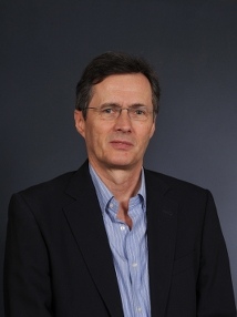 John M. Seddon合作论文数Department of Chemistry, Imperial College London14
 Dorian Haskard合作论文数National Heart & Lung Institute, Imperial College London;Faculty of Medicine, Imperial College London;Royal Postgraduate Medical School14
 Petros Nihoyannopoulos合作论文数National Heart & Lung Institute, Faculty of Medicine, Imperial College London;Hammersmith Hospital13
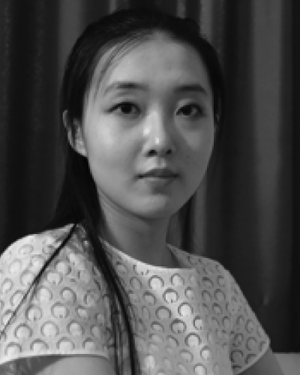 Jiaqi Zhu合作论文数Dept Bioengn, Imperial Coll London12Jenny Butler-Barnes合作论文数12
 Matthieu Toulemonde合作论文数11
 Richard J. Browning合作论文数Institute of Biomedical Engineering, University of Oxford11David O Cosgrove合作论文数11
 Chee Hau Leow合作论文数Imperial Coll London, Dept Bioengn, London, England10
 Kevin Chetty合作论文数Department of Security and Crime Science, Faculty of Engineering Sciences, University College London10
 Yeh James S M合作论文数10Adrian K. P. Lim合作论文数10M Blomley合作论文数8ellen mcconnell合作论文数8
 Yuanwei Li合作论文数8
 Jeff Bamber合作论文数Department of Physics, The Institute of Cancer Research and Royal Marsden NHS Foundation Trust8Sussan Nourshargh合作论文数8JA Butler-Barnes合作论文数7sinan li合作论文数7
 Laura Peralta合作论文数School of Biomedical Engineering & Imaging Sciences, Faculty of Life Sciences & Medicine, King’s College London7
 Dan Elson合作论文数Reproductive and Developmental Biology, Department of Metabolism, Digestion and Reproduction, Faculty of Medicine, Imperial College London7
 Jean Martial Mari合作论文数Univ Polynesie Francaise7Cheng Yi合作论文数7Paul S. Sidhu合作论文数7Martin J.K. Blomley合作论文数7AKP Lim合作论文数6A Bauer合作论文数6
 Antonio Stanziola合作论文数Medical Physics and Biomedical Engineering, Faculty of Engineering Sciences, University College London6
 Virginie Papadopoulou合作论文数Joint Department of Biomedical Engineering, College of Arts and Sciences, The University of North Carolina at Chapel Hill6




