Objective:Fasciocutaneous radial forearm free flaps (RFFF) are widely used for head and neck soft tissue reconstruction. Osteocutaneous radial forearm free flaps (OCRFFF) enable bony reconstruction, but concerns for donor site morbidity have limited their use. Long-term donor site outcomes following OCRFFF remain understudied. Study Design:Cross-sectional. Setting:Single-institution. Methods:Patients who underwent RFFF or OCRFFF between 2010 and 2023 were surveyed by phone. Upper extremity function was assessed using the Quick Disabilities of the Arm, Shoulder, and Hand (QuickDASH) survey (0-100 scale, higher scores indicate greater disability). Patients who were non-verbal or lacked follow-up after 2021 were excluded. QuickDASH scores were analyzed using Wilcoxon rank-sum test. Univariable linear regressions assessed clinical associations. Results:Survey response rate was 21.7%. The cohort included 26 RFFF, 14 OCRFFF, and 5 patients with multiple radial forearm flaps. At a median time of 3.25 years postoperatively (IQR: 2.2-5.6), OCRFFF patients had higher mean QuickDASH scores (21.6) than RFFF (10.7), suggesting greater functional limitation in OCRFFF cases (P = .12). Overall impairment remained low. OCRFFF patients reported greater difficulties compared to RFFF regarding jar opening (2.4 vs 1.6; P = 0.01), back washing (2.0 vs 1.3; P = .03), and recreational activities (2.5 vs 1.4; P = .02). History of depression was associated with higher QuickDASH scores (coef 12.7, 95% CI: 1.23-24.48). Conclusions:OCRFFF patients reported greater difficulty in certain task-specific activities compared to RFFF, but overall donor site impairment following forearm free flap reconstruction is minimal. History of depression is associated with worse self-reported upper extremity function.
BACKGROUND:Patterns of failure in early-stage oral tongue squamous cell carcinoma (OTSCC) managed with surgery alone remain understudied. METHODS:Retrospective cohort study from 2000 to 2022 at a single tertiary care center evaluated pT1-T2N0 OTSCC patients who underwent partial glossectomy with or without elective neck dissection (END), without any adjuvant therapy. Predictors of recurrence were analyzed via multivariate Cox proportional hazards model. RESULTS:The study included 201 OTSCC patients (131 pT1 and 70 pT2). Eighty patients (40%) underwent glossectomy alone; 121 (60%) underwent glossectomy with END. At a median follow-up of 56 months (IQR 23-82), 63 patients (31%) developed recurrence (21.4% local; 15.9% regional; 1% distant). Positive margins (HR 5.45 [1.9-15.4]), perineural invasion (HR 4.15 [1.8-9.9]), and increasing depth of invasion (DOI) (HR 1.32 [1.1-1.6]) were independently associated with recurrence. Upfront END reduced the risk of recurrence (HR 0.31 [95% CI: 0.15-0.6]), with benefit starting at tumor DOI 2 mm and greater. CONCLUSIONS:Upfront END decreases recurrence for pT1-T2N0 OTSCC treated with surgery alone starting at a DOI of 2 mm.
BACKGROUND:Dysphagia is a common and challenging long-term sequela following total laryngectomy (TL) that can negatively impact patient quality of life. Following TL, many patients require repeat esophageal dilations to maintain swallowing function. This study aimed to identify surgical and non-surgical risk factors associated with dysphagia following TL, defined as clinically diagnosed postoperative dysphagia based on patient-reported symptoms and dysphagia requiring dilation. METHODS:We conducted a retrospective single-institution cohort study of patients who underwent total laryngectomy from 1999 to 2024. Outcomes included clinically diagnosed dysphagia and dysphagia requiring esophageal dilations, with covariates including age, BMI, preoperative PEG tube, cricomyotomy, flap reconstruction type, pharyngectomy, glossectomy, base of tongue resection, postoperative complication, neoadjuvant radiation, and adjuvant radiation. Associations between dysphagia and covariates were assessed using univariable and multivariable logistic regressions. RESULTS:A total of 787 patients underwent total laryngectomy. Median follow-up was 28.5 months (Interquartile range [IQR]: 10.3-68.2 months). During the follow-up period, 212 patients (26.9 %) reported dysphagia, and 131 patients underwent dilation (16.6 %). Prior radiation (adjusted odds ratio [aOR]: 2.51, 95 % CI: 1.56-4.05, p < 0.001), adjuvant radiation (aOR: 1.86; 95 % CI 1.10 to 3.17; p = 0.021), and tongue base resection (aOR: 2.14, 95 % CI: 1.24-3.70, p = 0.006) were independently associated with postoperative clinical dysphagia. Total pharyngectomy (odds ratio [OR]: 1.83, 95 % CI 1.17-2.83, p = 0.008) was associated with dysphagia on univariable analysis. Pedicled reconstruction (OR: 1.38, 95 % CI 1.00-1.90, p = 0.053) showed a trend towards increased odds of clinical dysphagia. Only prior radiation was independently associated with dysphagia requiring esophageal dilation (aOR: 2.92, 95 % CI: 1.62-5.26, p < 0.001). CONCLUSIONS:Dysphagia is common following total laryngectomy. Prior radiation, adjuvant radiation, total pharyngectomy, tongue base resection, and pedicled reconstruction were factors influencing swallowing outcomes following TL. Salvage TL patients should be counseled on the possible need for post-laryngectomy esophageal dilations.
OBJECTIVE:Quantify the anatomic variation of the ansa cervicalis and evaluate neurostimulation of the infrahyoid musculature. STUDY DESIGN:Intraoperative physiology study. SETTING:Tertiary referral center. METHODS:Adult patients undergoing lateral neck dissection including level IV for head and neck cancer were recruited. Ansa cervicalis plexus branches were documented during surgical dissection. In a subset of participants, an electrode was placed on branches of the common sternothyroid trunk innervating the sternothyroid and sternohyoid muscles. Hyolaryngeal excursion with neurostimulation was recorded. RESULTS:Measurements were collected from 39 of 50 participants. Reasons for intraoperative exclusion included significant radiation fibrosis (n = 4), nodal disease burden (n = 3), and surgeon preference (n = 4). The mean lengths of the common sternothyroid trunk and sternothyroid branch were 37.6 ± 15.0 mm and 20.4 ± 8.1 mm, respectively. Their respective mean diameters were 2.1 ± 0.7 mm and 1.5 ± 0.5 mm. The distance from sternothyroid branch muscle insertion to the sternum varied substantially (12.8 ± 14.8 mm). Nine patients underwent neurostimulation of the common sternothyroid trunk. The amplitude of first observed muscle contraction was 0.35 ± 0.18 mA and maximal was 0.57 ± 0.40 mA, during which the hyolaryngeal complex descended by 13.6 ± 4.6 mm. In patients with anatomy amenable to neurostimulation of other nerve branches, stimulation of the common sternothyroid trunk produced the greatest hyolaryngeal descent (P < .05). CONCLUSION:The minor variability observed in ansa cervicalis nerve diameter across patients and branches supports the feasibility of a standardized electrode design for an implantable neurostimulation device. Stimulation of the common sternothyroid trunk resulted in the greatest hyolaryngeal descent, highlighting its value as a potential neuromodulation target.
ABSTRACT Background Time from surgery to initiation of postoperative radiation therapy (PORT) of less than 6 weeks was recently instituted as the first quality metric within head and neck cancer care. Methods We performed a retrospective single institution cohort study to investigate predictors of PORT delay and the impact of PORT delay on survival. Results PORT delay rate was 73.2%, with a median time to treatment initiation of 51 days. Outside radiation facility treatment was independently associated with increased likelihood of PORT delay (OR 1.94, 95% CI 1.03–3.74, p = 0.043). PORT delay and location of radiation treatment did not impact OS or PFS. Conclusions In this single institution study, most patients experienced PORT delay. Patients that were treated at outside radiation facilities were more likely to experience delay. However, PORT delay did not result in statistically significant difference in OS and PFS which contrasts with the current literature.
Objective:Determine relationships between pain and mental health disorders (MHDs) in patients undergoing microvascular free flap reconstruction for head and neck cancer (HNC). Study Design:Retrospective cohort. Setting:Tertiary Care Institution in the Southeastern United States. Methods:Clinical data were manually abstracted from digital health records to obtain demographic, MHD, clinical outcomes, and pain data for HNC patients who underwent free flap reconstruction from 2017 to 2023. Univariate and multivariable regression analyses were performed to delineate relationships between MHDs and postoperative pain. Results:The study cohort comprised 283 patients. Ninety-four patients (33%) had preoperative MHDs, which were more common in women (42% vs 30%, P = .04) and in patients with chronic pain (53% vs 32%, P < .01). Preoperative opioid use (P = .03) and preoperative MHD (P = .03) were predictive of higher postoperative day (POD) 5 pain score. Thirty-three patients (11.7%) were diagnosed with a new MHD postoperatively, and 58 patients (20.5%) were started on a new long-term psychiatric medication postoperatively. POD1 pain score was predictive of the need for a new psychiatric medication postoperatively (odds ratio [OR] = 1.27, 95% CI: 1.05-1.56, P = .02). Conclusion:Postoperative pain and MHDs are independently predictive of one another in patients with HNC undergoing microvascular free flap reconstruction. Higher POD5 pain is predicted by the presence of preoperative MHD, and the need for a new psychiatric medication postoperatively is predicted by higher POD1 pain. HNC surgeons should align themselves with psychiatrists, social workers, and other allied fields to meet the complex mental health needs of their patients both preoperatively and postoperatively.
BACKGROUND:We present a sustainable complex reconstructive program built through 12 years of surgical outreach work at Kijabe Hospital in Kenya. METHODS:Retrospective chart review and anecdotal experiences. RESULTS:In 2011, surgeons from a US-medical center performed Kijabe Hospital's first 3 successful free flap surgeries. Since then, they have returned 7 times, performing a total of 31 tumor excisions with microvascular reconstruction. One flap failure occurred that was reconstructed on a subsequent trip. In 2013, a US-trained missionary surgeon and a Kenyan-trained general surgeon began working with the visiting team with the goal of performing these surgeries independently. In 2016 they performed their first independent free flap reconstruction and have since performed 32 independent cases with only three flap losses. Establishing infrastructure, staff education, selective patient criteria, and continuous communication are the factors that enabled the success of this program. CONCLUSIONS:Establishing a successful microvascular reconstruction program in a resource-limited setting is feasible.
Background The current standard-of-care pathology report relies only on lengthy written text descriptions without a visual representation of the resected cancer specimen. This study demonstrates the feasibility of incorporating virtual, three-dimensional (3D) visual pathology reports to improve communication of final pathology reporting. Materials and methods Surgical specimens are 3D scanned and virtually mapped alongside the pathology team to replicate grossing. The 3D specimen maps are incorporated into a hybrid visual pathology report which displays the resected specimen and sampled margins alongside gross measurements, tumor characteristics, and microscopic diagnoses. Results Visual pathology reports were created for 10 head and neck cancer cases. Each report concisely communicated information from the final pathology report in a single page and contained significantly fewer words (293.4 words) than standard written pathology reports (850.1 words, p < 0.01). Conclusions We establish the feasibility of a novel visual pathology report that includes an annotated visual model of the resected cancer specimen in place of lengthy written text of standard of care head and neck cancer pathology reports.
PURPOSE:Multidisciplinary tumor boards (TB) are the standard for discussing complex head and neck cancer cases. During TB, imaging and microscopic pathology is reviewed, but there is typically no visualization of the resected cancer. METHODS:A pilot study was conducted to investigate the utility of visual pathology reports at weekly TB for 10 consecutive weeks. Faculty-level participants completed a pre-survey and post-survey to assess understanding of resected cancer specimens. RESULTS:Providers (n = 25) across seven medical specialties completed pre-survey and post-survey. Following intervention, providers reported significant improvement in understanding of anatomic orientation of the specimen and sites of margin sampling (mean 47.4-96.1, p < 0.001), ability to locate the site of a positive margin (mean 69.5-91.1, p < 0.001), and confidence in treatment plans created (mean 69.5-89.2, p < 0.001) with the addition of visual pathology reports. CONCLUSIONS:Visual pathology reports improve provider understanding of resected cancer specimens at multidisciplinary TB.
Background:Positive margins are frequently observed in total laryngectomy (TL) specimens. Effective communication of margin sampling sites and final margin status between surgeons and pathologists is crucial. In this study, we evaluate the utility of multimedia visual pathology reports to facilitate interdisciplinary discussion of margin status in laryngeal cancer surgery. Methods:Ex vivo laryngeal cancer surgical specimens were three-dimensional (3D) scanned before standard of care pathological analysis. Using computer-aided design software, the 3D model was annotated to reflect inking, sectioning, and margin sampling sites, generating a visual pathology report. These reports were distributed to head and neck surgeons and pathologists postoperatively. Results:Fifteen laryngeal cancer surgical specimens were 3D scanned and virtually annotated from January 2022 to December 2023. Most specimens (73.3%) were squamous cell carcinomas (SCCs). Among the cases, 26.7% had final positive surgical margins, whereas 13.3% had close margins, defined as <5 mm. The visual pathology report demonstrated sites of close or positive margins on the 3D specimens and was used to facilitate postoperative communication between surgeons and pathologists in 85.7% of these cases. Visual pathology reports were presented in multidisciplinary tumor board discussions (20%), email correspondences (13.3%), and teleconferences (6.7%), and were referenced in the final written pathology reports (26.7%). Conclusions:3D scanning and virtual annotation of laryngeal cancer specimens for the creation of visual pathology reports is an innovative approach for postoperative pathology documentation, margin analysis, and surgeon-pathologist communication.
Numerous challenges exist in determining surgical margin status. Communication between surgeons and pathologists is crucial for specimen orientation and accurate margin assessment.
Structured light three-dimensional (3D) scanning is a ubiquitous mainstay of object inspection and quality control in industrial manufacturing, and has recently been integrated into various medical disciplines. Photorealistic 3D scans can readily be acquired from fresh or formalin-fixed tissue and have potential for use within anatomic pathology (AP) in a variety of scenarios, ranging from direct clinical care to documentation and education. Methods for scanning and post-processing of fresh surgical specimens rely on relatively low-cost and technically simple procedures. Here, we demonstrate potential use of 3D scanning in surgical pathology in the form of a mixed media pathology report with a novel post-scan virtual inking and marking technique to precisely demarcate areas of tissue sectioning and details of final tumor and margin status. We display a sample mixed-media pathology report (3D specimen map) which integrates 3D and conventional pathology reporting methods. Finally, we describe the potential utility of 3D specimen modeling in both didactic and experiential teaching of gross pathology lab procedures.
Introduction: Sinonasal glomangiopericytoma (GPC) is an uncommon neoplasm, accounting for less than 0.5% of all sinonasal tumors. It is characterized as having low malignant potential, with complete surgical excision representing optimal treatment. Presenting symptoms are typically due to mass effect and vascularity of this tumor, often including unilateral nasal obstruction or epistaxis. Accounts of this tumor in the literature remain sparse. Methods: Single institution retrospective review. Results: Six cases of sinonasal GPC were identified from a review of the electronic medical records spanning from 2009 through 2021. Age at diagnosis ranged from 48 to 67 years, with a gender distribution of 5 males and 1 female. Most subjects presented with unilateral sinonasal obstruction of variable duration. Each underwent endoscopic resection of the mass with negative margins, and no adjuvant therapy was pursued. Pathologic specimens demonstrated a vascular patterned tumor with spindled cells surrounding vessels and were positive for smooth muscle actin and negative for cytokeratin. Active post-surgical follow-up ranged from 11 months to 10 years. All patients were without endoscopic evidence of recurrence, and 2 had post-operative imaging which showed no evidence of disease. Conclusions: This review of 6 cases of sinonasal GPC represents the largest known series of this rare pathology in the literature to date. Based on our experience, and in agreement with the available literature, this disease is reliably managed with complete surgical excision. Adjuvant therapy can be avoided in otherwise uncomplicated cases. Although rare, GPC should be considered in the differential diagnosis of all vascular sinonasal tumors.
PURPOSE:To examine the relationship between comorbidities and the development of immediate post-operative complications in patients undergoing oral cavity composite resection (OCCR) with free flap (FF) reconstruction. MATERIALS AND METHODS:Retrospective analysis was completed on all consecutive OCCRs with FF reconstruction performed at a single quaternary care facility between 1999 and 2020. Comorbidities, immediate post-operative complications, patient demographics, and tumor characteristics were collected. Odds ratios (OR) with 95 % confidence intervals were calculated for associations between comorbidities and immediate post-operative complications. RESULTS:320 patients who underwent OCCR with FF reconstruction were included. One hundred twenty-one (37.8 %) patients developed a post-operative complication during their initial hospital admission. The most common complications were non-pneumonia cardiopulmonary events (14.1 %), pneumonia (9.4 %), and wound infection (8.4 %). Other complications included flap compromise, bleeding, and fistula. On multivariate analysis, patients without comorbid conditions were less likely to develop a post-operative complication (OR 0.64; 0.41-0.98). Atrial fibrillation (OR 2.94; 1.17-7.39) and cerebrovascular disease (OR 2.28; 1.08-4.84) were associated with increased odds of developing any complications. Furthermore, cerebrovascular disease (OR: 2.33; 1.04-5.39) and peripheral vascular disease (OR: 2.7; 1.2-6.08) were independently associated with pneumonia. CONCLUSION:In this retrospective review of patients undergoing OCCR with FF reconstruction for oral cavity SCC, lack of identifiable comorbidities appeared to be protective for post-operative complications while atrial fibrillation and cerebrovascular disease were associated with increased odds of any complication. Pre-existing vascular disease was also associated with an increased risk of pneumonia.
Objectives Virtual 3D specimen mapping of oncologic surgical specimens provides a visual record of the specimen and margin sampling sites which can be utilized in a variety of cancer care settings. Our objective was to perform a retrospective review of head and neck surgical oncology cases where the specimen was mapped post‐operatively and to evaluate the utility of these 3D specimen maps amongst the multidisciplinary cancer care team. Methods A retrospective review of our 3D specimen model biorepository was performed. Surgical specimens were 3D scanned and then graphically annotated (or “mapped”) during routine pathologic processing. The resulting 3D specimen maps were distributed to the multidisciplinary oncologic care team. Final margin status and any use of the 3D specimen maps were recorded. Results A total of 28 cases were included. Virtual 3D specimen maps were utilized by the cancer care team in 8 cases (29%), including 2 positive margin cases, 2 close margin cases, and 4 indeterminate margin cases. 3D specimen maps were used to visualize positive margin sites for pathologist‐surgeon communication as a visual reference during tumor board discussions and to inform radiation treatment planning. Conclusion Post‐operative virtual 3D specimen mapping of oncologic specimens creates a permanent visual record of the specimen and the margins sampled and may serve as a beneficial tool for communication amongst the multidisciplinary cancer care team. Level of Evidence 4 Laryngoscope , 134:191–197, 2024
The North American Airway Collaborative (NoAAC) previously published a 3-year multi-institutional prospective cohort study showing variation in treatment effectiveness between 3 primary surgical techniques for idiopathic subglottic stenosis (iSGS). In this report, we update these findings to include 5 years of data evaluating treatment effectiveness. Patients in the NoAAC cohort were re-enrolled for 2 additional years and followed using the prespecified published protocol. Consistent with prior data, prospective observation of 487 iSGS patients for 5 years showed treatment effectiveness differed by modality. Cricotracheal resection maintained the lowest rate of recurrent operation (5%), followed by endoscopic resection with adjuvant medical therapy (30%) and endoscopic dilation (50%). These data support the initial observations and continue to provide value to providers and patients navigating longitudinal decision-making. Level of evidence: 2-prospective cohort study.
Objective To evaluate the rate at which carcinoma is present in the re‐resection specimen following initial positive margins during head and neck cancer surgery and its impact on oncologic outcomes. Study Design Retrospective chart review. Methods A single institution retrospective chart review of patients that underwent curative‐intent surgery for oral cavity cancer was performed. Final pathology reports were reviewed to identify patients with initial positive margins who underwent re‐resection during the same operation. Initial positive margin was defined as severe dysplasia, carcinoma in situ (CIS), or carcinoma. Cox proportional hazards and Kaplan–Meier analyses were used to assess for associations with survival outcomes. Results Among 1873 total patients, 190 patients (10.1%) had initial positive margins and underwent re‐resection during the same surgery. Additional carcinoma, CIS, or severe dysplasia was found in 29% of re‐resections, and 31% of patients with initial positive margins had final positive margins. Half of the patients with a final positive margin had a positive margin at an anatomic site different than the initial positive margin that was re‐resected. The median follow‐up was 636 days (range 230–1537). Re‐resection with cancer and final positive margin status was associated with worse overall survival (OS; p = 0.044 and p = 0.05, respectively). However, only age, T4 disease, and surgery for recurrent oral cavity cancer were independently associated with OS ( p < 0.001, p = 0.005, and p = 0.001, respectively). Conclusions Fewer than a third of oral cavity re‐resections contain further malignancy, which may suggest that surgeons have difficulty relocating the site of initial positive margin. Final positive margins are often at anatomic sites different than the initial positive margin. Level of Evidence 4 Laryngoscope , 134:717–724, 2024
PURPOSE:The study aimed to describe the prevalence, severity, and trajectory of internal lymphedema, external lymphedema, and fibrosis in patients with oral cavity or oropharyngeal (OCOP) cancer. METHODS AND MATERIALS:One hundred twenty patients with newly diagnosed OCOP cancer were enrolled in a prospective longitudinal study. Recruitment was conducted at a comprehensive medical center. Participants were assessed pretreatment; at end of treatment; and at 3, 6, 9, and 12 months post-cancer treatment. Validated clinician-reported measures and computed tomography were used to assess the study outcomes. RESULTS:Seventy-six patients who completed the 9- or 12-month assessments were included in this report. Examination of the external lymphedema and fibrosis trajectories revealed that the total severity score peaked between the end of treatment and 3 months posttreatment and then decreased gradually over time but did not return to baseline by 12 months posttreatment (P < .001). The longitudinal patterns of severity scores for patients treated with surgery only or with multimodality therapy were similar. Examination of the internal swelling trajectories revealed that all patients experienced a significant increase in sites with swelling immediately posttreatment. For patients treated with surgery only, swelling was minimal and returned to baseline by 9 to 12 months posttreatment. Patients receiving multimodal treatment experienced a gradual decrease in number of sites with swelling during the 12-month posttreatment period that remained significantly above baseline (P < .05). Computed tomography revealed different patterns of changes in prevertebral soft tissue and epiglottic thickness in the surgery-only and multimodality treatment groups during the 12-month posttreatment period. There were minimal changes in thickness in both regions in the surgery-only group. Patients with multimodal treatment had significant increases in thickness in both regions 3 months posttreatment that remained thicker at 12 months than at baseline (P < .001). CONCLUSIONS:Lymphedema and fibrosis are the common complications of OCOP cancer therapy. Routine assessment, monitoring, and timely treatment of lymphedema and fibrosis are critical.
OBJECTIVE:To determine the preoperative risk factors most predictive of prolonged length of stay (LOS) or admission to a skilled nursing facility (SNF) or inpatient rehabilitation center (IPR) after free flap reconstruction of the head and neck.STUDY DESIGN:Retrospective cohort study.SETTING:Tertiary academic medical center.METHODS:Retrospective review of 1008 patients who underwent tumor resection and free flap reconstruction of the head and neck at a tertiary referral center from 2002 to 2019.RESULTS:Of 1008 patients (65.7% male; mean age of 61.4 years, SD 14.0 years), 161 (15.6%) were discharged to SNF/IPR, and the median LOS was 7 days. In multiple linear regression analysis, Charlson Comorbidity Index (CCI; P < .001), American Society of Anesthesiologists (ASA) classification (P = .021), female gender (P = .023), and inability to tolerate oral diet preoperatively (P = .006) were statistically significantly related to increased LOS, whereas age, body mass index (BMI), modified frailty index (MFI), a history of prior radiation or chemotherapy, and home oxygen use were not. Multiple logistic regression analysis demonstrated that CCI (odds ratio [OR] = 1.119, confidence interval [CI] 1.023-1.223), age (OR = 1.082, CI 1.056-1.108), and BMI <19.0 (OR = 2.141, CI 1.159-3.807) were the only variables statistically significantly related to posthospital placement in an SNF or IPR.CONCLUSION:Common tools for assessing frailty and need for additional care may be inadequate in a head and neck reconstructive population. CCI appears to be the best of the aggregate metrics assessed, with significant relationships to both LOS and placement in SNF/IPR.
Background: Lack of reliable and valid tools significantly impacts early identification and timely treatment of lymphedema and fibrosis (LEF) in the head and neck cancer population. To address this need, we developed and reported a patient-reported outcome measure (Head and Neck Lymphedema and Fibrosis Symptom Inventory [HN-LEF SI]). This article reports the construct validity (convergent and divergent validity) testing of the tool. Materials and Methods: A prospective, longitudinal, instrument validation study was conducted in patients with a newly diagnosed oral cavity or oropharyngeal cancer. Participants completed the HN-LEF SI and six carefully selected self-report measures at pretreatment, end-of-treatment, and every 3 months up to 12 months after treatment. Spearman correlations were used. Results: A total of 117 patients completed the study. Patterns of correlations of the HN-LEF SI scores with the established self-report measure scores were consistent with expected convergent and divergent validity. Conclusion: Evidence from this work supports the construct validity of the HN-LEF SI.
 James Netterville合作论文数Vanderbilt Institute for Global Health, Vanderbilt University Medical Center;Department of Otolaryncology, Vanderbilt University Medical Center31
 Kyle Mannion合作论文数Department of Otolaryngology Head and Neck Surgery, Vanderbilt University30
 Rohde Sarah合作论文数30
 Langerman Alexander J合作论文数Department of Surgery, University of Chicago22
 Michael Topf合作论文数Med Ctr, Vanderbilt Univ16
 Barbara A. Murphy合作论文数Vanderbilt-Ingram Cancer Center, Vanderbilt University Medical Center16
 Anthony J Cmelak合作论文数13
 Eben Lloyd Rosenthal合作论文数Department of Otolaryngology - Head & Neck Surgery, School of Medicine, Vanderbilt University10
 Mary S. Dietrich合作论文数8
 Jie Deng合作论文数School of Nursing, University of Pennsylvania7
 Sheila Ridner合作论文数School of Nursing, Vanderbilt University7
 Brian Burkey合作论文数Department of Otolaryngology, Cleveland Clinic7
 Kavita Prasad合作论文数6
 Mehrad Mitra合作论文数Division of Women's and Perinatal Pathology, Department of Pathology, Brigham and Women's Hospital, Boston, MA 02115, USA.6
 David O. Francis合作论文数5Young Kim合作论文数4Jaclyn Lee合作论文数4
 Carey Burton Wood合作论文数4Kayvon F Sharif合作论文数4
 Dale C Ekbom合作论文数Department of Otolaryngology/Head and Neck Surgery, Mayo Clinic Rochester4
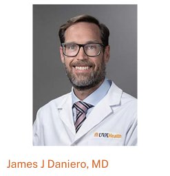 James J Daniero合作论文数Division of Pediatric Otolaryngology, Nemours/Alfred I. duPont Hospital for Children4Young J Kim合作论文数4
 Ongkasuwan Julina合作论文数Baylor College of Medicine4
 Kenneth A. Wallston合作论文数Vanderbilt Institute for Global Health, Vanderbilt University4Bryson Paul C合作论文数4Stevens Madelyn N合作论文数4
 Jan L. Kasperbauer合作论文数School of Continuous Professional Development, Mayo Clinic;Mayo Medical School4Joseph M. Aulino合作论文数the Department of Radiology, Medical College of Virginia4
 Fabien Maldonado合作论文数University Of Missouri Kansas City4
 Yu Shyr合作论文数Department of Biostatistics, Vanderbilt University Medical Center;Vanderbilt Center for Quantitative Sciences, Vanderbilt University Medical Center;School of Medicine, Vanderbilt University;Taipei Medical University4Kim S. Ely合作论文数4Robert R. Lorenz合作论文数4Sanjay M. Athavale合作论文数4
 Joshua S Schindler合作论文数4
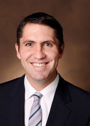 Alexander Gelbard合作论文数4
 Guri S. Sandhu合作论文数4Michael S. Benninger合作论文数4
 Otis B. Rickman合作论文数Division of Pulmonary and Critical Care Medicine4
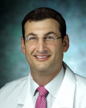 Alexander Hillel合作论文数Department of Otolaryngology, Johns Hopkins University;Johns Hopkins School of Medicine4
 David G. Lott合作论文数Mayo Clinic Comprehensive Cancer Center4Eric S Edell合作论文数4Marshall E Smith合作论文数Division of Otolaryngology/Head and Neck Surgery, University of Utah4Donald T. Donovan合作论文数4
 Christopher T Wootten合作论文数4Li-Ching Huang合作论文数3Kim A. Ely合作论文数3Rebecca J. Howell合作论文数3Michael J. Rutter合作论文数3
 Ahmed Soliman合作论文数3




