BACKGROUND:Anterior cruciate ligament (ACL) injuries are common and increasingly prevalent in the pediatric population. However, there remain sparse epidemiological data on the surgical treatment of these injuries. The objective of this study is to assess the trends in the rate of pediatric ACL reconstruction in Australia over the past 2 decades.METHODS:The incidence of ACL reconstruction from 2001 to 2020 in patients 5 to 14 years of age was analyzed using the Australian Medicare Benefits Schedule (MBS) database. Data were stratified by sex and year. An offset term was introduced using population data from the Australian Bureau of Statistics to account for population changes over the study period.RESULTS:A total of 3719 reconstructions for the management of pediatric ACL injuries were performed in Australia under the MBS in the 20-year period from 2001 to 2020. There was a statistically significant annual increase in the total volume and per capita volume of pediatric ACL reconstructions performed across the study period ( P <0.0001). There was a significant increase in the rate of both male and female reconstructions ( P <0.0001), with a greater proportion of reconstructions performed on males (n=2073, 56%) than females (n=1646, 44%). In 2020, the rate of pediatric ACL reconstructions decreased to a level last seen in 2015, likely due to the effects of COVID-19.CONCLUSIONS:The incidence of ACL reconstruction in skeletally immature patients has increased in Australia over the 20-year study period. This increase is in keeping with evidence suggesting poor outcomes with nonoperative or delayed operative management.
Purpose: Anterior cruciate ligament (ACL) reconstruction is an evolving field, though there remains sparse epidemiological data on the treatment of ACL ruptures. The objective of this study is to analyse the trends in the rate of ACL reconstruction (ACLR) in adult patients over the past two decades in the Australian population. Methods: The incidence of ACLR between 2001 and 2020 in patients 15 years and over was analysed using the Australian Medicare Benefits Schedule (MBS) database. The data reflect patients with private health coverage (45% of the Australian population). An offset term was introduced using national population data to account for population changes over the study period. ResultsA total of 160,353 ACLRs were performed in Australia under the MBS in the 20-year period from 2001 to 2020. An annual increase in the total volume and per capita rate of ACLRs was found (p < 0.05). The annual volume of cases increased by 82%, from 5512 in 2001 to a peak of 10,011 in 2017. This increase was seen across all age groups (p < 0.05) and both sexes (p < 0.05), with a greater proportion of reconstructions performed on males (n = 102,357, 64%) than females (n = 57,996, 36%). In 2020, the rate of adult ACLRs decreased to a level last seen in 2004, likely due to the effects of COVID-19. Conclusions: The incidence of ACLR in adult patients has increased in Australia over the 20-year study period. The trends noted provide information that can be used to guide resource allocation and health provision in the future. Level of Evidence: Level IV.
Introduction: We compared the 12-month effects of arthroscopic surgery and physiotherapist-led care for femoroacetabular impingement (FAI) syndrome on the time-varying magnitude of hip contact force and muscle contributions to hip contact force during walking. Methods: Secondary analysis was performed on 37 individuals with FAI syndrome who received biomechanical assessment before and 12 months after either arthroscopic surgery (n = 17) or physiotherapist-led care (personalized hip therapy [PHT]) (n = 20). At both time points, three-dimensional whole-body motions, ground reaction forces, and surface electromyograms (n = 14) were acquired during overground walking. A neuromusculoskeletal model was used to determine hip contact force and muscle contributions to hip contact force. Two-way repeated measures analyses of variance, implemented through statistical parametric mapping, were used to assess interactions between, and main effects of, treatment (arthroscopy vs PHT) and time (baseline vs follow-up) on time-varying magnitude of hip contact force and muscle contributions to hip contact force. Effects were reported as mean differences (normalized to bodyweight, BW) with 95% confidence intervals [95% CI, lower, upper bound]. Results: For both treatment groups, hip contact force was larger at 12 months compared with their respective baseline value (mean increase across stride, arthroscopy: 0.97 BW [95% CI, 0.49-1.46] P < 0.001; PHT: 1.05 BW [95% CI, 0.68-1.43] P < 0.001); however, no interaction effects were found. For both treatment groups, hip flexor, adductor, and abductor muscle groups made greater contributions to hip contact force after 12 months compared with baseline, whereas hip extensors made smaller contributions. Conclusions: Compared with baseline, both treatments resulted in 12-month increases in hip contact force during walking caused by larger flexor, adductor, and abductor muscle forces. At 12 months, hip contact force magnitude remained different from normative values reported for healthy individuals, indicating that neither treatment fully restored hip biomechanics.
Background:The influence of implant factors on patient-reported outcome measures (PROMS) after total knee arthroplasty (TKA) has previously been studied but findings are often inconsistent, restricted in scope and biased by confounding factors. This study aims to determine the association between implant-related factors and early post-operative PROMs after TKA. Methods:Using data from the Australian Orthopaedic Association National Joint Replacement Registry (AOANJRR) PROMs program, the study included 9487 primary TKA procedures performed in 43 hospitals from July 31, 2018 to December 31, 2020. Data included baseline demographic data, surgical details and PROMs collected pre- and 6 months post-operatively. Seven prosthesis characteristics were incorporated in the multivariable model, with proportional odds ordinal regression analysis used to assess their effects on post-operative Oxford knee score (OKS) and joint pain. Adjustments were made for pre-operative OKS and joint pain, as well as potential patient confounders. Results:At six months, fixed bearing implants showed higher odds of better OKS compared to mobile bearing implants (odds ratio [OR] = 1.20, 95 % confidence interval [CI] 1.06-1.37. p < 0.004). Similar findings were found with the use of image derived instrumentation (IDI) (OR = 1.27, 95 % CI 1.11-1.46, p < 0.001), robotic assisted vs. non-navigated knees (OR = 1.21, 95 % CI 1.06-1.38, p < 0.005) and no patella resurfacing (OR = 1.10, 95 % CI 1.01-1.20, p < 0.032). For lower pain scores at six months, the use of highly crosslinked polyethylene (OR = 1.12, 95 % CI 1.02-1.22, p < 0.015), cemented femoral components (OR = 1.11, 95 % CI 1.01-1.22, p < 0.024), IDI (OR = 1.20, 95 % CI 1.05-1.37, p < 0.009) or no patella resurfacing (OR = 1.10, 95 % CI 1.01-1.20, p < 0.034) were associated. Conclusion:Utilizing a fixed bearing or robotic assistance correlated with improved OKS scores, whereas highly crosslinked polyethylene or cemented femoral components were associated with improved pain scores. At six months, the use of IDI compared to non-navigated TKA, and patellar resurfacing showed both improved OKS and pain scores.
BACKGROUND:Early revision for total hip arthroplasty is a serious adverse outcome. There are multiple contributing risk factors for early revision. Risk factors can exist at the level of the surgeon and the level of the institution. The primary research question of this study was to determine the relative contribution of surgeon-level and hospital-level variance to rates of early revision (overall and for infection) after primary total hip arthroplasty. METHODS:This is a registry-based study from the Australian Orthopaedic Association National Joint Replacement Registry (AOANJRR). Data for the most commonly used stem (Exeter V40) were used to reduce prosthesis variation from the analysis. A mixed effects Cox Model (also known as a frailty model) with crossed random effects for surgeon and hospital was used. Outcomes were early revision (within 2 years) for all causes and for infection. This model allowed for the risk of early revision to be explained by the variability at the surgeon level or hospital level. RESULTS:There were 32 031 procedures performed by 735 surgeons across 250 hospitals between 1 January 2015 and 31 December 2019. Surgeon variability significantly contributed to overall variation in revision for any cause and revision for infection (P < 0.0001). There was no significant contribution of hospital-level variation to overall revision or for infection. CONCLUSIONS:Surgeon-level factors play a more important role than institution-level factors in early revision after primary total hip arthroplasty. If surgeons are identified as having a higher risk of revision, there is potential for surgeon-level practice change to reduce the risk of early revision.
BACKGROUND:Bony morphology is central to the pathomechanism of femoroacetabular impingement syndrome (FAIS), however isolated radiographic measures poorly predict symptom onset and severity. More comprehensive morphology measurement considered together with patient factors may better predict symptom presentation. This study aimed to determine the morphological parameter(s) and patient factor(s) associated with symptom age of onset and severity in FAIS.METHODS:99 participants (age 32.9 ± 10.5 years; body mass index (BMI 24.3 ± 3.1 kg/m2; 42% females) diagnosed with FAIS received standardised plain radiographs and magnetic resonance scans. Alpha angle in four radial planes (superior to anterior), acetabular version (AV), femoral torsion, lateral centre-edge, anterior centre-edge (ACEA) and femoral neck-shaft angles were measured. Age of symptom onset (age at presentation minus duration of symptoms), international Hip Outcome Tool-33 (iHOT-33) and modified UCLA activity scores were recorded. Backward stepwise regression assessed morphological parameters and patient factors (age, sex, BMI, symptom duration, annual income, private/public healthcare system accessed) to determine variables independently associated with onset age and iHOT-33 score.RESULTS:Earlier symptom onset was associated with larger superoanterior alpha angle (p = 0.007), smaller AV (p = 0.023), lower BMI (p = 0.010) and public healthcare system access (p = 0.041) (r2 = 0.320). Worse iHOT-33 score was associated with smaller ACEA (p = 0.034), female sex (p = 0.040), worse modified UCLA activity score (p = 0.010) and public healthcare system access (p < 0.001) (r2 = 0.340).CONCLUSIONS:Age of symptom onset was chiefly predicted by femoral and acetabular bony morphology measures, whereas symptom severity predominantly by patient factors. Factors measured explained a small amount of variance in the data; additional unmeasured factors may be more influential.
Patella fractures in children are rare accounting for 1 % of all paediatric fractures. Rarer still are transverse patella fractures in children with only 1 case previously described in the literature of a child less than the age of 6. We present the second case in the literature of a transverse patella fracture in a child, and the only case of a transverse fracture in a partially ossified patella in a 5-year-old. Our patient suffered an open patella fracture following fall with a flexed knee onto a broken tile. The case was successfully managed with adult fixation principles of patella fractures with a tension band wire construct. The patient had regular follow-up with knee radiographs showing a well healed fracture and a good range of motion of the knee at 3 months. The hardware was subsequently removed with no complications. In conclusion, we present an extremely rare case of an open transverse patella fracture in a child, utilising adult fixation principles of a patella which resulted in a successful outcome for the patient.
Acute instability of the first ray following trauma is a rare type of Lisfranc injury. Plain radiographs may demonstrate instability if there is a clear dislocation of the first ray, however subtle injuries are difficult to recognise. Magnetic resonance imaging (MRI) has been described in the literature as an adjunct in the diagnosis of subtle Lisfranc injuries. There has been no description of the use of MRI and the unique findings of instability of the first ray in the acute setting. We report 2 cases of traumatic instability of the first ray of the foot. Both cases underwent plain radiographs, where in one patient there was a suggestive Lisfranc injury and the other patient had normal radiographical findings. Further investigation utilising an MRI showed a distinct pattern of bone bruising in the navicular and medial cuneiform at the naviculocuneiform joint. These findings indicated disruption of the ligamentous construct stabilising the first ray to the foot. Operative management was undertaken and both patients had a good outcome. This case report is important as it is the first to describe the unique MRI findings of instability of the first ray which will enable other clinicians to appropriately diagnose and manage this injury.
Background: Femoroacetabular impingement syndrome is characterized by chondrolabral damage and hip pain. The specific biomechanics used by people with femoroacetabular impingement syndrome during daily activities may exacerbate their symptoms. Femoroacetabular impingement syndrome can be treated nonoperatively or surgically; however, differential treatment effects on walking biomechanics have not been examined. Purpose: To compare the 12-month effects of physical therapist–led care or arthroscopy on trunk, pelvis, and hip kinematics as well as hip moments during walking. Study Design: Secondary analysis of multi-centre, pragmatic, two-arm superiority randomized controlled trial subsample; Level of evidence, 1. Methods: A subsample of 43 participants from the Australian Full randomised controlled trial of Arthroscopic Surgery for Hip Impingement versus best cONventional (FASHIoN trial) underwent gait analysis and completed the International Hip Outcome Tool (iHOT-33) at both baseline and 12 months after random allocation to physical therapist–led care (personalized hip therapy; n = 22; mean age 35; 41% female) or arthroscopy (n = 21; mean age 36; 48% female). Changes in trunk, pelvis, and hip biomechanics were compared between treatment groups across the gait cycle using statistical parametric mapping. Associations between changes in iHOT-33 and changes in hip kinematics across 3 planes of motion were examined. Results: As compared with the arthroscopy group, the personalized hip therapy group increased its peak hip adduction moments (mean difference = 0.35 N·m/body weight·height [%] [95% CI, 0.05-0.65]; effect size = 0.72; P = .02). Hip adduction moments in the arthroscopy group were unchanged in response to treatment. No other between-group differences were detected. Improvements in iHOT-33 were not associated with changes in hip kinematics. Conclusion: Peak hip adduction moments were increased in the personalized hip therapy group and unchanged in the arthroscopy group. No biomechanical changes favoring arthroscopy were detected, suggesting that personalized hip therapy elicits greater changes in hip moments during walking at 12-month follow-up. Twelve-month changes in hip-related quality of life were not associated with changes in hip kinematics.
ABSTRACT Purpose The magnitude and location of hip contact force influence the local mechanical environment of the articular tissue, driving remodeling. We used a neuromusculoskeletal model to investigate hip contact force magnitudes and their regional loading patterns on the articular surfaces in those with femoroacetabular impingement (FAI) syndrome and controls during walking. Methods An EMG-assisted neuromusculoskeletal model was used to estimate hip contact forces in eligible participants with FAI syndrome (n = 41) and controls (n = 24), walking at self-selected speed. Hip contact forces were used to determine the average and spread of regional loading for femoral and acetabular articular surfaces. Hip contact force magnitude and region of loading were compared between groups using statistical parametric mapping and independent t-tests, respectively (P < 0.05). Results All of the following findings are reported compared with controls. Those with FAI syndrome walked with lower-magnitude hip contact forces (mean difference, −0.7 N·BW−1; P < 0.001) during first and second halves of stance, and with lower anteroposterior, vertical, and mediolateral contact force vector components. Participants with FAI syndrome also had less between-participant variation in average regional loading, which was located more anteriorly (3.8°, P = 0.035) and laterally (2.2°, P = 0.01) on the acetabulum but more posteriorly (−4.8°, P = 0.01) on the femoral head. Participants with FAI syndrome had a smaller spread of regional loading across both the acetabulum (−1.9 mm, P = 0.049) and femoral head (1 mm, P < 0.001) during stance. Conclusions Compared with controls, participants with FAI syndrome walked with lower-magnitude hip contact forces that were constrained to smaller regions on the acetabulum and femoral head. Differences in regional loading patterns might contribute to the mechanobiological processes driving cartilage maladaptation in those with FAI syndrome.
OBJECTIVES: To examine whether a hip brace can improve hip health quality-of-life (QoL) and is well-tolerated in people with femoroacetabular impingement syndrome (FAIS) or symptomatic labral tears after 6 weeks of wear. DESIGN: Parallel, two-arm, exploratory randomized trial. SETTING: Hospital and private clinics of orthopaedic surgeons. PARTICIPANTS: Individuals >18 years with FAIS or labral tears. INTERVENTIONS: Usual conservative care versus usual conservative care plus a hip brace. MAIN OUTCOMES: Patient-reported outcomes were assessed with the International Hip Outcome Tool (iHOT-33), and Copenhagen Hip and Groin Outcome Scores (HAGOS). Brace acceptability was measured using the Quebec User Evaluation of Satisfaction with Assistive Technology survey. Independent t-tests assessed between-group differences. RESULTS: Thirty-eight participants were recruited, 19 each group, 60% women, mean age 39.3 +/- 11.8 years, body mass index 25.3 +/- 4.4 kg/m(2), iHOT-33 36.6 +/- 24.8. Three participants dropped out (one usual care, 2 braced). The mean between-group difference for iHOT-33 was 19.4 (95% confidence interval [CI] 1.68-37.06, P = 0.03) favoring the brace. There were improvements in most HAGOS subscale scores favoring the brace. Issues with brace tolerability for some participants were perceived comfort and effectiveness. Three brace-related adverse events were reported. CONCLUSION: Between-group differences favored the braced group for hip health QoL, pain, symptoms, and function. Although these were promising results, the CIs for the estimates were wide, the small sample size likely a contributing factor. Our results suggest that further investigation of the brace is warranted, we calculated sample sizes and made recommendations for the design of a future trial.
Background: This study describes a new technique of computer navigation for total hip arthroplasty (THA) that uses a customized perspex board to register the coronal plane of a patient in the lateral position. Cup placement is then referenced to the coronal plane. Purposes: This paper looks at the outcome of this technique. Materials and Methods: Seventeen patients who underwent a total of 18 THA were included. During surgery, the patient was placed in a lateral decubitus position and care taken to ensure the patient was secured perpendicular to the table. The coronal plane was registered using a customized perspex board aligned with the long axis of the operating table. The intraoperative target for acetabular cup placement was 20-degree anteversion and 40-degree inclination with respect to the coronal plane. Radiographs were taken 6 weeks postoperatively. Results: The mean cup angle of anteversion was larger in the postoperative method (M=22.83, SD=4.436) than in the intraoperative method (M=22.39, SD=4.629). A 1-sample t test indicated the difference between means was not statistically significant. The mean cup angle of inclination was larger in the intraoperative method (M=44.83, SD=3.400) than in the postoperative method (M=41.56, SD=4.693). A 1-sample t test indicated the difference between means was significant although still within the prescribed safe zones. Conclusion: Using a customized perspex board to register the patient and targeting cup placement to a safe zone referenced to the coronal plane result in optimal cup placement when assessed by postoperative radiographs. This avoids the need to turn the patient after registration and obviates the effect of pelvic tilt on cup placement. Level of Evidence: Level IV—case series.
1. Bergqvist D, Svensson PJ. Treatment of mesenteric vein thrombosis. Semin Vasc Surg. 2010;23:65–8. 2. Acosta S, Alhadad A, Svensson P, Ekberg O. Epidemiology, risk and prognostic factors in mesenteric venous thrombosis. Br J Surg. 2008;95: 1245–51. 3. Bradbury MS, Kavanagh PV, Bechtold RE, Chen MY, Ott DJ, Regan JD, et al. Mesenteric venous thrombosis: diagnosis and noninvasive imaging. Radiographics. 2002;22:527–41. 4. Morasch MD, Ebaugh JL, Chiou AC, Matsumura JS, Pearce WH, Yao JS. Mesenteric venous thrombosis: a changing clinical entity. J Vasc Surg. 2001;34:680–4. 5. Gnanapandithan K, Feuerstadt P. Review article: mesenteric ischemia. Curr Gastroenterol Rep. 2020;22:17. 6. Bala M, Kashuk J, Moore EE, Kluger Y, Biffl W, Gomes CA, et al. Acute mesenteric ischemia: guidelines of the World Society of Emergency Surgery. World J Emerg Surg. 2017;12:38. 7. Brunaud L, Antunes L, Collinet-Adler S, Marchal F, Ayav A, Bresler L, et al. Acute mesenteric venous thrombosis: case for nonoperative management. J Vasc Surg. 2001;34:673–9. 8. Singal AK, Kamath PS, Tefferi A. Mesenteric venous thrombosis. Mayo Clin Proc. 2013;88:285–94. 9. Committee on Obstetric Practice. Committee Opinion No. 723: Guidelines for diagnostic imaging during pregnancy and lactation. Obstet Gynecol. 2017;130:e210–e6.
Background Arthroscopic surgery for femoroacetabular impingement syndrome (FAI) is known to lead to self-reported symptom improvement. In the context of surgical interventions with known contextual effects and no true sham comparator trials, it is important to ascertain outcomes that are less susceptible to placebo effects. The primary aim of this trial was to determine if study participants with FAI who have hip arthroscopy demonstrate greater improvements in delayed gadolinium-enhanced magnetic resonance imaging (MRI) of cartilage (dGEMRIC) index between baseline and 12 months, compared to participants who undergo physiotherapist-led management. Methods Multi-centre, pragmatic, two-arm superiority randomised controlled trial comparing physiotherapist-led management to hip arthroscopy for FAI. FAI participants were recruited from participating orthopaedic surgeons clinics, and randomly allocated to receive either physiotherapist-led conservative care or surgery. The surgical intervention was arthroscopic FAI surgery. The physiotherapist-led conservative management was an individualised physiotherapy program, named Personalised Hip Therapy (PHT). The primary outcome measure was change in dGEMRIC score between baseline and 12 months. Secondary outcomes included a range of patient-reported outcomes and structural measures relevant to FAI pathoanatomy and hip osteoarthritis development. Interventions were compared by intention-to-treat analysis. Results Ninety-nine participants were recruited, of mean age 33 years and 58% male. Primary outcome data were available for 53 participants (27 in surgical group, 26 in PHT). The adjusted group difference in change at 12 months in dGEMRIC was -59 ms (95%CI − 137.9 to - 19.6) ( p = 0.14) favouring PHT. Hip-related quality of life (iHOT-33) showed improvements in both groups with the adjusted between-group difference at 12 months showing a statistically and clinically important improvement in arthroscopy of 14 units (95% CI 5.6 to 23.9) ( p = 0.003). Conclusion The primary outcome of dGEMRIC showed no statistically significant difference between PHT and arthroscopic hip surgery at 12 months of follow-up. Patients treated with surgery reported greater benefits in symptoms at 12 months compared to PHT, but these benefits are not explained by better hip cartilage metabolism. Trial registration details Australia New Zealand Clinical Trials Registry reference: ACTRN12615001177549 . Trial registered 2/11/2015.
Background: Studies of walking in those with femoroacetabular impingement syndrome have found altered pelvis and hip biomechanics. But a whole body, time-contiuous, assessment of biomechanical parameters has not been reported. Additionally, larger cam morphology has been associated with more pain, faster progression to end-stage osteoarthritis and increased cartilage damage but differences in walking biomechanics between large compared to small cam morphologies have not been assessed. Research question: Are trunk, pelvis and lower limb biomechanics different between healthy pain-free controls and individuals with FAI syndrome and are those biomechanics different between those with larger, compared to smaller, cam morphologies? Methods: Twenty four pain-free controls were compared against 41 participants with FAI syndrome who were stratified into two groups according to their maximum alpha angle. Participants underwent three-dimensional motion capture during walking. Trunk, pelvis, and lower limb biomechanics were compared between groups using statistical parametric mapping corrected for walking speed and pain. Results: Compared to pain-free controls, participants with FAI syndrome walked with more trunk anterior tilt (mean difference 7.6 degrees, p < 0.001) as well as less pelvic rise (3 degrees, p < 0.001), hip abduction (-4.6 degrees, p < 0.05) and external rotation (-6.5 degrees, p < 0.05). They also had lower hip flexion (-0.06Nm.kg(-1), p < 0.05), abduction (-0.07Nm.kg(-1), p < 0.05) and ankle plantarflexion moments (-0.19Nm.kg(-1), p < 0.001). These biomechanical differences occurred throughout the gait cycle. There were no differences in walking biomechanics according to cam morphology size. Significance: Results do not support the hypothesis that larger cam morphology is associated with larger differences in walking biomechanics but did demonstrate general differences in trunk, pelvis and lower limb biomechanics between those with FAI sydrome and pain-free controls. Altered external biomechanics are likely the result of complex sensory-motor strategy resulting from pain inhibition or impingement avoidance. Future studies should examine internal loading in those with FAI sydnrome.
BACKGROUND:Total hip arthroplasty (THA) provides excellent pain relief and improved function in patients with painful arthritis. The aim of this study was to identify rates and predictors of dissatisfaction following THA.METHODS:Data were collected prospectively from the Australian Arthroplasty Clinical Outcomes Registry National (ACORN) database between 2014 and 2016 from 2096 patients who underwent THA. Data included baseline demographics, patient-reported outcome measures (PROMs) and postoperative clinical outcomes. Patients were dichotomized into two groups based on their 6-month response to the satisfaction question answered on a Likert scale. Eighteen predefined variables were analyzed. PROMs included full Oxford Hip Score, EQ-5D, and patient satisfaction. A Bayesian model averaging approach was used to build the best predictive model for dissatisfaction. Multiple logistic regression techniques were applied to quantify the effect size of the best model.RESULTS:At 6 months following THA, 95.4% of patients (n = 2000) were satisfied with surgical outcome and 4.6% (n = 96) were dissatisfied. The only variable that was significantly associated with dissatisfaction after THA was "complications after discharge." This result was consistent for both the complete and imputed dataset (odds ratio 4.78, 95% confidence interval 2.60-8.80, P < 0.001 and odds ratio 3.8, 95% confidence interval 2.60-5.60, P < 0.001, respectively).CONCLUSION:Our study confirms the high rates of patient satisfaction following THA, with postoperative complications being the only determinant of dissatisfaction. Optimization of patients prior to surgery, reducing postoperative complications, may further improve satisfaction rates after THA.
Objective Accurate insertion of a guidewire under image intensifier guidance is a fundamental skill required by orthopaedic surgeons. This study investigated how image intensifier distortion, which is composed of pin-cushion and sigmoidal components, changed the apparent trajectory of a guidewire, and the resulting deviation between the intended and actual guidewire tip position. Materials and methods Intraoperative image intensifier images for 220 consecutive patients with hip fractures were retrospectively corrected for distortion using a global polynomial method. The deviation between the intended and actual guidewire tip positions was calculated. Additional distortion parameters were tested using an image intensifier produced by a different manufacturer, and a flat-panel c-arm. Results Deviation was approximately 1 cm if the guidewire was aimed from the extremity of the image and almost 0 if the entry point was only 20% from the centre (p < 0.001). The direction of deviation was different for left and right hips, with average deviations measuring 3 mm proximal and 5 mm distal respectively (p < 0.001). The flat-panel c-arm almost completely eliminated distortion. Conclusions Image intensifier distortion significantly altered the intended trajectory of a guidewire, with guidewires aimed from the image periphery more affected than guidewires aimed from the centre. Furthermore, for right hips, guidewires should be aimed distal to their intended position, and for left hips they should be aimed proximal to achieve their desired position. The flat-panel c-arm eliminated the effect of distortion; hence, it may be preferable if precision in guidewire positioning is vital.
Background: Posterior cruciate ligament (PCL) injuries of the knee are common and sometimes difficult to diagnose. Magnetic resonance imaging (MRI), performed using standard orthogonal plane views, is the investigation of choice. It can be particularly difficult to differentiate acute partial and complete tears and identify elongation of chronic healed tears. The aim of the paper is to describe a new method of positioning the patient with the knee flexed at 90 degrees, allowing the PCL to be visualised in a position of greatest length and tension which may assist in differentiating and identifying these injuries. Methods: Four symptomatic patients with suspected PCL injuries, two acute and two chronic, were MRI scanned using a routine protocol with the knee in extension before performing oblique sagittal fast spin-echo (FSE) proton-density (PD) sequences with the knee positioned in 90 degrees of flexion. The appearance of the PCLs were then qualitatively assessed. Results: MRI scanning with the knee in flexion identified more extensive PCL injury than standard imaging. In the two patients with acute injuries, partial tears on the standard orthogonal plane views were found to be complete ruptures. In the two patients with chronic injuries, elongation of the PCL not identifiable on the standard orthogonal plane views was apparent. Conclusion: MRI scanning of the PCL with the knee flexed at 90 degrees may help in differentiating partial and complete ruptures of the PCL and identifying elongation of the PCL in chronic injuries. (C) 2018 Elsevier B.V. All rights reserved.
Femoroacetabular impingement syndrome (FAI), a hip disorder affecting active young adults, is believed to be a leading cause of hip osteoarthritis (OA). Current management approaches for FAI include arthroscopic hip surgery and physiotherapy-led non-surgical care; however, there is a paucity of clinical trial evidence comparing these approaches. In particular, it is unknown whether these management approaches modify the future risk of developing hip OA. The primary objective of this randomised controlled trial is to determine if participants with FAI who undergo hip arthroscopy have greater improvements in hip cartilage health, as demonstrated by changes in delayed gadolinium-enhanced magnetic resonance imaging (MRI) of cartilage (dGEMRIC) index between baseline and 12 months, compared to those who undergo physiotherapy-led non-surgical management.
OBJECTIVES:To evaluate the ionizing radiation dose received by the eyes of orthopaedic surgeons during various orthopaedic procedures. Secondary objective was to compare the ionizing radiation dose received between differing experience level.DESIGN:Prospective comparative study between January 2013 and May 2014.SETTING:Westmead Hospital, a Level 1 Trauma Centre for Greater Western Sydney.PARTICIPANTS:A total of 26 surgeons volunteered to participate within the study.INTERVENTION:Experience level, procedure performed, fluoroscopy time, dose area product, total air kerma, and eye dose received was recorded. Participants were evaluated on procedure and experience level.MAIN OUTCOME MEASUREMENTS:Radiation dose received at eye level by the primary surgeon during an orthopaedic procedure.RESULTS:Data from a total of 131 cases was recorded and included for analysis. The mean radiation dose detected at the eye level of the primary surgeon was 0.02 mSv (SD = 0.05 mSv) per procedure. Radiation at eye level was only detected in 31 of the 131 cases. The highest registered dose for a single procedure was 0.31 mSv. Femoral nails and pelvic fixation procedures had a significantly higher mean dose received than other procedure groups (0.04 mSv (SD = 0.07 mSv) and 0.04 mSv (SD = 0.06 mSv), respectively). Comparing the eye doses received by orthopaedic consultants and trainees, there was no significant difference between the 2 groups.CONCLUSIONS:The risk of harmful levels of radiation exposure at eye level to orthopaedic surgeons is low. This risk is greatest during insertion of femoral intramedullary nails and pelvic fixation, and it is recommended that in these situations, surgeons take all reasonable precautions to minimize radiation dose. The orthopaedic trainees in this study were not subjected to higher doses of radiation than their consultant trainers. On the basis of these results, most of the orthopaedic surgeons remain well below the yearly radiation dose of 20 mSv as recommended by the International Commission on Radiological Protection.
 Libby Spiers合作论文数Department of Physiotherapy, Melbourne School of Health Sciences, Faculty of Medicine, Dentistry and Health Sciences, The University of Melbourne8
 Phong Tran合作论文数7
 Nicholas Murphy合作论文数7
 John O'Donnell合作论文数7
 Jillian Eyles合作论文数School of Health Sciences, Faculty of Medicine and Health, The University of Sydney7
 Kim Bennell合作论文数Centre for Health, Exercise and Sports Medicine, School of Health Sciences, University of Melbourne7
 Michelle Hall合作论文数Department of Physiotherapy, Melbourne School of Health Sciences, Faculty of Medicine, Dentistry and Health Sciences, The University of Melbourne6
 David G Lloyd合作论文数School of Allied Health Sciences, Griffith University6
 David Hunter合作论文数Faculty of Medicine and Health, The University of Sydney;Royal North Shore Hospital6
 Sunny Randhawa合作论文数5
 Parminder J. Singh合作论文数5David J Saxby合作论文数5Diamond Laura E合作论文数Centre for Health, Exercise &, The University of Melbourne;Sports Medicine, Department of Physiotherapy, School of Health Sciences, Parkville, Victoria, Australia.;School of Health Sciences, The University of Melbourne5
 Camdon Fary合作论文数Department of Orthopaedics, The Royal Melbourne Hospital5
 Claudio Pizzolato合作论文数Menzies Health Institute Queensland, Griffith University4
 Symes Michael合作论文数Discipline of Physiotherapy, University of Sydney.4Dickenson Edward合作论文数4
 Trevor N. Savage合作论文数4Tim V. Wrigley合作论文数4
 Damian R. Griffin合作论文数Clinical Sciences Research Institute, Warwick University4Young Jo Kim合作论文数3Michael D O'Sullivan合作论文数3
 Ryan Campbell合作论文数Dept Med, Univ New South Wales3
 William Du Moulin合作论文数3Alexander Burns合作论文数3
 Rachel Louise O'Connell合作论文数NHMRC Clin Trials Ctr, Univ Sydney3
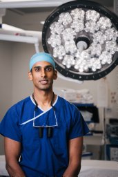 Sivakumar Brahman合作论文数Department of Orthopedic Surgery, The Prince Charles Hospital3
 James Linklater合作论文数Australian Institute of Musculoskeletal Research;University of Sydney;Australian Institute of Musculoskeletal Research, University of Sydney3
 Sam Adie合作论文数South West Sydney Clinical School, University of New South Wales3
 Ian Harris AM合作论文数Orthopaedic Department, Liverpool Hospital;South West Sydney Clinical School, University of New South Wales;Royal Prince Alfred Hospital;Fairfield Hospital;St George and Sutherland Hospitals3Mehr Vather合作论文数2Adrian Hughes合作论文数2Antony Nguyen合作论文数2Edin K Suwarganda合作论文数2Deanne E Jenkin合作论文数Ingham Inst Appl Med Res2
 Vincent An合作论文数2
 Stuart Grieve合作论文数School of Health Sciences, Faculty of Medicine and Health, The University of Sydney2
 Nadine Foster合作论文数School of Medicine, Keele University;Clinical Trials Center, University of Queensland;School of Health and Rehabilitation Sciences, University of Queensland2Le Brian T N合作论文数2Michelle F. Lorimer合作论文数2
 Thor Besier合作论文数Auckland Bioengineering Institute, Faculty of Engineering, The University of Auckland2
 Stephan Reichenbach合作论文数MRC Health Services Research Collaboration, University of Bristol2Royal North Shore Hospital合作论文数1Emeka Oragui合作论文数1Jan Peter Van Meirhaeghe合作论文数1Ben Schwarz合作论文数1Sonika Virk合作论文数1
 Chinmoy Mukerjee合作论文数1Thomas Ward合作论文数1




