Intratumoral injections often lack visibility, leading to unpredictable outcomes such as incomplete tumor coverage, off-target drug delivery and systemic toxicities. This study investigated an ultrasound (US) and x-ray imageable thermosensitive hydrogel based on poloxamer 407 (POL) percutaneously delivered in a healthy swine model. The primary objective was to assess the 2D and 3D distribution of the hydrogel within tissue across three different needle devices and injection sites: liver, kidney, and intercostal muscle region. Secondly, pharmacokinetics of POL loaded with doxorubicin (POLDOX) were evaluated and compared to free doxorubicin injection (DOXSoln) with a Single End Hole Needle. Utilizing 2D and 3D morphometrics from US and x-ray imaging techniques such as Computed Tomography (CT) and Cone Beam CT (CBCT), we monitored the localization and leakage of POLDOX over time. Relative iodine concentrations measured with CBCT following incorporation of an iodinated contrast agent in POL indicated potential drug diffusion and advection transport. Furthermore, US imaging revealed temporal changes, suggesting variations in acoustic intensity, heterogeneity, and echotextures. Notably, 3D reconstruction of the distribution of POL and POLDOX from 2D ultrasound frames was achieved and morphometric data obtained. Pharmacokinetic analysis revealed lower systemic exposure of the drug in various organs with POLDOX formulation compared to DOXSoln formulation. This was demonstrated by a lower area under the curve (852.1 ± 409.1 ng/mL·h vs 2283.4 ± 377.2 ng/mL·h) in the plasma profile, suggesting a potential reduction in systemic toxicity. Overall, the use of POL formulation offers a promising strategy for precise and localized drug delivery, that may minimize adverse effects. Dual modality POL imaging enabled analysis of patterns of gel distribution and morphology, alongside of pharmacokinetics of local delivery. Incorporating hydrogels into drug delivery systems holds significant promise for improving the predictability of the delivered drug and enhancing spatial conformability. These advancements can potentially enhance the safety and precision of anticancer therapy.
Intratumoral injections have the potential for enhanced cancer treatment efficacy while reducing costs and systemic exposure. However, intratumoral drug injections can result in substantial off-target leakage and are invisible under standard imaging modalities like ultrasound (US) and x-ray. A thermosensitive poloxamer-based gel for drug delivery was developed that is visible using x-ray imaging (computed tomography (CT), cone beam CT, fluoroscopy), as well as using US by means of integrating perfluorobutane-filled microbubbles (MBs). MBs content was optimized using tissue mimicking phantoms and ex vivo bovine livers. Gel formulations less than 1% MBs provided gel depositions that were clearly identifiable on US and distinguishable from tissue background and with minimal acoustic artifacts. The cross-sectional areas of gel depositions obtained with US and CT imaging were similar in studies using ex vivo bovine liver and postmortem in situ swine liver. The gel formulation enhanced multimodal image-guided navigation, enabling fusion of ultrasound and x-ray/CT imaging, which may enhance targeting, definition of spatial delivery, and overlap of tumor and gel. Although speculative, such a paradigm for intratumoral drug delivery might streamline clinical workflows, reduce radiation exposure by reliance on US, and boost the precision and accuracy of drug delivery targeting during procedures. Imageable gels may also provide enhanced temporal and spatial control of intratumoral conformal drug delivery.
Publicly available audio data presents a unique opportunity for the development of digital health technologies with large language models (LLMs). In this study, YouTube was mined to collect audio data from individuals with self-declared positive COVID-19 tests as well as those with other upper respiratory infections (URI) and healthy subjects discussing a diverse range of topics. The resulting dataset was transcribed with the Whisper model and used to assess the capacity of LLMs for detecting self-reported COVID-19 cases and performing variant classification. Following prompt optimization, LLMs achieved accuracies of 0.89, 0.97, respectively, in the tasks of identifying self-reported COVID-19 cases and other respiratory illnesses. The model also obtained a mean accuracy of 0.77 at identifying the variant of self-reported COVID-19 cases using only symptoms and other health-related factors described in the YouTube videos. In comparison with past studies, which used scripted, standardized voice samples to capture biomarkers, this study focused on extracting meaningful information from public online audio data. This work introduced novel design paradigms for pandemic management tools, showing the potential of audio data in clinical and public health applications.
Liver cancer ranks as the fifth leading cause of cancer-related death globally. Direct intratumoral injections of anti-cancer therapeutics may improve therapeutic efficacy and mitigate adverse effects compared to intravenous injections. Some challenges of intratumoral injections are that the liquid drug formulation may not remain localized and have unpredictable volumetric distribution. Thus, drug delivery varies widely, highly-dependent upon technique. An x-ray imageable poloxamer 407 (POL)-based drug delivery gel was developed and characterized, enabling real-time feedback. Utilizing three needle devices, POL or a control iodinated contrast solution were injected into an ex vivo bovine liver. The 3D distribution was assessed with cone beam computed tomography (CBCT). The 3D distribution of POL gels demonstrated localized spherical morphologies regardless of the injection rate. In addition, the gel 3D conformal distribution could be intentionally altered, depending on the injection technique. When doxorubicin (DOX) was loaded into the POL and injected, DOX distribution on optical imaging matched iodine distribution on CBCT suggesting spatial alignment of DOX and iodine localization in tissue. The controllability and localized deposition of this formulation may ultimately reduce the dependence on operator technique, reduce systemic side effects, and facilitate reproducibility across treatments, through more predictable standardized delivery.
Heat-based local ablation techniques are effective treatments for specific oligometastatic and localized cancers and are being studied for their potential to induce immunogenic cell death and augment systemic immune responses to immunotherapies. The diverse technologies associated with thermal therapy have an unmet need for method development to enable device-specific experimentation, optimization, calibration and refinement of the parameter space to optimize therapeutic intent while minimizing side effects or risk to the patient. Quality assurance, training, or comparing thermal dose among different modalities or techniques using animal models is time and resource intensive. Therefore, the application and use of tissue mimicking thermosensitive, thermochromic liquid crystal and thermochromic paint phantom models may reduce costs and hurdles associated with animal use. Further, their homogenous composition may enable more precise assessment of ablative techniques. This review utilized SciFinder, Web of Science, PubMed and EMBASE to systematically evaluate the literature describing the background and applications of thermochromic liquid crystal, thermochromic paint and tissue-mimicking thermochromic phantoms used to characterize the thermal effects of ablation devices with a focus on facilitating their use across the medical device development life cycle. Graphical Abstract
Although systemic immunotherapy has achieved durable responses and improved survival for certain patients and cancer types, low response rates and immune system-related systemic toxicities limit its overall impact. Intratumoral (intralesional) delivery of immunotherapy is a promising technique to combat mechanisms of tumor immune suppression within the tumor microenvironment and reduce systemic drug exposure and associated side effects. However, intratumoral injections are prone to variable tumor drug distribution and leakage into surrounding tissues, which can compromise efficacy and contribute to toxicity. Controlled release drug delivery systems such as in situ-forming hydrogels are promising vehicles for addressing these challenges by providing improved spatio-temporal control of locally administered immunotherapies with the goal of promoting systemic tumor-specific immune responses and abscopal effects. In this review we will discuss concepts, applications, and challenges in local delivery of immunotherapy using controlled release drug delivery systems with a focus on intratumorally injected hydrogel-based drug carriers.
To develop and characterize an x-ray-imageable gel for image-guided hepatic intra-tumoral drug delivery and to assess performance and 3D distribution post-injection in an ex-vivo bovine liver model
To develop and characterize a polymeric platform imageable under both ultrasound (US) and x-ray for image-guided intratumoral injection or transcatheter embolization in agar phantoms and ex vivo livers
Intratumoral injections pressurize fluid that can distribute a drug over a large volume, but the distribution is highly variable because fluid may rupture tissue and take a path of least resistance. This study was designed to inform technique in a clinical trial injecting a recombinant immunotoxin with a prescribed 4mL injection volume. A secondary goal was to determine if drug distribution may be approximated from contrast distribution on CT to facilitate future intra-procedural evaluation.
The success of artificial intelligence in clinical environments relies upon the diversity and availability of training data. In some cases, social media data may be used to counterbalance the limited amount of accessible, well-curated clinical data, but this possibility remains largely unexplored. In this study, we mined YouTube to collect voice data from individuals with self-declared positive COVID-19 tests during time periods in which Omicron was the predominant variant , while also sampling non-Omicron COVID-19 variants, other upper respiratory infections (URI), and healthy subjects. The resulting dataset was used to train a DenseNet model to detect the Omicron variant from voice changes. Our model achieved 0.85/0.80 sensitivity/specificity in separating Omicron samples from healthy samples and 0.76/0.70 sensitivity/specificity in separating Omicron samples from symptomatic non-COVID samples. In comparison with past studies, which used scripted voice samples, we showed that leveraging the intra-sample variance inherent to unscripted speech enhanced generalization. Our work introduced novel design paradigms for audio-based diagnostic tools and established the potential of social media data to train digital diagnostic models suitable for real-world deployment.
In low-income countries, up to 80% of women diagnosed with cervical dysplasia do not return for follow-up care, primarily due to treatment being inaccessible. Here, we describe development of a low-cost, portable treatment suitable for such settings. It is based on injection of ethyl cellulose (EC)-ethanol to ablate the transformation zone around the os, the site most impacted by dysplasia. EC is a polymer that sequesters the ethanol within a prescribed volume when injected into tissue, and this is modulated by the injected volume and delivery parameters (needle gauge, bevel orientation, insertion rate, depth, and infusion rate). Salient injection-based delivery parameters were varied in excised swine cervices. The resulting injection distribution volume was imaged with a wide-field fluorescence imaging device or computed tomography. A 27G needle and insertion rate of 10 mm/s achieved the desired insertion depth in tissue . Orienting the needle bevel towards the outer edge of the cervix and keeping infusion volumes ≤ 500 µL minimized leakage into off-target tissue. These results guided development of a custom hand-held injector, which was used to locate and ablate the upper quadrant of a swine cervix in vivo with no adverse events or changes in host temperature or heart rate. After 24 h, a distinct region of necrosis was detected that covered a majority (> 75%) of the upper quadrant of the cervix, indicating four injections could effectively cover the full cervix. The work here informs follow up large animal in vivo studies, e.g. in swine, to further assess safety and efficacy of EC-ethanol ablation in the cervix.
It is difficult to retain tumoricidal doses of ethanol in large or unencapsulated tumours without causing intoxication or damaging surrounding tissue. Ethyl cellulose-ethanol ablation (ECEA) overcomes this limitation by trapping ethanol intratumorally. To evaluate the safety of ECEA and to develop a clinically feasible workflow, a single-arm pilot study was performed in cats with lingual/sublingual squamous cell carcinoma (SCC). Six cats underwent intratumoral injection of 6% ethyl cellulose in ethanol. Subjects were observed overnight. There was mild bleeding and transient hyperthermia, and injection site pain and swelling that improved with anti-inflammatory drugs. Serum ethanol was minimally elevated; the mean concentration peaked 1 hour after injection (129 +/- 15.1 nM). Cats were rechecked at weeks 1 and 2; booster treatments were given in cats (n = 3) with stable quality of life and partial response to therapy. Recheck examinations were then performed monthly. The longest tumour dimension increased in each animal (progressive disease via cRECIST); however, estimated tumour volume was reduced in 3 of 6 cats, within 1 week of ECEA. All cats were euthanized (median survival time 70 days) because of local tumour progression and/or lingual dysfunction that was likely hastened by ECEA. ECEA is not a viable treatment for feline lingual/sublingual SCC; tumour volume was effectively reduced in some cats, but the simultaneous loss of lingual function was poorly tolerated. Further optimization may make ECEA a useful option for SCC at other oral sites in the cat, and for head and neck malignancies in other species.
Ethanol provides a rapid, low-cost ablative solution for liver tumors with a small technological footprint but suffers from uncontrolled diffusion in target tissue, limiting treatment precision and accuracy. Incorporating the gel-forming polymer ethyl cellulose to ethanol localizes the distribution. The purpose of this study was to establish a non-invasive methodology based on CT imaging to quantitatively determine the relationship between the delivery parameters of the EC-ethanol formulation, its distribution, and the corresponding necrotic volume. The relationship of radiodensity to ethanol concentration was characterized with water–ethanol surrogates. Ex vivo EC-ethanol ablations were performed to optimize the formulation (n = 6). In vivo ablations were performed to compare the optimal EC-ethanol formulation to pure ethanol (n = 6). Ablations were monitored with CT and ethanol distribution volume was quantified. Livers were removed, sectioned and stained with NADH-diaphorase to determine the ablative extent, and a detailed time-course histological study was performed to assess the wound healing process. CT imaging of ethanol–water surrogates demonstrated the ethanol concentration-radiodensity relationship is approximately linear. A concentration of 12% EC in ethanol created the largest distribution volume, more than eight-fold that of pure ethanol, ex vivo. In vivo, 12% EC-ethanol was superior to pure ethanol, yielding a distribution volume three-fold greater and an ablation zone six-fold greater than pure ethanol. Finally, a time course histological evaluation of the liver post-ablation with 12% EC-ethanol and pure ethanol revealed that while both induce coagulative necrosis and similar tissue responses at 1–4 weeks post-ablation, 12% EC-ethanol yielded a larger ablation zone. The current study demonstrates the suitability of CT imaging to determine distribution volume and concentration of ethanol in tissue. The distribution volume of EC-ethanol is nearly equivalent to the resultant necrotic volume and increases distribution and necrosis compared to pure ethanol.
Focal tumor ablation with ethanol could provide benefits in low-resource settings because of its low overall cost, minimal imaging technology requirements, and acceptable clinical outcomes. Unfortunately, ethanol ablation is not commonly utilized because of a lack of predictability of the ablation zone, caused by inefficient retention of ethanol at the injection site. To create a predictable zone of ablation, we have developed a polymer-assisted ablation method using ethyl cellulose (EC) mixed with ethanol. EC is ethanol-soluble and water-insoluble, allowing for EC-ethanol to be injected as a liquid and precipitate into a solid, occluding the leakage of ethanol upon contact with tissue. The aims of this study were to compare the 1) safety, 2) release kinetics, 3) spatial distribution, 4) necrotic volume, and 5) overall survival of EC-ethanol to conventional ethanol ablation in a murine breast tumor model. Non-target tissue damage was monitored through localized adverse events recording, ethanol release kinetics with Raman spectroscopy, injectate distribution with in vivo imaging, target-tissue necrosis with NADH-diaphorase staining, and overall survival by proxy of tumor growth. EC-ethanol exhibited decreased localized adverse events, a slowing of the release rate of ethanol, more compact injection zones, 5-fold increase in target-tissue necrosis, and longer overall survival rates compared to the same volume of pure ethanol. A single 150 μL dose of 6% EC-ethanol achieved a similar survival probability rates to six daily 50 μL doses of pure ethanol used to simulate a slow-release of ethanol over 6 days. Taken together, these results demonstrate that EC-ethanol is safer and more effective than ethanol alone for ablating tumors.
OBJECTIVE:Ethanol ablation, the injection of ethanol to induce necrosis, was originally used to treat hepatocellular carcinoma, with survival rates comparable to surgery. However, efficacy is limited due to leakage into surrounding tissue. To reduce leakage, we previously reported incorporating ethyl cellulose (EC) with ethanol as this mixture forms a gel when injected into tissue. To further develop EC-ethanol injection as an ablative therapy, the present study evaluates the extent to which salient injection parameters govern the injected fluid distribution.METHODS:Utilizing ex vivo swine liver, injection parameters (infusion rate, EC%, infusion volume) were examined with fluorescein added to each solution. After injection, tissue samples were frozen, sectioned, and imaged.RESULTS:While leakage was higher for ethanol and 3%EC-ethanol at a rate of 10 mL/hr compared to 1 mL/hr, leakage remained low for 6%EC-ethanol regardless of infusion rate. The impact of infusion volume and pressure were also investigated first in tissue-mimicking surrogates and then in tissue. Results indicated that there is a critical infusion pressure beyond which crack formation occurs leading to fluid leakage. At a rate of 10 mL/hr, a volume of 50 μL remained below the critical pressure.CONCLUSIONS:Although increasing the infusion rate increases stress on the tissue and the risk of crack formation, injections of 6%EC-ethanol were localized regardless of infusion rate. To further limit leakage, multiple low-volume infusions may be employed.SIGNIFICANCE:These results, and the experimental framework developed to obtain them, can inform optimizing EC-ethanol to treat a range of medical conditions.
Abstract In low-resource settings, surgery or radiation are inaccessible to 90% of cancer patients due to inadequate infrastructure and lack of trained personnel. Resource appropriate technologies must be developed to make cancer treatments accessible. Ablative techniques, such as radiofrequency ablation are often used as a minimally invasive alternative to surgery. However, energy-based ablation techniques require expensive machinery and a steady supply of electricity and expensive consumables. To address these shortcomings, we have developed a low-cost ablation technique that stabilizes tumor growth with a simple injection of ethanol in combination with a polymer, ethylcellulose (EC). The EC injected with ethanol creates a slow-release depot of ethanol at the injection site by undergoing a solvent induced phase-transition initiated by water in tissues. EC-ethanol improves upon traditional ethanol ablation by providing increased precision while remaining low-cost and simple to administer. We evaluated the precision of EC-ethanol as well as its vascular sclerosing capabilities as a strategy to avert tumor growth in rodent models of breast cancer. EC-ethanol injected at 1 mL/hr with 6% w/w EC was used as this demonstrated to be the optimal formulation and rate for causing tumor necrosis (Morhard et al., Scientific Reports, 2017) and enhanced the anti-tumor immunostimulatory response (Nief et al., 2019, Under Review). We demonstrate the precision delivery of EC-ethanol in orthotopic 67NR mammary tumor as with small-animal MRI. Further, ethanol devascularization is monitored with photoacoustic imaging 24 hours post ablation. Local tumor control was demonstrated both in 67NR mammary tumor and in 7,12-Dimethylbenz[a]anthracene (DMBA) induced rat mammary tumor models. Repeated ablations on significantly decrease primary tumor growth compared to untreated tumors in both the mouse and rat models (P<0.05). In rats weekly 200 µL EC-ethanol injections began when tumors reached 2 mL in volume (n=5), and in 30 days tumors decreased 20% ± 3% untreated controls grew an average of 721%. For mice (n=5), 50 µL was given daily for 6 days on day 3 after tumor implantation to account for small body weight and rapidly growing tumors. Mice treated with EC-ethanol lived significantly longer (P<0.05 log-rank test) than untreated mice and mice injected with the same volume of pure ethanol. EC-ethanol is a low-cost, non-invasive tumor destruction method that shows a clear control of tumor growth when applied as a fractionated dose. We utilized a mouse and rat tumor model to ensure that the EC-ethanol's success is not dependent on the model. Ongoing studies will examine the impact of varying injected volumes at well-defined fractionation schedules on ethanol distribution, necrosis, tumor growth, and survival. Citation Format: Corrine A. Nief, Erika Chelales, Robert Morhard, Maomao Chem, Jeffrey Everitt, Jenna Mueller, Junjie Yao, Mark W. Dewhirst, Nirmala Ramanujam. Averting tumor growth in rodent breast cancer models with a liquid ablation approach [abstract]. In: Proceedings of the Annual Meeting of the American Association for Cancer Research 2020; 2020 Apr 27-28 and Jun 22-24. Philadelphia (PA): AACR; Cancer Res 2020;80(16 Suppl):Abstract nr 5243.
While surgery is at the foundation of cancer treatment, its access is limited in low-income countries. Here, we describe development of a low-cost alternative therapy based on intratumoral ethanol injection suitable for resource-limited settings. Although ethanol-based tumor ablation is successful in treating hepatocellular carcinomas, the necessity for multiple treatments, injection of large fluid volumes, and decreased efficacy in treatment of non-capsulated tumors limit its applicability. To address these limitations, we investigated an enhanced ethanol ablation strategy to retain ethanol within the tumor through the addition of ethyl cellulose. This increases the viscosity of injected ethanol and forms an ethanol-based gel-phase upon exposure to the aqueous tumor environment. This technique was first optimized to maximize distribution volume, using tissue-simulating phantoms. Then, chemically-induced epithelial tumors in the hamster cheek pouch were treated. As controls, pure ethanol injections of either four times or one-fourth the tumor volume induced complete regression of 33% and 0% of tumors, respectively. In contrast, ethyl cellulose-ethanol injections of one-fourth the tumor volume induced complete regression in 100% of tumors. These results contribute to proof-of-concept for enhanced ethanol ablation as a novel and effective alternative to surgery for tumor treatment, with relevance to resource-limited settings.
Inducing angiogenesis is one hallmark of cancer. Tumor induced neovasculature is often characterized as leaky, tortuous and chaotic, unlike a highly organized normal vasculature. Additionally, in the course of carcinogenesis, angiogenesis precedes a visible lesion. Tumor cannot grow beyond 1-2 mm in diameter without inducing angiogenesis. Therefore, capturing the event of angiogenesis may aid early detection of pre-cancer –important for better treatment prognoses in regions that lack the resources to manage invasive cancer. In this study, we imaged the neovascularization in vivo in a spontaneous hamster cheek pouch carcinogen model using a, non-invasive, label-free, high resolution, reflected-light spectral darkfield microscope. Hamsters’ cheek pouches were painted with 7,12-Dimethylbenz[a]anthracene (DMBA) to induce pre-cancerous to cancerous changes, or mineral oil as control. High resolution spectral darkfield images were obtained over the course of pre-cancer development and in control cheek pouches. The vasculature was segmented with a multi-scale Gabor filter with an 85% accuracy compared with manually traced masks. Highly tortuous vasculature was observed only in the DMBA treated cheek pouches as early as 6 weeks of treatment. In addition, the highly tortuous vessels could be identified before a visible lesion occurred later during the treatment. The vessel patterns as determined by the tortuosity index were significantly different from that of the control cheek pouch. This preliminary study suggests that high-resolution darkfield microscopy is promising tool for pre-cancer and early cancer detection in low resource settings.
In this study, we propose a low-cost cross-polarized dark field microscopy system for in vivo vascular imaging to detect head and neck cancer. A simple-to-use Gabor-filter-based image processing technique was developed to objectively and automatically quantify several important vascular features, including tortuosity, length, diameter and area fraction, from vascular images. Simulations were performed to evaluate the accuracies of vessel segmentation and feature extraction for our algorithm. Sensitivity and specificity for vessel segmentation of the Gabor masks both remained above 80% at all contrast levels when compared to gold-standard masks. Errors for vascular feature extraction were under 5%. Moreover, vascular contrast and vessel diameter were identified to be the two primary factors which affected the segmentation accuracies. After our algorithm was validated, we monitored the blood vessels in an inducible hamster cheek pouch carcinogen model over 17 weeks and quantified vascular features during carcinogenesis. A significant increase in vascular tortuosity and a significant decrease in vessel length were observed during carcinogenesis.
 Bradford Wood合作论文数Center for Interventional Oncology, National Institutes of Health, U.S. Department of Health & Human Services;Clinical Center, National Institutes of Health, U.S. Department of Health & Human Services11
 Nimmi Ramanujam合作论文数Department of Biomedical Engineering, Pratt School of Engineering, Duke University;Department of Pharmacology & Cancer Biology, Duke University8
 Andrew S. Mikhail合作论文数Institute Biomaterials and Biomedical Engineering, University of Toronto8
 Jenna Mueller合作论文数7
 John W. Karanian合作论文数7William F. Pritchard合作论文数FDA7
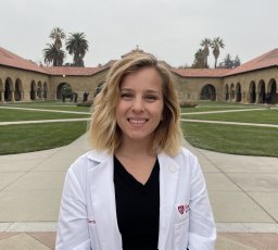 Corrine Nief合作论文数Stanford Medical School, Stanford University6
 Varble Nicole合作论文数Toshiba Stroke and Vascular Research Center, State University of New York6
 Erika Chelales合作论文数5Jose F. Delgado合作论文数4Jocelyne Rivera合作论文数4J. Delgado合作论文数4
 Antonio Arrichiello合作论文数4
 Jeffrey Everitt合作论文数Department of Pathology, Duke University School of Medicine4
 David F. Katz合作论文数Department of Biomedical Engineering, Edmund T. Pratt, Jr. School of Engineering, Duke University4Ayele H. Negussie合作论文数Department of Chemistry, University of Oslo4J. Owen合作论文数3M. Kassin合作论文数3Joshua W Owen合作论文数3Jeffrey Yang合作论文数3Tabea Borde合作论文数3Brian T Crouch合作论文数Department of Biomedical Engineering, Pratt School of Engineering, Duke University3Hu Fangyao合作论文数3
 Mark Wesley Dewhirst合作论文数Department of Radiation Oncology, School of Medicine, Duke University3M. Mauda-Havakuk合作论文数2Lindsey Hazen合作论文数2Anna S. Christou合作论文数2
 Helen A Murphy合作论文数2Miranda Song合作论文数2
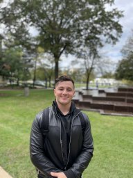 Alec Peltekian合作论文数2James T. Anibal合作论文数Laboratory of Integrative Cancer Immunology, National Cancer Institute2
 Tania L. Lopez-Silva合作论文数Dept Chem, Rice Univ2S. Xu合作论文数2
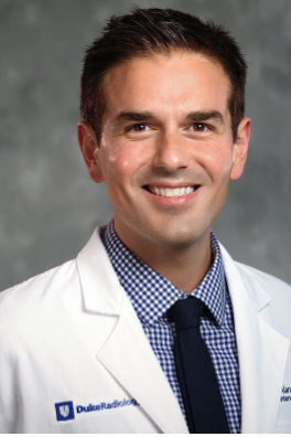 Alan A. Sag合作论文数Department of Radiology, Duke University School of Medicine2
 David Clifton合作论文数Department of Engineering Science, University of Oxford;Reuben College, University of Oxford;National Institute for Health and Care Research2
 Heather E. Jeffery合作论文数2Balasubramanian Aditya合作论文数Department of Materials Science and Engineering, Carnegie Mellon University2Alvarez Daniel Adrianzen合作论文数Department of Electrical and Computer Engineering, Boston University2
 Joel P. Schneider合作论文数Chemical Biology Laboratory, National Cancer Institute2Sheng Xu (徐胜)合作论文数2
 Christopher Wai-Kei Lam (林偉基)合作论文数Macau Institute for Applied Research in Medicine and Health,Macau University of Science and Technology2
 Christopher J. Bettinger合作论文数Department of Biomedical Engineering, College of Engineering, Carnegie Mellon University;Laboratory for Therapeutic Biodegradable Microsystems, Carnegie Mellon University;Defense Advanced Research Projects Agency2
 Ulaş Bağcı合作论文数ISTP Research Fellow
NATIONAL INSTITUTES OF HEALTH (NIH)2
Ulaş Bağcı合作论文数ISTP Research Fellow
NATIONAL INSTITUTES OF HEALTH (NIH)2 Alistair McEwan合作论文数School of Biomedical Engineering, Faculty of Engineering, The University of Sydney;ARC Training Centre for Innovative BioEngineering, The University of Sydney2Michael G. DeSoto合作论文数1Hannah Huth合作论文数1Maomao Chem合作论文数1Michal Mauda-Havakuk合作论文数1Ming Li合作论文数1




