Cervical squamous cell carcinoma (SCC) is classified by the World Health Organization based on its association with human papillomavirus (HPV) into HPV-associated (HPVA) and HPV-independent (HPVI) categories. HPVI SCCs can be p53 wild-type or p53 abnormal, the latter harboring a driver alteration in TP53 and portending worse survival. Recent developments have expanded the spectrum of squamous intraepithelial lesions (SILs) in the cervix. In the HPVA category, seborrheic keratosis-like lesions are now established as having an aetiological association with HPV42, a common low-risk HPV type. Papillary immature metaplasia, a further low-risk HPVA lesion, also falls into this category. More recently, potential HPVI squamous precursor lesions have been described; these form a wide morphologic spectrum and are difficult to diagnose, with the use of p16, p53, and HPV testing mandatory. When these ancillary tests are used, the HPVI SILs, similar to their invasive counterparts, stratify into p53 abnormal and p53 wild-type categories. Different genomic alterations are seen within the two groups, supporting their neoplastic nature. In this review, we provide a historical perspective and comprehensive description of all cervical SILs, with a focus on emerging entities and premalignant lesions, and appraise the terminology used over past years and that recently proposed for new entities. Key clinical, colposcopic, and morphologic features, differential diagnosis, treatment, follow-up, and prognosis are discussed. Special attention is paid to ancillary studies that assist in resolving differential diagnoses and their interpretation in light of recent scientific publications and international guidelines. We also discuss the clinical impact of a pathologic diagnosis of HPVA versus HPVI SILs.
Papillary immature metaplasia, originally described in the uterine cervix, is increasingly recognized as a distinctive squamous intraepithelial lesion rather than a true metaplastic process. Accordingly, the term papillary squamous intraepithelial lesion (PSIL) has been proposed. PSIL occurring outside the uterine cervix is exceedingly rare. In this study, we characterize the clinicopathologic, immunophenotypic, and virologic features of 25 cases of PSIL involving non-cervical sites (vagina, vulva, anus, and endometrium). All lesions demonstrated exophytic papillary architecture lined by uniform immature squamous cells with limited surface maturation. Mitotic figures were frequently identified in the lower half of the epithelium. Low-risk HPV E6/E7 mRNA was detected in 76% of cases, whereas none harbored high-risk HPV. HPV transcripts exhibited a distinctive punctate distribution throughout the epithelium, contrasting with the superficial clustered pattern seen in condyloma/LSIL. Non-block p16 immunostaining pattern was seen in all PSILs, while Ki-67 labeling indices were increased (median 50%), predominantly involving the lower half to two-thirds of the epithelium. Clinical follow-up (median 32 months) demonstrated recurrence in 43% of cases, most commonly with incomplete excision. Our study shows that non-cervical PSIL shares morphologic, immunophenotypic, and virologic features with cervical PSIL. Despite the absence of block-type p16 expression, PSIL demonstrates high proliferative activity, frequently leading to diagnostic confusion with HSIL. Given its distinctive papillary architecture, unusual HPV distribution pattern, and tendency for recurrence, PSIL represents a unique low-risk HPV-associated intraepithelial lesion distinct from conventional LSIL and HSIL. Adoption of the term papillary squamous intraepithelial lesion is recommended in accordance with LAST terminology.
OBJECTIVE:Access to homologous recombination testing remains limited in many centers. We aim to correlate the morphology and immunophenotype of high-grade serous ovarian carcinoma with homologous recombination statuses. METHODS:A retrospective analysis of a high-grade serous ovarian carcinoma tumors with known homologous recombination status. A pathological review of morphology was performed for each tumor, along with immunohistochemical profiling. Tumor morphology was classified as (1) solid, pseudo-endometrioid, or transitional (2) micropapillary or nested. RESULTS:Overall, 81 tumors were included. The median age was 62 (interquartile range; 52-71). Of those, 27 (33.3%) tumors were BRCA1mut, 19 (23.5%) were BRCA2mut, 15 (18.5%) tumors had no BRCA1 or BRCA2 mutations but exhibited a genomic instability score ≥42 and were classified as BRCA1/2-wild-type with homologous recombinant deficient. The remainder 20 (24.7%) cases were homologous recombinant proficient. The proportion of tumors with solid transitional-like morphology was higher in BRCA1 (12/21, 57%) and BRCA2 (12/18, 67%) compared to the tumors with homologous recombinant proficient (3/17, 18%), p =.019. When stratified by genomic instability score, tumors with low score (genomic instability score <26) exhibited 0% solid transitional-like morphology versus 43% solid transitional-like morphology in high-score (genomic instability score >26), p =.03. PAX8 diffuse expression was detected in 71% of BRCA1, 65% of BRCA2, 92% of BRCA-wild-type homologous recombinant deficient tumors, and 100% of homologous recombinant proficient tumors, p =.071. The proportion of diffuse expression was higher in homologous recombinant proficient (100%) versus BRCA2 (65%) (Bonferroni-adjusted pairwise comparisons). CONCLUSIONS:Homologous recombinant deficient tumors are associated with the solid transitional-like morphology, with the BRCA1/2-mutated homologous recombinant deficient cases showing the strongest correlation. Genomic instability score alone may not fully capture the spectrum of homologous recombinant deficient-related phenotypes. The variation in solid transitional-like morphology features among BRCA1- or BRCA2-mutated, BRCA1/2- wild-type with homologous recombinant deficient, and homologous recombinant proficient cases may reflect the diverse biological spectrum of different homologous recombination alterations.
AIMS:Papillary squamous intraepithelial lesion (PSIL), also known as papillary immature metaplasia, is a rare anogenital lesion associated with low-risk human papillomavirus (HPV) infection. The molecular pathogenesis of PSIL remains unclear. This study aims to investigate the activation of YAP1, a Hippo pathway co-activator, in PSIL and compare it to condyloma and papillary HSIL. METHODS AND RESULTS:YAP1 activation was evaluated by immunohistochemistry using an antibody specific for activated/unphosphorylated YAP1 (act-YAP1) in 36 PSIL cases, 20 condyloma and 2 papillary HSIL cases. HPV genotyping and chromogenic in situ hybridization (CISH) were performed to determine HPV status. Low-risk HPV, predominantly HPV6, was detected in 78.6% of PSILs. CISH revealed a diffuse fine punctate distribution of low-risk HPV E6/E7 transcripts in PSIL, distinct from the large focal clusters seen in condyloma, but similar to high-risk HPV in HSIL. All PSILs showed non-block p16 expression. Act-YAP1 immunostaining demonstrated YAP1 activation in 86.1% of PSILs (72.2% diffuse, 13.9% focal), resembling the pattern in papillary HSIL but differing from condyloma, which exhibited nuclear act-YAP1 only in basal and granular layers. No significant associations were found between YAP1 activation and HPV status or immunoprofiles of p16, p53, Rb1 and CK7. CONCLUSIONS:PSILs demonstrate prominent YAP1 activation, implicating Hippo pathway disruption as a potential driver of PSIL pathogenesis. These findings support a distinct molecular profile of PSIL and provide insight into the biological mechanisms underlying this rare lesion.
Papillary immature metaplasia, originally described in the uterine cervix, is increasingly recognized as a distinctive squamous intraepithelial lesion rather than a true metaplastic process. Accordingly, the term papillary squamous intraepithelial lesion (PSIL) has been proposed. PSIL occurring outside the uterine cervix is exceedingly rare. In this study, we characterize the clinicopathologic, immunophenotypic, and virologic features of 25 cases of PSIL involving non-cervical sites (vagina, vulva, anus, and endometrium). All lesions demonstrated exophytic papillary architecture lined by uniform immature squamous cells with limited surface maturation. Mitotic figures were frequently identified in the lower half of the epithelium. Low-risk HPV E6/E7 mRNA was detected in 76% of cases, whereas none harbored high-risk HPV. HPV transcripts exhibited a distinctive punctate distribution throughout the epithelium, contrasting with the superficial clustered pattern seen in condyloma/LSIL. Non-block p16 immunostaining pattern was seen in all PSILs, while Ki-67 labeling indices were increased (median 50%), predominantly involving the lower half to two-thirds of the epithelium. Clinical follow-up (median 32 months) demonstrated recurrence in 43% of cases, most commonly with incomplete excision. Our study shows that non-cervical PSIL shares morphologic, immunophenotypic, and virologic features with cervical PSIL. Despite the absence of block-type p16 expression, PSIL demonstrates high proliferative activity, frequently leading to diagnostic confusion with HSIL. Given its distinctive papillary architecture, unusual HPV distribution pattern, and tendency for recurrence, PSIL represents a unique low-risk HPV–associated intraepithelial lesion distinct from conventional LSIL and HSIL. Adoption of the term papillary squamous intraepithelial lesion is recommended in accordance with LAST terminology.
Verruciform acanthotic vulvar intraepithelial neoplasia (vaVIN) is a rare type of human papillomavirus (HPV)-independent vulvar squamous cell carcinoma (SCC) precursor lacking aberrant p53 expression. Our recent study demonstrated GLUT1 overexpression in vulvar SCC and identified 2 distinct patterns in HPV-associated high-grade squamous intraepithelial lesions and HPV-independent differentiated vulvar intraepithelial neoplasia. Herein, we expand on our study by assessing GLUT1 expression in 24 cases of vaVIN and 8 cases of associated invasive SCC, compared with 48 cases of benign vulvar squamous lesions, including 40 cases of non-HPV benign vulvar lesions and 8 cases of condyloma associated with low-risk HPV infection. GLUT1 immunohistochemistry (IHC) demonstrated consistent diffuse GLUT1 immunostaining in all vaVIN cases. The GLUT1 immunostaining pattern in vaVIN is characterized by strong membranous staining in the basal layer with suprabasal extension to the full thickness of intermediate layers. In vulvar SCC associated with vaVIN, GLUT1 expression was prominent at the periphery of tumor nests without expression in the central portion. GLUT1 IHC revealed weak peri-papillae or intensified peri-papillae patterns in non-neoplastic vulvar lesions and HPV-related patterns in condyloma. The GLUT1 immunostaining patterns in vaVIN and associated SCC are different from benign mimickers, but similar to those of differentiated vulvar intraepithelial neoplasia and associated SCC, despite distinct molecular alterations and pathways. The latter suggests that upregulation of GLUT1 is likely a common metabolic pathway shared by 2 types of HPV-independent VINs, regardless of p53 status. GLUT1 IHC is highly sensitive in detecting vaVIN. However, the specificity rests on recognizing GLUT1-staining patterns in benign mimickers of vaVIN. Along with HPV chromogenic in situ hybridization and other well-characterized biomarkers, GLUT1 immunochemistry can be a helpful adjunct in assisting the diagnosis of vaVIN.
Introduction Endometrial endometrioid carcinomas can show multiple lines of differentiation, including pilomatrix-like high-grade endometrioid carcinoma, a recently described tumor with similarity to cutaneous pilomatrix carcinoma and associated with very aggressive clinical behavior. Methods We present a 56-year-old woman with an endometrial tumor associated with secondary involvement of both ovaries, left tubo-ovarian ligament and obturator lymph nodes. The diagnosis of high-grade endometrioid carcinoma in a previously performed curettage was confirmed in the hysterectomy specimen. Results Microscopically, the tumor exhibited a solid, nested/insular pattern with basaloid cells, predominantly seen at the periphery, ghost cell keratinization towards the center of the nests, and extensive geographic necrosis. No low-grade endometrioid carcinoma component was identified throughout the primary tumor or metastases after extensive sampling. Immunohistochemical assessment showed aberrant cytoplasmic and nuclear expression of β-catenin, and focal CDX2 expression. Tumor cells were negative for PAX8, and estrogen and progesterone receptors (ER/PR). The next-generation sequencing (NGS) analysis found a CTNNB1 pathogenic mutation (p.Ser37Phe, c.110C > T; variant allele frequency: 18.6%). Based on these morphologic, immunohistochemistry and NGS analysis, a diagnosis of pilomatrix-like high-grade endometrioid carcinoma was established. Conclusion The absence of a low-grade endometrioid carcinoma component makes this pilomatrix-like high-grade endometrioid carcinoma, a very rare tumor, even more special. This and the absence of PAX8 and ER/PR expression in an unusual morphological context proved to be diagnostically challenging. This patient's presentation at high stage is concordant with the literature's description of this tumor as aggressive. It is not yet known whether standard adjuvant therapies for high-risk endometrial carcinomas are effective.
Human papillomavirus (HPV)-independent cervical squamous cell carcinomas (HPVI SCCs) represent a poorly characterized entity. We aimed to explore the clinicopathological and survival features in the largest series of HPVI SCCs and compare them to HPV-associated (HPVA) SCCs. Eighty-nine cases of SCC previously tested negative for high-risk and low-risk HPV were collected from 22 institutions. A total of 363 HPVA SCCs were retrieved from a previously published database. Demographic and clinicopathological features, including p16 and p53 immunohistochemistry, and follow-up data were recorded. Forty-nine of 89 cases were classified as "true" HPVI SCC (HPV-/ p16-), whereas 40 were equivocal HPVI SCC (HPV-/p16 & thorn; or HPV-/p16 not performed) and were excluded. The median age of true HPVI SCCs was 68 (range, 36-88) years and 38 (77.6%) patients were diagnosed aged 60 years or older. Seven (14.2%) had a history of uterine prolapse. Compared with HPVA SCC, patients with HPVI SCCs were older (P < .001), had larger tumors (P < .001), were diagnosed at higher Federation of Gynecology and Obstetrics stage (P < .001), recurred more
Objective To assess the influence of geographies and race on the survival outcomes in patients diagnosed with cervical squamous cell carcinoma (CSCC) across three continents. Methods This multicontinental retrospective study was conducted in 8 hospitals across Asia, Europe, and North America (NA). Clinicopathologic data of 595 patients with presumed early stages of CSCC, treated surgically, with curative intent was collected. Descriptive analysis and Cox regression models were produced. Results A total of 595 patients, consisting of 445 (74.8 %) white, 75 (12.6 %) Blacks, and 75 (12.6 %) Asian patients were included. Geographical distribution comprised 69 % of patients from NA, 22 % from Europe, and 9 % from Asia. The median age at diagnosis was 46 years. The median overall survival (OS) and relapse-free survival (RFS) were 22.09 years and 21.19 years, respectively. Patient characteristics varied significantly across geographical regions, except for consensus tumor grade. Patients in Europe from middle-income countries with limited CC screening had a substantially higher risk of death than those in NA (HR, 1.79; 95 % CI, 1.13 to 2.79; p = 0.015). Patients from single center in Japan had higher risk of relapse than those from the four heterogeneous NA centers (sub-distribution hazard ratio, 2.19; 95 % CI, 1.22 to 3.95; p = 0.009), although OS did not differ significantly. Race remained statistically insignificant for survival outcomes across the three continents but seemed to influence survival outcomes in NA centers. Conclusion Our study highlights impact of geographies and races on CSCC survival outcomes, emphasizing the need of considering these factors when developing targeted interventions against CSCC.
Objectives. To evaluate clinical, laboratory, and radiological variables from preoperative contrast-enhanced computed tomography (CECT) for their ability to distinguish ovarian clear cell carcinoma (OCCC) from nonOCCC and to develop a nomogram to preoperatively predict the probability of OCCC.Methods. This IRB-approved, retrospective study included consecutive patients who underwent surgery for an ovarian tumor from 1/1/2000 to 12/31/2016 and CECT of the abdomen and pelvis & LE;90 days before primary debulking surgery. Using a standardized form, two experienced oncologic radiologists independently analyzed imaging features and provided a subjective 5-point impression of the probability of the histological diagnosis. Nomogram models incorporating clinical, laboratory, and radiological features were created to predict histological diagnosis of OCCC over non-OCCC. Results. The final analysis included 533 patients with surgically confirmed OCCC (n = 61) and non-OCCC (n = 472); history of endometriosis was more often found in patients with OCCC (20% versus 3.6%; p < 0.001), while CA-125 was significantly higher in patients with non-OCCC (351 ng/mL versus 70 ng/mL; p < 0.001). A nomogram model incorporating clinical (age, history of endometriosis and adenomyosis), laboratory (CA-125) and imaging findings (peritoneal implant distribution, morphology, laterality, and diameter of ovarian lesion and of the largest solid component) had an AUC of 0.9 (95% CI: 0.847, 0.949), which was comparable to the AUCs of the experienced radiologists' subjective impressions [0.8 (95% CI: 0.822, 0.891) and 0.9 (95% CI: 0.865, 0.936)].Conclusions. A presurgical nomogram model incorporating readily accessible clinical, laboratory, and CECT variables was a powerful predictor of OCCC, a subtype often requiring a distinctive treatment approach. & COPY; 2023 Elsevier Inc. All rights reserved.
PDF - 64K, Supplementary Table 4. Evaluation of EMP2 IgG1 toxicity compared to control IgG following 7 weeks of treatment delivered systemically IP. Supplementary Table 5. Evaluation of EMP2 IgG1 toxicity compared to control IgG following systemic IP treatment of up to 40mg/kg.
The complexity of ovary and fallopian tube pathology is striking, with dozens of tumor types. There is a greater appreciation now of the importance of hereditary cancer syndromes in ovarian and tubal malignancies, as two common autosomal dominant hereditary cancer predisposition syndromes include carcinomas of ovary or fallopian tube. This book is organized following the structure and terminology of the World Health Organization (WHO) classification of Female Genital Tumors, 5th edition. It includes some new entities that have been described since that book’s publication in 2020 and provides detailed discussion of differential diagnosis, relevant ancillary tests, and strategies to approach diagnosis. In addition, discussions of the underlying biology of these tumors, as it relates to their diagnosis, are also presented. This fascicle provides an opportunity for deeper reading and quiet reflection about the progress that has been made in our understanding of tumors of the ovary since the previous landmark edition was published in 1998, as well as a respect for the numerous challenges that remain.
PDF - 54K, Supplementary Table 2. Clinical Data for Invasive Breast Cancer Patients used in the TMA.
With the advancement of diagnostic molecular technology and the molecular classification of endometrial endometrioid carcinoma (EEC), it remains to be seen whether conventional International Federation of Gynecology and Obstetrics (FIGO) grading retains clinical significance in certain molecular subtypes of EECs. In this study, we explored the clinical significance of FIGO grading in microsatellite instability-high (MSI-H) and POLE-mutant EECs. A total of 162 cases of MSI-H EECs and 50 cases of POLE-mutant EECs were included in the analysis. Significant differences in tumor mutation burden (TMB), progression-free survival, and disease-specific survival were seen between the MSI-H and POLE-mutant cohorts. Within the MSI-H cohort, there were statistically significant differences in TMB and stage at presentation across FIGO grades, but not survival. Within the POLE-mutant cohort, there was significantly greater TMB with increasing FIGO grade, but there were no significant differences in stage or survival. In both the MSI-H and POLE-mutant cohorts, log-rank survival analysis showed no statistically significant difference in progression-free and disease-specific survival across FIGO grades. Similar findings were also seen when a binary grading system was utilized. Since FIGO grade was not associated with survival, we conclude that the intrinsic biology of these tumors, characterized by their molecular profile, may override the significance of FIGO grading.
We present the case of a 71-year-old patient, with vaginal bleeding, dyspnea, headache, loss of appetite and weakness. Clinical examination revealed a pediculated vaginal mass of 25 mm diameter, of dark-red color and soft spongy consistency, with an ulcerated surface and originating from the anterior wall, which was surgically removed. The morphology was dominanted by large, round to polygonal tumor cells, arranged in a predominantly tubulo-cystic architecture, surrounding numerous blood vessels that dominated the appearance, suggesting a perivascular epithelioid cell tumor (PEComa) or hemangioblastoma but the presence of pleomorphic nuclei, numerous mitoses together with immunohistochemistry helped for a correct diagnosis of vaginal .
OBJECTIVES:Clear cell carcinoma is a high-risk subtype of endometrial cancer. Some patients have a mixture of clear cell carcinoma with other histologic types (endometrioid or serous) or cannot be neatly assigned to one of these types. Protocol GOG-8032 within GOG-210 was designed to determine whether these tumors differ from pure clear cell carcinoma in stage at diagnosis, initial pattern of spread, or patient survival. METHODS:The term "mixed" was applied to tumors with multiple identifiable components, and "indeterminate" was applied to tumors with features intermediate between different histologic types. Three hundred eleven women with pure, mixed, or indeterminate clear cell carcinoma were identified in a larger cohort of patients undergoing hysterectomy for endometrial cancer in GOG-210. Histologic slides were centrally reviewed by expert pathologists. Baseline and follow-up data were analyzed. RESULTS:One hundred thirty-six patients had pure clear cell carcinoma and 175 had a mixed or indeterminate clear cell pattern. Baseline clinicopathologic characteristics were similar except for a small difference in age at presentation. Univariate survival analysis confirmed the significance of typical endometrial cancer prognostic factors. Patients in the mixed categories had disease-free and overall survival similar to pure clear cell carcinoma, but the indeterminate clear cell/endometrioid group had longer survival. CONCLUSION:In clear cell endometrial cancer, the presence of a definite admixed endometrioid or serous component did not correlate with a significant difference in prognosis. Patients whose tumors had indeterminate clear cell features had better prognosis. Some of these tumors may be endometrioid tumors mimicking clear cell carcinoma.
 Nadeem R. Abu-Rustum合作论文数Weill Cornell Medical College;Memorial Sloan Kettering Cancer Center118
 Esther Oliva合作论文数Department of Pathology, Massachusetts General Hospital, Harvard Medical School80
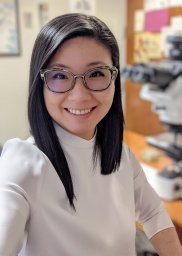 Kay J. Park合作论文数Department of Pathology, Memorial Sloan-Kettering Cancer Center80
 Richard Barakat合作论文数Institute of Cancer Research, Feinstein Institutes for Medical Research;Applied Surgical Robotics;Donald and Barbara Zucker School of Medicine at Hofstra/Northwell73
 Douglas A Levine合作论文数Merck65
 Britta Weigelt合作论文数Gynecology DMT Research Laboratory, Memorial Sloan Kettering Cancer Center60
 Simona Stolnicu合作论文数56
 Carol Aghajanian合作论文数Memorial Sloan Kettering Cancer Center55
 Martee L. Hensley合作论文数Memorial Sloan Kettering Cancer Center54Deborah Delair合作论文数53
 Blake Gilks合作论文数Department of Pathology and Laboratory Medicine, Faculty of Medicine, University of British Columbia52
 Rajmohan Murali合作论文数Sydney Medical School, University of Sydney48
 Lien Hoang合作论文数Department of Pathology and Laboratory Medicine, Faculty of Medicine, The University of British Columbia;Vancouver Coastal Health Research Institute46Mario M. Leitao合作论文数46
 Dennis S. Chi合作论文数Department of Surgery, Memorial Sloan Kettering Cancer Center43Sarah Chiang合作论文数38
 Teri A Longacre合作论文数Department of Pathology, School of Medicine, Stanford University;Stanford Cancer Institute, School of Medicine, Stanford University38
 Jennifer Mueller合作论文数Department of Surgery, Memorial Sloan Kettering Cancer Center34
 Alexia Iasonos合作论文数Department of Epidemiology and Biostatistics, Memorial Sloan-Kettering Cancer Center31Kaled M. Alektiar合作论文数31M. M. Leitao合作论文数30Oliver Zivanovic合作论文数29
 Martin Koebel合作论文数Department of Pathology and Laboratory Medicine, Cumming School of Medicine, University of Calgary;Arnie Charbonneau Cancer Institute, University of Calgary28Anna Pesci合作论文数28
 Cheng-Han Lee合作论文数University of British Columbia25
 Marc Ladanyi合作论文数Memorial Sloan Kettering Cancer Center25Ginger J. Gardner合作论文数25
 Narciso Olvera合作论文数Department of Surgery, Memorial Sloan-Kettering Cancer Center24
 David Huntsman合作论文数Department of Pathology and Laboratory Medicine, Faculty of Medicine, The University of British Columbia;Department of Obstetrics & Gynaecology, Faculty of Medicine, The University of British Columbia;Department of Molecular Oncology, BC Cancer Research Centre23
 Basile Tessier Cloutier合作论文数22Cadoo Karen A合作论文数Weill Medical College, Cornell University22K. Garg合作论文数21
 Arnaud Da Cruz Paula合作论文数Mem Sloan Kettering Canc Ctr, 1275 York Ave, New York, NY 10021 USA21Takako Kiyokawa合作论文数21Makker Vicky合作论文数Medical Oncology, Memorial Sloan Kettering Cancer Center21
 Jinru Shia合作论文数Department of Pathology, Memorial Sloan Kettering Cancer Center21Amir Momeni Boroujeni合作论文数Department of Pathology and Laboratory Medicine, Memorial Sloan Kettering Cancer Center20
 Glenn McCluggage合作论文数Queen’s University of Belfast;Department of Pathology, Belfast Health and Social Care Trust20
 Kauff Noah D合作论文数Mem Sloan Kettering Canc Ctr20
 Jorge S. Reis-Filho合作论文数AstraZeneca20Carmen Tornos合作论文数20Karuna Garg合作论文数20
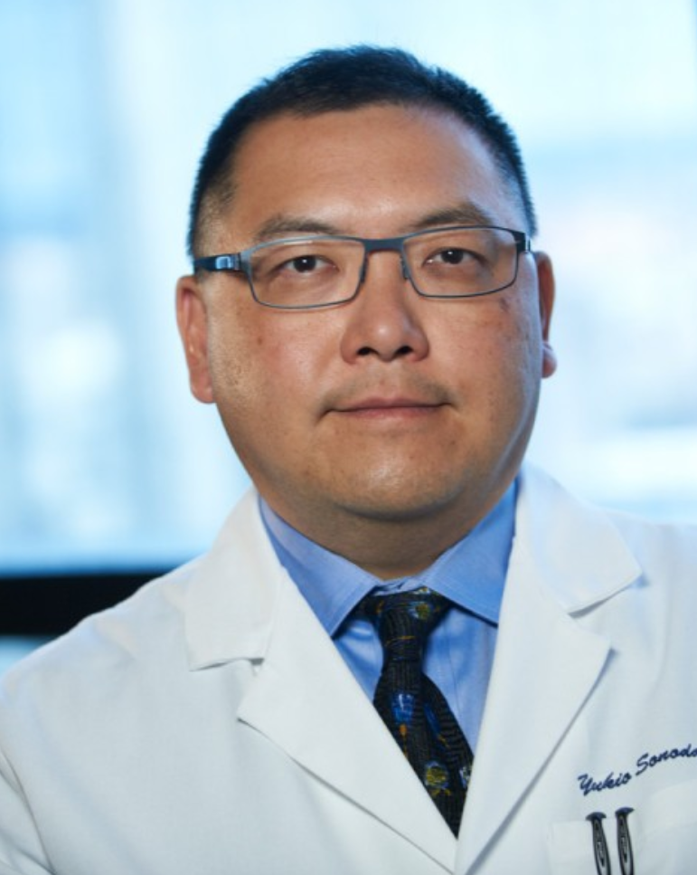 Yukio Sonoda合作论文数Memorial Sloan Kettering Cancer Center19
 Isabel Alvarado-Cabrero合作论文数19M. Leitao合作论文数18Pier Selenica合作论文数Mem Sloan Kettering Canc Ctr, Dept Pathol, 1275 York Ave, New York, NY 10021 USA17Cristina Terinte合作论文数15O'Cearbhaill Roisin E合作论文数Memorial Sloan-Kettering Cancer Center15
 Parra-Herran Carlos合作论文数15




