Abstract Tumor-reactive human CD8 T cells are found predominantly in the tumor microenvironment with an exhausted phenotype (extremely high levels of CD39, PD-1, CTLA-4, and TIM-3). The tumor-reactive cells can be enriched from patients’ tumors by sorting CD8+ TIL that express both CD39 and CD103. Our unique expansion method allowed these phenotypically exhausted cells to grow from thousands into billions. After expansion the CD39 and CD103 Double Positive (DP) CD8 TIL can recognize/kill autologous tumor cells in vitro. We tested whether the CD8 DP TIL could recognize/kill autologous tumor in vivo using a xenograft model with immune-compromised mice that constitutively secrete human IL-2 (NOG-hIL-2). The CD8 DP TIL were able to persist long-term, traffic to the tumor site, recognize autologous tumor, and facilitate tumor regression. Human IL-2 was necessary for their long-term survival in the periphery and for tumor regression. These preclinical data were the basis for a human clinical trial design for the adoptive transfer of CD8 DP TIL (product termed, AGX148). We recently opened a Phase 1 clinical trial using the CD39/103 DP CD8 TIL in a first-in-human protocol for adults with solid tumors (NCT05902520). The trial design has three cohorts based on giving IL-2 for 2 weeks, 3 weeks, or 4 weeks after adoptive transfer with six patients/cohort (3 receiving DP TIL alone and 3 receiving DP TIL combined with PD-1 siRNA knockdown with the INTASYLTM compound PH-762). The first 3 patients have been treated and received 27, 40, and 8 billion DP TIL, respectively. All 3 patients had failed standard therapy prior to their enrollment, including checkpoint blockade. No serious adverse events were observed in the first three patients. Two of three patients had partial responses 29 days after treatment and the third patient had stable disease for >56 days. Blood draws and tumor biopsies were obtained prior to and after treatment and we are currently assessing the CD8 T cell repertoire (TCRseq) and phenotype from these patient samples. In conclusion, this is the first study showing that CD8 T cells with an exhausted phenotype isolated from human tumors can be expanded to billions and induce tumor regression in vivo. Citation Format: Andrew Weinberg, Brendan Curti, Ryan Montler, Colin Thalhofer, Elaine Ballinger, Jessica Cross, Eric Tran, Nelson Sanjuan, Matthew Taylor, Christopher Fountain, Roxanne Payne, James Cardia, Nicholas Morris. Tumor-reactive CD8 TIL with an exhausted phenotype can be expanded and regress human tumors [abstract]. In: Proceedings of the American Association for Cancer Research Annual Meeting 2024; Part 2 (Late-Breaking, Clinical Trial, and Invited Abstracts); 2024 Apr 5-10; San Diego, CA. Philadelphia (PA): AACR; Cancer Res 2024;84(7_Suppl):Abstract nr LB067.
A patient with progressive metastatic pancreatic cancer was treated with a single infusion of 16.2×10⁹ autologous T cells that had been genetically engineered to clonally express two allogeneic HLA-C*08:02-restricted T-cell receptors (TCRs) targeting mutant KRAS G12D expressed by the tumors. The patient had regression of visceral metastases (overall partial response of 72% according to the Response Evaluation Criteria in Solid Tumors, version 1.1); the response was ongoing at 6 months. The engineered T cells constituted more than 2% of all the circulating peripheral-blood T cells 6 months after the cell transfer. In this patient, TCR gene therapy targeting the KRAS G12D driver mutation mediated the objective regression of metastatic pancreatic cancer. (Funded by the Providence Portland Medical Foundation.).
Background A pilot study of stereotactic body radiation therapy (SBRT) followed by high-dose interleukin-2 (IL-2) showed a higher than anticipated objective response rate (ORR) among patients with metastatic melanoma (MM). We performed a prospective randomized study to determine if the ORR of SBRT + IL-2 was greater than IL-2 monotherapy in patients with advanced melanoma. Methods Patients with MM who had adequate physiological reserve for IL-2 and at least one site suitable for SBRT were eligible. There was a 1:1 randomization to SBRT + IL-2 or IL-2 monotherapy. Patients received one or two doses of SBRT (20 Gy per fraction) with the last dose administered 3 days before starting the first cycle of IL-2. IL-2 (600,000 IU per kg via intravenous bolus infusion) was given every 8 hours for a maximum of 14 doses with a second cycle after a 2-week rest. Responding patients received up to six IL-2 cycles. Patients assigned to IL-2 monotherapy who exhibited progression of melanoma after cycle 2 were allowed to crossover and receive SBRT and additional IL-2. Response Evaluation Criteria in Solid Tumors 1.1 criteria were applied to non-irradiated lesions for response assessment. Results 44 patients were included in the analysis. The ORR in the SBRT + IL-2 group was 54%: 21% complete response (CR), 33% partial response (PR), 21% stable disease (SD) and 25% progressive disease (PD). The ORR in patients receiving IL-2 monotherapy was 35%: 15% CR, 20% PR, 25% SD and 40% PD. Seven patients assigned to IL-2 subsequently received SBRT + IL-2. One CR and two PRs were observed in the crossover group. There was no difference in progression-free or overall survival (OS). At 5 years the OS was 26% in the SBRT + IL-2 group and 25% in the IL-2 monotherapy group. The disease control rate (DCR) was higher in the SBRT + IL-2 group (75% vs 60%, p=0.34). Conclusions SBRT + IL-2 induced more objective responses with a higher DCR compared to IL-2 monotherapy in MM. IL-2 monotherapy resulted in a significantly higher ORR than anticipated. Some patients in the crossover group also achieved objective responses. Trial registration number NCT01416831 .
BACKGROUND:High-dose interleukin-2 (IL-2) has been FDA-approved for over 20 years, but it is offered only at a small number of centers with expertise in its administration. We analyzed the outcomes of patients receiving high-dose IL-2 in relation to the severity of toxicity to ascertain if response or survival were adversely affected. METHODS:A retrospective analysis of the outcomes of 500 patients with metastatic renal cell carcinoma (RCC) (n = 186) or melanoma (n = 314) treated with high-dose IL-2 between 1997 and 2012 at Providence Cancer Center was performed. IL-2 was administered at a dose of 600,000 international units per kg by IV bolus every 8 hours for up to 14 doses. A second cycle was administered 16 days after the first and patients with tumor regression could receive additional cycles. Survival and anti-tumor response were analyzed by diagnosis, severity of toxicity, number of IL-2 cycles and subsequent therapy. RESULTS:The objective response rate in melanoma was 28% (complete 12% and partial 16%), and in RCC was 24% (complete 7% and partial 17%). The 1-, 2- and 3-year survivals were 59%, 41% and 31%, for melanoma and 75%, 56% and 44%, for RCC, respectively. The proportion of patients with complete or partial response in both melanoma and RCC was higher in patients who a) required higher phenylephrine doses to treat hypotension (p < 0.003), b) developed acidosis (bicarbonate < 19 mmol (p < 0.01)), or c) thrombocytopenia (<50, 50-100, >100,000 platelets; p < 0.025). The proportion achieving a complete or partial response was greater in patients with melanoma who received 5 or more compared with 4 or fewer IL-2 cycles (p < 0.0001). The incidence of death from IL-2 was less than 1% and was not higher in patients who required phenylephrine. CONCLUSIONS:High-dose IL-2 can be administered safely; severe toxicity including hypotension is reversible and can be managed in a community hospital. The tumor response and survival reported here are superior to the published literature and support treating patients to their individualized maximum tolerated dose. IL-2 should remain part of the treatment paradigm in selected patients with melanoma and RCC.
Preclinical models suggest that focal high-dose radiation can make tumors more immunogenic. We performed a pilot study of stereotactic body radiation therapy (SBRT) followed by high-dose interleukin-2 (IL-2) to assess safety and tumor response rate and perform exploratory immune monitoring studies. Patients with metastatic melanoma or renal cell carcinoma (RCC) who had received no previous medical therapy for metastatic disease were eligible. Patients received one, two, or three doses of SBRT (20 Gy per fraction) with the last dose administered 3 days before starting IL-2. IL-2 (600,000 IU per kilogram by means of intravenous bolus infusion) was given every 8 hours for a maximum of 14 doses with a second cycle after a 2-week rest. Patients with regressing disease received up to six IL-2 cycles. Twelve patients were included in the intent-to-treat analysis, and 11 completed treatment per the study design. Response Evaluation Criteria in Solid Tumors criteria were used to assess overall response in nonirradiated target lesions. Eight of 12 patients (66.6%) achieved a complete (CR) or partial response (PR) (1 CR and 7 PR). Six of the patients with PR on computed tomography had a CR by positron emission tomography imaging. Five of seven (71.4%) patients with melanoma had a PR or CR, and three of five (60%) with RCC had a PR. Immune monitoring showed a statistically significantly greater frequency of proliferating CD4(+) T cells with an early activated effector memory phenotype (CD3(+)CD4(+)Ki67(+)CD25(+)FoxP3(-)CCR7(-)CD45RA(-)CD27(+)CD28(+/-)) in the peripheral blood of responding patients. SBRT and IL-2 can be administered safely. Because the response rate in patients with melanoma was significantly higher than expected on the basis of historical data, we believe that the combination and investigation of CD4(+) effector memory T cells as a predictor of response warrant further study.
 Brendan D. Curti合作论文数Providence Health & Services;Earle A. Chiles Research Institute, Providence Cancer Institute5
 Walter Urba合作论文数Earle A. Chiles Research Institute;Providence Cancer Institute of Oregon;Sisters of Providence Health System4
 Eric Tran合作论文数National Cancer Institute (NCI),, National Institutes of Health3
 Elaine Ballinger合作论文数2
 Colin Thalhofer合作论文数2Nelson Sanjuan合作论文数2Nicholas Morris合作论文数2Jessica Cross合作论文数2James Cardia合作论文数2
 Matthew H. Taylor合作论文数Providence Cancer Institute Franz Clinic2Glenn Lyn合作论文数2Andrew D. Weinberg合作论文数2Marka Crittenden合作论文数2Steven K. Seung合作论文数2Ryan Montler合作论文数2Nelson Sanjuan Silva合作论文数1Helena Hoen合作论文数1David Sprott合作论文数1Christopher B Fountain合作论文数1Lyn B. Dobrunick合作论文数1Huayu Huang合作论文数1Jw Smith合作论文数1
 Chunhong Zheng (郑春红)合作论文数School of Basic Medical Sciences, Peking University Health Science Center;Peking University International Cancer Institute1Shu-Ching Chang合作论文数Providence St Joseph Hlth, Med Data Res Ctr, Portland, OR USA1Julie Cramer合作论文数1
 Leidner Rom S合作论文数Division of General Internal Medicine, University of Washington1Coffey Todd合作论文数1Bageac Alexandru合作论文数1Robert B. Lufkin合作论文数1
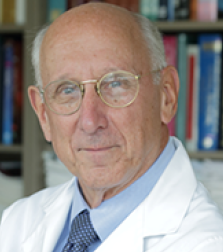 Steven A. Rosenberg合作论文数Center for Cancer Research, National Cancer Institute, National Institutes of Health;Uniformed Services University of Health Sciences;School of Medicine, George Washington University;Department of Laboratory Medicine, Karolinska Institutet1Janet Siebert合作论文数Analytical Services, CytoAnalytics1
 Bernard A. Fox合作论文数Robert W. Franz Cancer Research Center, Earle A. Chiles Research Institute, Providence Portland Medical Center1Yi-Ping Shih合作论文数1Edwin B. Walker合作论文数1Todd S. Crocenzi合作论文数Providence Portland Medical Center;Providence Cancer Center1




