BACKGROUND AND AIMS:Enteral feeding intolerance is a frequent and clinically significant complication in patients with traumatic brain injury (TBI), yet its mechanisms remain poorly understood and predictive tools are lacking. We investigated a prospective study to determine whether elevated circulating glucagon-like peptide-1(GLP-1) is associated with feeding intolerance after TBI and to assess its predictive utility. METHODS:A prospective cohort study in a tertiary neuro-ICU including 109 adults aged 18-80 years with TBI was conducted. A standardised enteral feeding strategy targeted 25 kcal/kg/d and was adjusted according to tolerance. Participants were classified by average caloric adequacy over 14 days: tolerance (≥70% of goal) vs intolerance (<70%). Blood was sampled during post-injury day (PID) windows 1-3, 4-7, 8-10, and 11-14. Longitudinal trajectories of circulating GLP-1, insulin, glucose, and glial fibrillary acidic protein. The Primary outcome was feeding intolerance within 14 days. Associations of biomarkers with feeding intolerance were evaluated, and discrimination was quantified using multivariable logistic regression and receiver operating characteristic analysis. RESULTS:Insulin (P = 0.49) and glucose (P = 0.47) did not differ between intolerance (n = 50) and tolerance (n = 59) groups, whereas GLP-1 was higher in the intolerance group (P = 0.024). GLP-1 increased with injury severity (P = 0.034) and was greatest in patients with GCS 3-5 vs 6-12 (P < 0.001). GLP-1 correlated with glial fibrillary acidic protein over 14 days (r = 0.259; P < 0.001); intestinal permeability markers did not differ between groups. In multivariable models, GLP-1 at PID8-10 (OR, 1.029; P = 0.001), IL-6 at PID8-10 (OR, 1.033; P = 0.035), insulin at PID1-3 (OR, 1.025; P = 0.006), and admission SOFA score (OR, 1.646; P = 0.023) were independently associated with feeding intolerance. Combining insulin at PID1 and SOFA offered early risk stratification (AUC, 0.786; 95% CI, 0.693-0.879; P < 0.001; specificity 71.4%; sensitivity 75%). CONCLUSIONS:Elevated plasma GLP-1 levels are associated with enteral feeding intolerance in patients with TBI. These findings are hypothesis-generating and suggest that altered GLP-1 signalling may contribute to feeding intolerance. STATEMENT OF SIGNIFICANCE:We propose that disrupted central GLP-1 signalling diminishes afferent satiety input and efferent anorexigenic output, yielding central GLP-1 resistance with secondary GLP-1 accumulation in plasma; higher circulating GLP-1 may therefore indicate central GLP-1 resistance in TBI and associate with FI. Elevated circulating GLP-1 may serve as a pragmatic predictor of feeding intolerance after TBI and could inform feeding strategies.
Somatic genome editing in mouse models has increased our understanding of the in vivo effects of genetic alterations. However, existing models have a limited ability to create multiple targeted edits, hindering our understanding of complex genetic interactions. Here we generate transgenic mice with Cre-regulated and constitutive expression of enhanced Acidaminococcus sp. Cas12a (enAsCas12a), which robustly generates compound genotypes, including diverse cancers driven by inactivation of trios of tumour suppressor genes or an oncogenic translocation. We integrate these modular CRISPR RNA (crRNA) arrays with clonal barcoding to quantify the size and number of tumours with each array, as well as the impact of varying the guide number and position within a four-guide array. Finally, we generate tumours with inactivation of all combinations of nine tumour suppressor genes and find that the fitness of triple-knockout genotypes is largely explainable by one- and two-gene effects. These Cas12a alleles will enable further rapid creation of disease models and high-throughput investigation of coincident genomic alterations in vivo. Multiplexed editing using Cas12a transgenic mice facilitates the study of disease models with complex genotypes.
Most cancers are diagnosed in people over 60 years of age, but little is known about how age impacts tumorigenesis. While aging is accompanied by mutation accumulation (widely understood to contribute to cancer risk) it is associated with numerous other cellular and molecular changes likely to impact tumorigenesis. Moreover, cancer incidence decreases in the oldest part of the population, suggesting that very old age may reduce carcinogenesis. Here we show that aging represses oncogenic KRAS-driven tumor initiation and growth in genetically engineered mouse models of human lung cancer. Moreover, aging dampens the impact of inactivating many tumor suppressor genes with the impact of inactivating PTEN, a negative regulator of the PI3K–AKT pathway, weakened disproportionately. Single-cell transcriptomic analysis revealed that neoplastic cells in aged mice retain age-related transcriptomic changes, showing that the impact of age persists through oncogenic transformation. Furthermore, the consequences of PTEN inactivation were strikingly age-dependent, with PTEN deficiency reducing signatures of aging in cancer cells and the tumor microenvironment. Our findings underscore the interconnectedness of the pathways involved in aging and tumorigenesis and document tumor-suppressive effects of aging that may contribute to the deceleration in cancer incidence with age. The mechanisms that impact tumorigenesis during aging are incompletely understood. Here Shuldiner et al. show that in mice, aging represses KRAS-driven lung tumorigenesis and dampens the impact of inactivating many tumor suppressor genes, which may contribute to the deceleration in cancer incidence with extreme age in humans.
Objective: We aimed to determine whether quantitative electroencephalography (QEEG) measures have predictive value for cerebral edema (CED) and clinical outcomes in acute ischemic stroke (AIS) patients with anterior circulation large vessel occlusion who underwent mechanical thrombectomy (MT). Methods: A total of 105 patients with AIS in the anterior circulation were enrolled in this prospective study. The occurrence and severity of CED were assessed through computed tomography conducted 24 h after MT. Clinical outcomes were evaluated based on early neurological deterioration (END) and 3-month functional status, as measured by the modified Rankin scale (mRS). Electroencephalography (EEG) recordings were performed 24 h after MT, and QEEG indices were calculated from the standard 16 electrodes and 2 frontal channels (F3-C3, F4-C4). The delta/alpha ratio (DAR), the (delta + theta) / (alpha + beta) ratio (DTABR), and relative delta power were averaged over all electrodes (global) and the F3- C3 and F4-C4 channels (frontal). The predictive effect and value of QEEG indices for CED and clinical outcomes were assessed using ordinal and logistic regression models, as well as receiver operating characteristic (ROC) curves. Results: Significantly, both global and frontal DAR were found to be associated with the severity of CED, END, and poor functional outcomes at 90 days, while global and frontal DTABR and relative delta power were not associated with outcomes. In ROC analysis, the best predictive effect was observed in frontal DAR, with an area under the curve of approximately 0.80. It exhibited approximately 75% sensitivity and 71% specificity for radiological and clinical outcomes when a threshold of 3.3 was used. Conclusions: QEEG techniques may be considered an efficient bedside monitoring method for assessing treatment efficacy, identifying patients at higher risk of severe CED and END, and predicting long-term functional outcomes. Significance: QEEG can help identify patients at risk of severe neurological complications that can impact long-term functional recovery in AIS patients who underwent MT. (c) 2024 Published by Elsevier B.V. on behalf of International Federation of Clinical Neurophysiology.
Most cancers are diagnosed in persons over the age of sixty, but little is known about how age impacts tumorigenesis. While aging is accompanied by mutation accumulation - widely understood to contribute to cancer risk - it is also associated with numerous other cellular and molecular changes likely to impact tumorigenesis. Moreover, cancer incidence decreases in the oldest part of the population, suggesting that very old age may reduce carcinogenesis. Here we show that aging represses tumor initiation and growth in genetically engineered mouse models of human lung cancer. Moreover, aging dampens the impact of inactivating many, but not all, tumor suppressor genes with the impact of inactivating PTEN, a negative regulator of the PI3K/AKT pathway, weakened to a disproportionate extent. Single-cell transcriptomic analysis revealed that neoplastic cells from tumors in old mice retain many age-related transcriptomic changes, showing that age has an enduring impact that persists through oncogenic transformation. Furthermore, the consequences of PTEN inactivation were strikingly age-dependent, with PTEN deficiency reducing signatures of aging in cancer cells and the tumor microenvironment. Our findings suggest that the relationship between age and lung cancer incidence may reflect an integration of the competing effects of driver mutation accumulation and tumor suppressive effects of aging.
Summary of GSEA analysis on Sik-targeted tumors using gene sets from previous publications centered on transcriptional profiling of the Lkb1-deficient state.
(Updated, with minor text changes and rearrangements.) Figure S1-S4 (related to Figure 1) provide details on the selected genes and method. Figure S5 (related to Figure 2) shows the percentiles of all sgRNAs. Figure S6-S8 (related to Figure 3) present Stag2 validation and mechanistic investigations. Figure S9 (related to Figure 4) presents the percentiles of all genotypes at 26-week stage. Figure S10-S11 (related to Figure 5) suggest that genes that regulate tumor number are consistent across datasets. Figure S12 (related to Figure 6) provides additional evidence for the genes that regulate rare large tumors. Figure S13-S14 measure the influences of crowding and sex on Tuba-seq. Figure S15-S16 and Table S3 evaluate the specificity and sensitivity of Tuba-seq. Figure S17 (related to Figure 7) shows the complementarity of Tuba-seq to human datasets.
Table S2. The information of sgRNA vectors in the high content Lenti-sgTS102/Cre pool.
Expression levels of immunomodulatory genes in KT;H11LSL-Cas9 sgSik relative to KT tumors.
Oncogenic KRAS mutations occur in approximately 30% of lung adenocarcinoma. Despite several decades of effort, oncogenic KRAS-driven lung cancer remains difficult to treat, and our understanding of the regulators of RAS signalling is incomplete. Here to uncover the impact of diverse KRAS-interacting proteins on lung cancer growth, we combined multiplexed somatic CRISPR/Cas9-based genome editing in genetically engineered mouse models with tumour barcoding and high-throughput barcode sequencing. Through a series of CRISPR/Cas9 screens in autochthonous lung cancer models, we show that HRAS and NRAS are suppressors of KRASG12D-driven tumour growth in vivo and confirm these effects in oncogenic KRAS-driven human lung cancer cell lines. Mechanistically, RAS paralogues interact with oncogenic KRAS, suppress KRAS–KRAS interactions, and reduce downstream ERK signalling. Furthermore, HRAS and NRAS mutations identified in oncogenic KRAS-driven human tumours partially abolished this effect. By comparing the tumour-suppressive effects of HRAS and NRAS in oncogenic KRAS- and oncogenic BRAF-driven lung cancer models, we confirm that RAS paralogues are specific suppressors of KRAS-driven lung cancer in vivo. Our study outlines a technological avenue to uncover positive and negative regulators of oncogenic KRAS-driven cancer in a multiplexed manner in vivo and highlights the role RAS paralogue imbalance in oncogenic KRAS-driven lung cancer. Using somatic genome editing and Tuba-seq, Tang et al. uncover a previously uncharacterized role for HRAS and NRAS in impairing KRAS–KRAS interaction to suppress lung tumour growth.
AIM: To explore the predictive value of red blood cell distribution width(RDW) in early poor neurologic improvement after intravenous thrombolysis in acute ischemic stroke(AIS). METHODS: A total of 102 patients with acute ischemic stroke who received intravenous thromblysis with alteplase within 4.5 hours of onset were analyzed retrospectively. RDW level was measured before thrombolysis. According to the percentage change in NIHSS at 24 hours, the patients were divided into two groups: good neurological improvement(≥30%) group(n=53) and poor neurological improvement(<30%) group(n=49). The univariate and multivariate Logistic regression analysis were used to investigate whether RDW level is an independent factor affecting patients’ neurological improvement. The receiver operating characteristic(ROC)curve was used to analyze the cut-off value of RDW to predict poor early neurological improvement after thrombolysis. RESULTS: Compared with the good neurological improvement group, higher proportion of atrial fibrillation(24.5% vs. 9.4%, P=0.042), diabetes mellitus(57.1% vs. 30.2%, P=0.006), hemorrhagic transformation(10.2% vs. 0%,P=0.023) in the poor neurological improvement group. The level of RDW in poor neurological improvement group was significantly higher than that in good neurological improved group[(14.09±0.77)vs.(13.31±0.63), P=0.000]. Logistic regression analysis showed that elevated RDW(OR=4.614, 95%CI:2.263-9.408, P=0.000) and history of diabetes mellitus(OR=2.606, 95%CI: 1.034-6.573, P=0.042) were independently associated with early poor neurological improvement. The ROC curve analysis showed that the optimal cut-off value of RDW to predict poor early neurological improvement after thrombolysis was 13.56%(AUC=0.782, 95%CI: 0.690-0.874; sensitivity 76%; specificity 74%). CONCLUSION: Elevated RDW is of a certain value in predicting the poor early neurological improvement of AIS patients after thrombolysis.
Introduction: Whether central glucagon-like peptide 1 (GLP-1)/GLP-1 receptor system mediated peripheral glucose homeostasis in patients with traumatic brain injury (TBI) is not clear. We aim to determine if plasma GLP-1 level could distinguish the non-survivors from the survivors during the first 14 days after TBI that could prognose 6 months mortality. Methods: Metabolic, inflammatory, and hematologic profiles were examined in 73 patients with TBI in neurological intensive care unit. Factors that discriminate non-survivors from survivors were determined by two-way ANOVA. Biomarkers associated with mortality were determined by binary logistic regression and Cox proportional hazard regression. Results: The non-survivors had higher infectious SOFA scores (p < 0.001), lower first 3 days’ body temperature (p = 0.017), greater chance of cerebral hernia (p = 0.048), and decompressive craniectomy (p = 0.001) than the survivors. Higher 14-day plasma GLP-1 (p < 0.0001), glucose (p = 0.002), and IL-6 (p = 0.005) levels, in contrast with lower insulin level at days 4–7 (p = 0.020) were found in non-survivors than in survivors. Except the survivors who had an increased 14-day platelet number (p < 0.001), the two groups did not differ in hematological profile and intestinal barrier function. Although GLP-1 correlated closely with IL-6 in both the groups, it correlated with neither insulin nor glucose in each group. GLP-1 on days 8–10 and IL-6 on days 1–3 were positively, while insulin on days 4–7 was negatively associated with mortality. Conclusion: Persistent higher GLP-1 level in non-survivors over the survivors may present more severe central resistance to endogenous GLP-1 in non-survivors, which may be associated with progressive hyperglycemia with increased mortality in TBI.
Summary of gene sets generated from previously published analyses of the LKB1-deficient state in human lung adenocarcinoma cell lines and tumors as well as genetically engineered mouse models.
Summaries of GSEA and ssGSEA analyses of human lung adenocarcinoma data sets using previously published signatures of mucinous adenocarcinoma.
Summary of differences in Sik1-3 mRNA abundance between Lkb1-proficient and deficient contexts.
Summary of published human lung adenocarcinoma datasets for which genotype and histological subtype annotations are available.
Lung cancer is a lethal and genomically-complex disease. Structural genomics has largely advanced our knowledge of genomic alterations, yet the function of a majority of altered genes remains less clear. Previous in silico and in vitro functional genomics data often lead to contradictory conclusions on gene functions. Genetically-engineered mouse models are reliable approaches for in vivo functional analyses, but development of these models are lagging behind due to the throughput limit. To overcome this throughput limit, we developed tumor barcoding and ultradeep barcode sequencing (Tuba-seq) that precisely quantifies the growth metrics of hundreds of tumor genotypes, which is a huge leap forward. Through this approach, we have begun a journey to create a quantitative functional taxonomy of tumor suppression in oncogenic KRAS-driven lung cancer. For example, STAG2 and CDKN2C emerged as novel functional tumor suppressor genes in the lung, when they were often overlooked by computational analyses due to relatively low mutation prevalence. Interestingly, STK11 and PTEN, both playing an important role in tumor growth, exhibit distinct roles in tumor initiation. These findings suggest that structural genomics is not sufficient to predict cancer driver genes, and calls for closer investigation of tumor suppressor functions in specific tumorigenesis stages. Furthermore, the quantitative nature of our data has enabled systematic characterization of interactions between tumor suppressor genes. For instance, RNF43 exhibits different tumor suppression modes in the presence or absence of STK11 or TRP53, while TRP53 can play opposite roles in PTEN- and RB1-deficient tumors. In addition, Foggetti et al. (2021) reported that tumor suppressors can play opposite roles in the contexts of different oncogenes. Collectively, these findings suggest that cooccurring mutations shift the functional landscape of tumor suppressors even in the same pathological subtype of cancer. Given the genomic diversity of lung cancer patients, driver genes may change case by case. We are now investigating the molecular mechanisms underlying these tumor suppressors and their genetic interactions. Our findings underscore the necessity of determining the consequences of enormous combinations of genomic alterations in their natural environment, which is challenging but critical for understanding cancer evolution, interpreting clinical cancer genome sequencing data, and directing approaches to limit tumor initiation and progression. Citation Format: Hongchen Cai, Su Kit Chew, Chuan Li, Christopher W. Murray, Laura Andrejka, Jess D. Hebert, Min K. Tsai, Rui Tang, Nicholas W. Hughes, Emily G. Shuldiner, Emily L. Ashkin, Shi Ya C. Lee, Maryam Yousefi, Dmitri A. Petrov, Charles Swanton, Monte W. Winslow. A journey to deconvolute the multifaceted functions and context-dependency of cancer driver genes [abstract]. In: Proceedings of the American Association for Cancer Research Annual Meeting 2022; 2022 Apr 8-13. Philadelphia (PA): AACR; Cancer Res 2022;82(12_Suppl):Abstract nr 827.
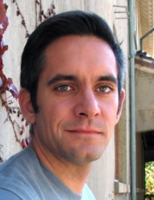 Monte Winslow合作论文数Winslow Lab, Department of Genetics, Stanford University24
 Min K Tsai合作论文数23Laura Andrejka合作论文数19Christopher W. Murray合作论文数19
 Chuan Li合作论文数14
 Christian Kunder合作论文数Stanford University;GynePath Laboratory, Inc13
 Hongchen Cai (蔡宏琛)合作论文数School of Pharmaceutical Sciences, Xiamen University12Pauline Chu合作论文数10
 Jennifer J Brady合作论文数10
 Dmitri Petrov合作论文数Stanford University10Shiyou Chen (陈世友)合作论文数Department of Physiology & Pharmacology, The University of Georgia10Emily Shuldiner合作论文数9
 Nicholas W Hughes合作论文数Department of Genetics, Stanford University School of Medicine9
 Rosanna Ma合作论文数Stanford Univ, Sch Med, Dept Genet, Stanford, CA 94305 USA9
 Winters Ian P合作论文数9Leo C Chen合作论文数7Min DU合作论文数7Zhi Gao合作论文数7ZongShu Ji (纪宗淑)合作论文数7Jess D Hebert合作论文数6
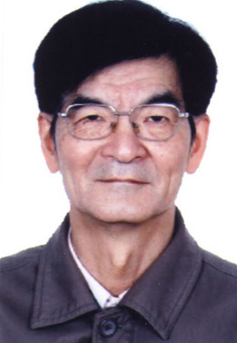 Junyuan Chen (陈均远)合作论文数Nanjing Institute of Geology and Palaeontology, Chinese Academy of Sciences;Nanjing University School of Life Sciences6
 Liu Haiyan合作论文数Division of Cardiology and Center for Cardiovascular Research, Washington University School of Medicine6Zhang Gui合作论文数6
 Le Cong (丛乐)合作论文数Department of Pathology, School of Medicine, Stanford University6
 Ke Zen合作论文数5Emily Ashkin合作论文数5Su Kit Chew合作论文数5
 Yousefi Maryam合作论文数5
 Charles Swanton合作论文数Cancer Evolution and Genome Instability Laboratory, The Francis Crick Institute5Min Zhou (周敏)合作论文数4Wen-Yang Lin合作论文数4Yanyan Yang合作论文数4
 Saswati Karmakar合作论文数Dept Biochem & Mol Biol, Univ Nebraska Med Ctr4
 king l hung合作论文数Univ Washington, Dept Biochem, Seattle, WA 98195 USA4
 Christopher D. McFarland合作论文数Graduate Program in Biophysics, Harvard University;Graduate Program in Biophysics, Harvard University4Linde Ian L合作论文数4
 Chenyu Zhang (张辰宇)合作论文数State Key Laboratory of Pharmaceutical Biotechnology, School of Life Sciences, Nanjing University4
 Edgar Engleman合作论文数Department of Pathology, School of Medicine, Stanford University4
 Aaron D. Gitler合作论文数Department of Genetics, School of Medicine, Stanford University;Bio-X, Stanford University;Wu Tsai Neurosciences Institute, Stanford University4He-Yang You合作论文数3
 Paul Tafforeau合作论文数3h sugiyama合作论文数3Wonjae Lee合作论文数Dept Neurosurg, Stanford Sch Med3
 Yung-Chun Wang合作论文数Dept Genet, Washington Univ3
 Michael Hadfield合作论文数Department of Biology, University of Hawaiʻi at Manoa3Yuning J. Tang合作论文数3
 Lyu Zhonglin合作论文数3
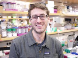 Nicholas J Kramer合作论文数Department of Genetics, Stanford University School of Medicine3Hongchao Liang合作论文数3




