Organ chips offer a disruptive innovation to study human diseases with tissue-specific resolution within a predictable and tunable in vitro environment. However, these platform technologies have for the most part failed to translate to broad use in the private sector due to a lack of high-throughput, user-friendly platforms. Here we present an automated high-throughput organ chip seeded with iPSC-derived cardiomyocytes transduced with GCaMP6f and interface with translational technologies to bridge the current academia-industry gap. Cardiomyocytes were seeded on-chip fully hands-free using an entry-level fluid handling robot to significantly reduce user handling requirements. Pipette interfaces were paramount to facilitating seeding and feeding through improved tolerances for establishing a functional connection to dispense and collect small fluidic volumes. Following successful seeding, GCaMP6f activity on-chip was monitored with our automated, non-invasive fiber-optic sensing platform. We show a significant decrease in cardiomyocyte beat rate in response to decreased ambient culture temperature using data collected with our optical sensing platform. This study provides a potential translational blueprint for academia-industry partnership toward broad adoption of organ chip technology in drug development and disease modeling.
Our engineered fiber optic-based sensing platform was validated for spatiotemporal monitoring of barrier function in our in-house fabricated gut-on-a-chip, offering a non-invasive approach for characterizing the on-chip microenvironment.
Microphysiological systems hold great potential for fundamental discovery and accelerating the drug discovery pipeline through simplifying complex tissues to their first principles and enabling real-time, high-resolution monitoring. Hydrophilic biomaterials, such as hydrogels, are important for microphysiological system innovations due to their ability to emulate the native extracellular matrix and tunable mechanical properties. Furthermore, hydrogels can be tailored to improve tissue maturity as well as the efficacy of instrumentation. However, many biopolymers are non-conductive, presenting complications for modeling excitable tissue environments like the heart. In this work, we show that an 8% (w/v) Gelatin Methacryloyl (GelMA) + 3.5% (v/v) Choline Acrylate hydrogel, nicknamed Gel-Amin, can amplify extracellular voltage recordings from a culture of cardiomyocytes (CMs) from commercial microelectrode arrays. Our laser-cut and assembly method for manufacturing 3D microphysiological systems allowed direct comparisons of CM contractile activity in Gel-Amin compared to control GelMA cultures in a single system. This innovative material supported in vitro CM cultures with improved synchronicity and greater signal-to-noise ratios (SNRs), suggesting potential improvements over conventional biomaterial limitations. Here, we developed a cost-effective in vitro cardiac tissue model that allows real-time electrical activity monitoring.
Microphysiological systems (MPSs) hold great potential for fundamental discovery and accelerating the drug discovery pipeline through simplifying complex tissues to their first principles and enabling real-time, high-resolution monitoring. Hydrophilic biomaterials, such as hydrogels, are important for MPS innovations due to their ability to emulate the native extracellular matrix and tunable mechanical properties. Furthermore, hydrogels can be tailored to improve tissue maturity as well as the efficacy of instrumentation. However, many biopolymers are non-conductive, presenting complications for modeling excitable tissue environments like the heart. In this work, we show that an 8% (w/v) Gelatin Methacryloyl (GelMA) + 3.5% (v/v) Choline Acrylate hydrogel, nicknamed Gel-Amin, can amplify extracellular voltage recordings from a culture of cardiomyocytes (CMs) from commercial microelectrode arrays. Our laser cut and assemble method for manufacturing 3D MPSs allowed direct comparisons of CM signal propagation in Gel-Amin compared to control GelMA cultures in a single system. This innovative material supported in vitro CM cultures with improved synchronicity and greater signal-to-noise ratios (SNRs), suggesting potential improvements over conventional biomaterial limitations. Here, we developed a cost-effective in vitro cardiac tissue model that allows real-time electrical activity monitoring. ### Competing Interest Statement The authors have declared no competing interest. Northeastern University PEAK Fellowship Program NIH NIGMS, R35GM142741 NASA, 80ARC023CA005
Organs-on-a-chip (OOC) are an emergent technology that bridge the gap between current in vitro and in vivo models used to inform drug discovery and investigate disease pathophysiology. These systems offer improved bio-relevance and controlled complexity through the integration of physical and/or chemical stimuli matched to physiologically relevant conditions. Although significant advancements have been made toward recreating organ-specific physiology on chip, the methods available to study structure and function of the cell microenvironment are still limited. Established analysis approaches, including fluorescence microscopy, rely on laborious offline workflows that yield limited time-point data. As the OOC field continues to evolve, there is a unique opportunity to engineer improved characterization methods into organ-chip devices. This review provides an overview of current integrated sensing approaches that address current limitations and enable real-time readout of relevant physiological parameters in OOC.
Objective. The use of conductive materials for aiding peripheral nerve regeneration is a promising method to recapitulate native conductance of nerve tissue and facilitate the delivery of exogeneous stimulation for enhanced recovery. This study systematically investigated the effects of applying electrical (ES) or magnetic stimulation (MS) to neurons within new ionically conductive hydrogels.Approach. The material properties of ionically conductive Gel-Amin hydrogels (Gelatin methacryloyl (GelMA) + Choline acrylate) were compared to those of GelMA hydrogels. Neonatal rat dorsal root ganglia (DRG) were encapsulated in both hydrogel formulations, subjected to ES or MS, and evaluated for differences in neuronal extension. Peripheral glia, Schwann cells (SCs), were subjected to the same stimuli and their secretion of various neurotrophic analytes were investigated.Main results. Gel-Amin hydrogels are 4x more ionically conductive than GelMA hydrogels. The application of electrical stimulation to the encapsulated cells led to a significant decrease (76%) in DRG outgrowth when encapsulated in GelMA versus the Gel-Amin hydrogel. In contrast, MS led to directional neurite extension in a direction perpendicular to the magnetic field gradient.Significance. We present here the first report of a controlled, direct comparison of ES and MS on whole DRG in synthetic materials. The combination of ES and MS decreased total neurite outgrowth but led to more directional growth. Aspects of the material and type of stimuli were noted to reduce several cytokine secretion levels from primary SC cultures. These results highlight the importance of understanding material and biophysical interactions to enhance peripheral nerve regeneration.
Enteric neurons, diverse in function and great in number, are heavily involved in homeostasis within the small intestine and their dysregulation has been implicated in gastrointestinal disorders and neurodegenerative diseases. Innovations in biofabrication have resulted in advances for in vitro models of the gut, however the majority lack enteric innervation, limiting therapeutic screening and discovery. Here, we present a high-throughput co-cultured microphysiological system (MPS), or organ chip, that supports a primary epithelial monolayer that directly interfaces with a three-dimensional hydrogel containing a primary enteric neuron culture, mimicking the close proximity present in vivo. The acrylic MPS device was fabricated with our established and cost-effective laser cut and assemble method. We have expanded this technology to include up to twelve 3D MPSs per device within the footprint of a traditional well-plate, supporting high-throughput experimentation. The inclusion of this 3D microtissue does not hinder physiologically relevant flow, standard measures of barrier function, and microscopy techniques. The device features gravity-driven flow to induce physiological shear stress on the epithelium culture and provide continuous nutrient presentation. Results show the intestinal and neural tissue maintained expected morphologies over an experimental timeline of ten days. Proximal enteric neurons extend neurites through the 3D hydrogel towards the epithelial monolayer. Barrier function was confirmed with both Transepithelial Electrical Resistance (TEER) and Lucifer Yellow diffusion on-chip. TEER confirmed a significantly more substantial barrier integrity in co-cultures compared to baseline values (1.25-fold) in epithelial cell-only. Lucifer yellow permeability assays performed in parallel supported the TEER results, with an 11.8% lower permeability of the co-cultured group than the epithelium only. The presence of the ENS on chip results in a significant (1.4 fold) reduction in epidermal growth factor (EGF). This is the first high-throughput, innervated gut on a chip device that demonstrates the importance of the autonomic nervous system on EGF expression and possibly epithelial renewal in vitro. Innervation is essential to create more biomimetic and physiologically relevant in vitro models for biological and pharmacological assays.
Enteric neurons are critical in maintaining organ homeostasis within the small intestine, and their dysregulation are implicated in gastrointestinal disorders and neurodegenerative diseases. Most in vitro models lack enteric innervation, limiting basic discovery and disease modeling research. Here, a high-throughput 3D microphysiological system (MPS), or organ chip is presented that supports a primary epithelial monolayer interfacing directly with encapsulated primary enteric neurons. The device features twelve 3D MPSs per device and gravity-driven flow via a laboratory rocker to induce biomimetic shear stress on the epithelium culture and provide continuous nutrient presentation. Intestinal and neural tissue exhibited expected morphologies. Neural gene upregulation in the epithelium suggests RNA contamination from proximal enteric neurons extending neurites toward the epithelial monolayer. With the enteric nervous system (ENS), barrier integrity significantly increased for both TEER and permeability assays, a 1.25-fold greater resistance and 10% lower permeability as compared to epithelium cultured alone. The presence of the ENS resulted in a significant (1.4-fold) reduction in epidermal growth factor (EGF). Additionally, several key epithelial genes are compared between duodenal tissue and epithelial monolayers with and without neurons present. Results demonstrated changes in cytokine gene expression and WNT pathways, highlighting innervation is essential to create more biomimetic and physiologically relevant in vitro models.
Neurosensory circuits of the gastrointestinal tract sense microbial and nutrient changes in the gut; however, studying these circuits in vivo is hindered by invasive techniques and ethical concerns. Here, an in vitro model of enteroendocrine cells (EECs) and calcium reporting enteric neurons (ENs) is established and validated for functional signaling. Both mechanical and sucrose stimulation of co-cultures increased the percentage of neurons undergoing a calcium flux, indicating an action potential. Neuronal activation is blocked with either a piezo or insulin receptor blocker. At baseline, a flow only stimulus elicited 51.9% of neurons to activate in co-culture, which is decreased to 15.1% with a piezo blocker. Piezo blocked and sucrose stimulated EECs increased neuronal activation to 43.9%, and an insulin blocker reduced response to 12.4%. Since a cell line is used to model the EEC in the previous experiments, primary rat duodenal epithelium enriched for EECs are also stimulated and found to produced measurable insulin. This work shows the ability of EECs to produce insulin and for ENs to sense insulin. These results inspire further work on how insulin production outside the pancreas effects diabetes, insulin as a neurotransmitter, and exploration of additional nutritional and microbiotic stimuli on enteroendocrine-to-neuronal signaling.
Axonal growth is heavily influenced by topography and biophysical stimuli including magnetic and electrical fields. Despite extensive investigation, the degree of influence and the underlying genetic mechanisms remain poorly understood. Here, a novel approach to guide neurite growth is undertaken using an innovative ferromagnetic composite material - glass-coated magnetic microwire - to furnish a synergistic combination of magnetic and topographical cues. Whole rat dorsal root ganglia (DRG) are cultured under five different conditions: control, static magnetic field, magnetic microwire, static magnetic field + glass fiber, and static magnetic field + magnetic microwire. DRG outgrowth responses under each condition, including total neurite outgrowth and directionality, are compared. The combination of both magnetic stimulation and topography significantly increases total neurite outgrowth compared to the controls. The combination of magnetic stimulation and magnetic microwire lead to a strong directional bias of growth along the microwire, double what is observed with the glass fiber. Next generation RNA sequencing of DRG exposed to static magnetic field + magnetic microwire reveals the downregulation of genes relating to the immune response, interleukin signaling, and signal transduction. These results set the stage for contemplating future biophysical stimulation for axonal guidance and improved understanding of material-tissue interactions.
Mesenchymal stem cells (MSCs) are a promising source of stem cells for treating peripheral nerve injuries. Here, we present the first investigation of differentiation of olfactory mucosa-derived MSC (OM-MSC) towards a Schwann Cell (SC)-like phenotype. OM-MSCs are an advantageous potential source of SCs for peripheral nerve repair, as isolation can be accomplished with a minimally invasive procedure compared to autologous nerve harvest and isolation. Here, Schwann Cell Conditioned Media (SCCM) or a defined growth factor supplemented media (GF) was applied to OM-MSC for twenty-one days. The differentiation process and resulting populations were characterized by immunocytochemistry and RT-qPCR. Functionality of differentiated populations was assessed in an in vitro co-culture model to evaluate interaction with sensory neurons (dorsal root ganglia) juxtaposed to native SCs. Compared to undifferentiated MSCs, differentiation protocols resulted in significant changes in morphology, gene expression, and functionality using SCCM and GF media, representing key characteristics of SCs. Specifically, differentiated populations exhibit elongated, spindle-like morphologies, a high degree of eccentricity, increased S-100, CD44, and NGF expression, and colocalization of myelin basic proteins with neurites in the co-culture model. In conclusion, this work highlights the potential of OM-MSCs to be expanded and differentiated to SCs to improve synthetic scaffolds or for use in decellularized allografts for nerve repair. ### Competing Interest Statement The authors have declared no competing interest.
Giant plasma membrane vesicles (GPMVs) have been utilized as a model to study phase separation in the plasma membrane. Additionally, GPMVs have been employed as vehicle for delivering molecular cargo, including small molecule drugs and nanoparticles. Nearly all examples of GPMV production use a defined salt buffer that is a stark contrast to typical cell culture medium. In this study, we demonstrate that the addition of formaldehyde and dithiothreitol to a standard culture medium was capable of generating GPMVs at a concentration equal to or higher than the traditional production buffer. These methods were evaluated for two human cell lines: kidney endothelial and Schwann cells (SCs). Morphological properties of the resultant GPMVs exhibited no significant differences between the two formulations. Factors such as pH and seeding density significantly influenced the production of GPMVs in both mediums. The cell type and seeding density was shown to influence the number of GPMVs to the greatest extent. SCs yield more GPMVs at higher seeding densities compared to endothelial cells. Stability of the membrane of the GPMVs produced in both mediums was evaluated by monitoring passive diffusion of two fluorescently tagged dextrans (3 and 10 kDa). Regardless of the production formulation or cell type, approximately 85% GPMVs are impermeable to either dextran. Cold storage for on-demand use and shipping are essential for broader use of GPMVs. Toward this aim, we have evaluated the GMPV number and morphologies following storage at -80 degrees C and in liquid nitrogen. A significant loss of the GPMV number, similar to 30%, was observed following storage across production formulations as well as cell types. Our results indicate that smaller GMPVs, <5 mu m are more stable for preservation. In conclusion, GPMVs can be produced in a broad range of formulations, exhibit a high degree of stability, and can undergo cold storage for further adoption.
The field of tissue engineering has benefited greatly from the broad development of natural and synthetic polymers. Extensive work in neural engineering has demonstrated the value of conductive materials to improve spontaneous neuron activity as well as lowering the necessary field parameters for exogenous electrical stimulation. Further, cell fate is directly coupled to the mechanical properties of the cell culture substrate. Increasing the conductivity of hydrogel materials often necessitates the addition of dopant materials that facilitate electron mobility. However, very little electron transfer is observed in native cell signaling and most of these materials are opaque, severely limiting microscopy applications commonly employed to assess cell culture morphology and function. To overcome these shortcomings, the inclusion of an ionic liquid, choline acrylate, into the backbone of a modified collagen polymer increases the bulk conductivity 5-fold at a 1:1 ratio while maintaining optical transmission of visible light. Here, we explore how the inclusion of choline acrylate influences bulk material properties including the mechanical, swelling, and optical properties of our hydrogels, referred to as Gel-Amin hydrogels, as a material for tissue culture. Despite an increase in swelling over traditional GelMA materials, the conductive hydrogels support whole dorsal root ganglia encapsulation and outgrowth. Our results indicate that our Gel-Amin system holds potential for neural engineering applications and lowering the required charge injection for the application of exogenous electrical stimulation. This is this first time an ionic liquid-hydrogel system has been used to culture and support primary neurons in vitro.
Hydrogen sulfide (H2S) is a gaseous microbial metabolite whose role in gut diseases is debated, largely due to the difficulty in controlling its concentration and the use of non-representative model systems in previous work. Here, we engineered E. coli to titrate H2S controllably across the physiological range in a gut microphysiological system (chip) supportive of the co-culture of microbes and host cells. The chip was designed to maintain H2S gas tension and enable visualization of co-culture in real-time with confocal microscopy. Engineered strains colonized the chip and were metabolically active for two days, during which they produced H2S across a sixteen-fold range and induced changes in host gene expression and metabolism in an H2S concentration-dependent manner. These results validate a novel platform for studying the mechanisms underlying microbe-host interactions, by enabling experiments that are infeasible with current animal and in vitro models.
Objective. Regeneration of damaged nerves is required for recovery following nervous system injury. While neural cell behavior may be modified by neuromodulation techniques, the impact of static direct current (DC) magnetic stimulation remains unclear. Approach. This study quantifies the effects of DC magnetostimulation on primary neuronal outgrowth in vitro. The extension of neurites of dorsal root ganglia (DRG) subjected to two different low-strength (mT) static magnetic flux configurations was investigated. Main results. After 3 d of 1 h in-plane (IP) magnetic field stimulation, a 62.5% increase in neurite outgrowth area was seen relative to unstimulated controls. The combined action of in-plane + out-of-plane (IP + OOP) magnetic field application produced a directional outgrowth bias parallel to the IP field direction. At the same time, the diverse magnetic field conditions produced no changes in two soluble neurotrophic factors, nerve growth factor and brain-derived neurotrophic factor, released from resident glia. Significance. These results demonstrate the potential for DC magnetostimulation to enhance neuronal regrowth and improve clinical outcomes.
Introduction Under conditions of limited iron availability, plants and microbes have evolved mechanisms to acquire iron. For example, metal deficiency stimulates reprogramming of carbon metabolism, increasing activity of enzymes involved in the Krebs cycle and the glycolytic pathway. Resultant carboxylates/hydroxycarboxylates then function as ligands to complex iron and facilitate solubilization and uptake, reversing the metal deficiency. Similarly, human intestinal epithelial cells may produce lactate, a hydroxycarboxylate, during absolute and functional iron deficiency to import metal to reverse limited availability. Methods Here we investigate (1) if lactate can increase cell metal import of epithelial cells in vitro, (2) if lactate dehydrogenase (LDH) activity in and lactate production by epithelial cells correspond to metal availability, and (3) if blood concentrations of LDH in a human cohort correlate with indices of iron homeostasis. Results Results show that exposures of human epithelial cells, Caco-2, to both sodium lactate and ferric ammonium citrate (FAC) increase metal import relative to FAC alone. Similarly, fumaric, isocitric, malic, and succinic acid coincubation with FAC increase iron import relative to FAC alone. Increased iron import following exposures to sodium lactate and FAC elevated both ferritin and metal associated with mitochondria. LDH did not change after exposure to deferoxamine but decreased with 24 h exposure to FAC. Lactate levels revealed decreased levels with FAC incubation. Review of the National Health and Nutrition Examination Survey demonstrated significant negative relationships between LDH concentrations and serum iron in human cohorts. Conclusions Therefore, we conclude that iron import in human epithelial cells can involve lactate, LDH activity can reflect the availability of this metal, and blood LDH concentrations can correlate with indices of iron homeostasis.
Excitation-coupling (ECC) is paramount for coordinated contraction to maintain sufficient cardiac output. The study of ECC regulation has primarily been limited to cardiomyocytes (CMs), which conduct voltage waves via calcium fluxes from one cell to another, eliciting contraction of the atria followed by the ventricles. CMs rapidly transmit ionic flux via gap junction proteins, predominantly connexin 43. While the expression of connexin isoforms has been identified in each of the individual cell populations comprising the heart, the formation of gap junctions with nonmuscle cells (i.e., macrophages and Schwann cells) has gained new attention. Evaluating nonmuscle contributions to ECC in vivo or in situ remains difficult and necessitates the development of simple, yet biomimetic in vitro models to better understand and prevent physiological dysfunction. Standard 2D cell culture often consists of homogenous cell populations and lacks the dynamic mechanical environment of native tissue, confounding the phenotypic and proteomic makeup of these highly mechanosensitive cell populations in prolonged culture conditions. This review will highlight the recent developments and the importance of new microphysiological systems to better understand the complex regulation of ECC in cardiac tissue.
Recent evidence implicates a gut-first pathogenesis in the enteric nervous system (ENS) within a portion of PD patients, yet in vitro investigations have primarily focused on the central nervous system. Here, the preformed fibril (PFF) PD model is applied with co-administered groups of butyrate and lipopolysaccharide to model the effects of the local gut microbiome. Significant PFF uptake and retention occur in isolated rat enteric neurons compared to untreated controls resulting in increasing immunostained aggregate conformation-specific, alpha-synuclein (a-Syn) average intensity between 6 µg PFF and untreated controls. Cortical neurons significantly retain PFFs with an increase in aggregated a-Syn average intensity within all dosages. Differences in growth cone morphology but not dynamics in PFF-treated ENS cultures occur. Electrophysiological recordings via a microelectrode array (MEA) indicate no overall difference in spontaneous spike rate. However, only untreated controls respond to PD-relevant dopamine stimulus, while 1 µg PFF and control populations respond to stimulus with ENS-abundant acetylcholine. Finally, no differences in substance P levels—correlated with PD and neurodegeneration—are observed. Overall, these findings suggest the ENS retains PFF dosage absent acute loss in function, however, does experience changes in growth cone morphology and dopamine-stimulated activity.
Gastrointestinal organoids provide an accessible model for studying human development and disease. In this issue of Cell Stem Cell, Eicher et al. (2022) direct human pluripotent stemcells to incorporate three germlayers into gastric organoids, recapitulating the structure and function of human gut tissue in an in vitro model.
 Abigail N Koppes合作论文数46
 Jonathan R Soucy合作论文数Harvard Med Sch, Ophthalmol13
 Polina Anikeeva合作论文数NeuroBionics;Department of Brain and Cogni6ve Sciences, Massachusetts Institute of Technology;Department of Materials Science and Engineering , Massachuse:s Ins6tute of Technology9Douglas M. Swank合作论文数9
 Sanjin Hosic合作论文数7
 Adam Bindas合作论文数7Katelyn Neuman合作论文数Department of Chemical Engineering, Northeastern University7
 Tess Torregrosa合作论文数5
 Nasim Annabi合作论文数Department of Chemical and Biomolecular Engineering, Samueli School of Engineering, University of California, Los Angeles5
 Nathan Schiele合作论文数Dept Biomed Engn, Rensselaer Polytech Inst5
 Froriep Ulrich P合作论文数Massachusetts Institute of Technology, Massachusetts Institute of Technology5
 David Corr合作论文数Departments of Biomedical Engineering, Rensselaer Polytechnic Institute;Center for Modeling, Simulation and Imaging in Medicine, Rensselaer Polytechnic Institute5Kyla Nichols合作论文数Department of Chemical Engineering, Northeastern University4
 Jennifer Selvidge合作论文数Dept Mat, Univ Calif Santa Barbara4
 Chi Lu合作论文数4Ryan Brady合作论文数4
 Andres Canales合作论文数MIT, Dept Mat Sci & Engn, Cambridge, MA 02139 USA4
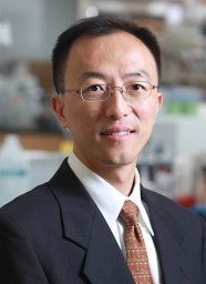 Guohao Dai合作论文数Department of Bioengineering, College of Engineering, Northeastern University4
 Xiaoting Jia合作论文数School of Life Sciences, Sun Yat-Sen University4
 Yoel Fink合作论文数Department of Materials Science & Engineering, Massachusetts Institute of Technology3
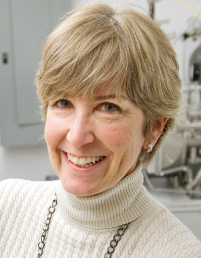 Laura H. Lewis合作论文数Department of Chemical Engineering, College of Engineering, Northeastern University;Department of Mechanical and Industrial Engineering, College of Engineering, Northeastern University3
 Douglas B. Chrisey合作论文数School of Science & Engineering, Tulane University;Department of Physics and Engineering Physics, Tulane University;Department of Biomedical Engineering, Tulane University3
 Walter Herzog合作论文数Faculty of Kinesiology, University of Calgary;Mccaig Institute for Bone and Joint Health, University of Calgary3
 Shashi Murthy合作论文数Nanite3
 David Breault合作论文数the Division of Endocrinology, Children's Hospital, Harvard Medical School, Boston;3Paul J. Kingham合作论文数2Nihal E. Vrana合作论文数2Kristen L. Lee合作论文数2Kacey G. Marra合作论文数2Paul J. Reier合作论文数2George J. Christ合作论文数2Guohao Dai合作论文数2Cailey M Denoncourt合作论文数2Jody Askaryan合作论文数2Will Lake合作论文数2Asel Primbetova合作论文数2Guojun Sheng合作论文数2
 Andrew Spencer合作论文数Holistick Medical, Paris, 75003, France.2
 Christina Tringides合作论文数Department of Materials Science and Nanoengineering, George R. Brown School of Engineering and Computing, Rice University2
 Marissa Puzan合作论文数Dept Chem Engn, Northeastern Univ2
 Ehsan Shirzaei Sani合作论文数Andrew and Peggy Cherng Department of Medical Engineering, California Institute of Technology2
 Rebecca L Carrier合作论文数Department of Chemical Engineering, College of Engineering, Northeastern University;TRET Therapeutics2
 Sabrina Lee合作论文数Neuromuscular Biomechanics Laboratory, Department of Physical Therapy and Human Movement Sciences, Feinberg School of Medicine, Northwestern University2
 Charles J Heckman合作论文数Department of Neuroscience, Feinberg School of Medicine, Northwestern University2
 Seongjun Park合作论文数School of Transdisciplinary Innovations, Seoul National University2
 James Wakeling合作论文数Neuromuscular Mechanics Laboratory, Department of Biomedical Physiology and Kinesiology, Simon Fraser University2
 David Reynaldo Diaz Rascon合作论文数2
 Snyder Jessica合作论文数2
 Achyuta Anil Kumar H合作论文数2




