Electron-pulse probing of fast laser-induced processes has allowed the direct observation of the structural dynamics in matter with a high spatiotemporal resolution. A thin gold film has appeared to be a convenient photocathode, and photoelectron emission has been induced by femtosecond ultraviolet radiation with a photon energy of about 4.65–4.75 eV (in particular, ħ ω ≅ 4.65 eV for the third harmonic of the Ti:sapphire laser). For the linear photoelectric effect, this energy contradicts the reference work function WAu ≅ 5.1–5.3 eV of pure metal. Reasons for such contradiction have been analyzed and good agreement with experimental data has been reached with a model proposed for the generation of photoelectron pulses.
The behavior of a thin-film GeTe crystal induced by intense femtosecond laser pulses ( λ = 0.8 1pt μm) has been studied using a pulsed electron diffractometer. The sample is an annealed 20-nm GeTe film on a copper grid with a carbon coating. It has been found that laser ablation results in the formation of an ultrathin GeTe crystal (assumingly, GeTe monolayer) with a high radiation resistance. Possible reasons for the detected nanosize effect are discussed.
Advances in the development of pulsed lasers provided a further breakthrough in the study of the structural dynamics of nuclei and electrons. As a result of this progress, the use of powerful femtosecond laser pulses, both for exciting a sample and for generating ultrashort (down to femto- and even subfemtosecond) photoelectron bunches synchronized with optical pulses for sensing matter, made it possible to observe the coherent dynamics of nuclei and electrons in samples at the required spatiotemporal scales. The possibility of direct observations of reaction processes is a major breakthrough in chemical physics. The many-particle potential is so complex that the degree of the interaction nonadiabaticity cannot be determined with an accuracy sufficient for predicting reaction paths. How can this information and a new look at the reaction dynamics be used in the future? This question arises in connection with the development of a new conceptual foundation of natural sciences incorporating the convergence of experimental and theoretical tools in studies of systems of any complexity with atomic resolution. In this approach, an ‘atomic–molecular’ movie is obtained by using mutually complementary information extracted from simultaneous studies of ultrafast electron (or X-ray) diffraction, spectroscopy, and the quantum dynamic theory of matter.
Coherent dynamics of lattice oscillations in a ∼20-nm-thick film obtained by thermal sputtering of bismuth is studied using ultrashort electron bunches synchronised with femtosecond laser pulses irradiating the sample. The Fourier analysis of ultrafast electron diffraction (UED) data shows that the observed modulation of the signal is due to the ensemble of modes corresponding to optical phonons with frequencies of approximately 3, 6 and 9 THz. A conclusion is made that these peaks correspond to the manifestation of the bismuth A1g mode (three-terahertz peak), as well as its first and second overtones, which is probably due to quantum confinement in a 20-nm Bi nanostructure. The possibility of a detailed study of the quantum-size effect in bismuth with the help of a transmission UED is analysed.
A common strategy aimed at achieving a high temporal resolution in ultrafast electron microscopy and diffraction is based on the use of strong electrostatic fields in the accelerating gap to form fast electron pulses of ultrashort duration. In this work, the dynamics of the propagation of ultrashort photoelectron bunches is studied taking into account the time-of-flight chromatic aberration: the spread-out of electron pulses at the exit from the region of the accelerating electric field. The results of the calculation of the duration of photoelectron pulses taking into account Coulomb repulsion are presented. According to the analysis, the use of strong electrostatic fields in the accelerating gap for the formation of fast electron pulses of ultrashort duration is not a necessary condition for achieving the final high temporal resolution in the method of ultrafast electron microscopy. The modes of operation of ultrafast transmission electron microscope were found for which the temporal resolution increases with the growth of time-of-flight chromatic aberration.
The structural dynamics of matter, induced by powerful and most often femtosecond laser pulses, is manifested in an atomic-molecular ‘movie’, a collection of processes, the exploration of which is of immense interest for today’s natural sciences. Studying this global phenomenon requires an ultra-high spatio-temporal resolution that involves the development of dedicated research methods. Laser spectroscopy may be applied to determine energy states of samples and track their evolution in time; however, the structural dynamics of matter can only be disclosed from that information by indirect methods. We present here direct techniques for studying laser-perturbed substances in the spatio-temporal continuum where matter is probed with ultrashort electron and X-ray pulses. We also describe the first series of experiments performed with the aid of a femtosecond electron diffractometer and picosecond transmission electron microscope at the Institute of Spectroscopy of the Russian Academy of Sciences based in Troitsk, Moscow.
We present a new concept for scanning probe microscopy characterization of molecular microstructures. It is based on a thin capillary using as a sharp tip to probe and map the morphology of a surface. In our experiment a collimated ion beam is formed by tungsten ions passing through a quartz tapered capillary with a 100-nm aperture and enters a 2D position-sensitive detector. We demonstrate that such ions are capable of producing the image of a dielectric nanoaperture in the case of low-dose ion beam. Ion transmission through a nanoscale capillary opens the door to observing photodesorption of large organic molecular ions with high spatially-element resolution using the combination of a hollow-tip vacuum scanner with time-of-flight mass spectrometer.
The possibilities of a new type of scanning probe microscopy (SPM) for two different samples are experimentally demonstrated. The method is based on the use of a pointed capillary, which can simultaneously act as a 'classical' SPM probe and also as a controlled thin channel for transporting charged particles emitted by the surface to the detector. In the experiment, photoelectrons pass through a dielectric hollow cone probe with an aperture radius of 1 mm and detected by microchannel plates at different points of the investigated conducting surface irradiated by the second harmonic of a femtosecond Ti : sapphire laser. As a result, the sample's surface profile is visualised with a subwavelength spatial resolution. This method makes it possible to control spatially localised beams of electrons, ions, neutral atoms (molecules) and soft X-ray radiation, as well as opens a possibility for research in the field of nanoscale photodesorption of molecular ions.
We demonstrate the use of a conical capillary in a scanning probe microscopy for surface analysis. The probe can measure photoemission from a substrate by transmitting photoelectrons along the capillary as a function of probe position. The technique is demonstrated on a model substrate consisting of a gold reflecting layer on a compact disc which has been illuminated by an unfocused laser beam with a wavelength 400nm, from a femtosecond laser with a beam size of 4mm. A quartz capillary with a 2-µm aperture has been used in the experiments. The period of gold microstructure, shown to be 1.6µ, was measured by the conical probe operating in shear force mode. In shear force regime, the dielectric capillary has been used as a “classical” SPM tip, which provided images reflecting the surface topology. In a photoelectron regime photoelectrons passed through hollow tip and entered a detector. The spatial distribution of the recorded photoelectrons consisted of periodic mountain-valley strips, resembling the surface profile of the sample. Submicron spatial resolution has been achieved. This approach paves the way to study pulsed photodesorption of large organic molecular ions with high spatial and element resolution using the combination of a hollow-tip scanner with time-of-flight technique.
A new type of microscopy based on scanning in vacuum by a beam of charged particles transmitted through a hollow probe has been implemented. This approach provides controllable motion of spatially localized ion, electron, molecular (atomic), and soft X-ray beams and investigation of the surface in the shear force mode. In the photoelectron mode, in which electrons are transmitted through a 2-μm quartz capillary, a surface profile of gadolinium irradiated by 400-nm femtosecond laser pulses has been visualized with a subwave spatial resolution. The new method of microscopy opens an opportunity of investigations in the field of nanometer local photodesorption of molecular ions (one of the last ideas of V.S. Letokhov).
Acting as complementary research tools, high time-resolved spectroscopy and diffractometry techniques proceeding from various physical principles open up new possibilities for studying matter with necessary integration of the 'structure–dynamics–function' triad in physics, chemistry, biology and materials science. Since the 1980s, a new field of research has started at the leading research laboratories, aimed at developing means of filming the coherent dynamics of nuclei in molecules and fast processes in biological objects ('atomic and molecular movies'). The utilization of ultrashort laser pulse sources has significantly modified traditional electron beam approaches to and provided high space–time resolution for the study of materials. Diffraction methods using frame-by-frame filming and the development of the main principles of the study of coherent dynamics of atoms have paved the way to observing wave packet dynamics, the intermediate states of reaction centers, and the dynamics of electrons in molecules, thus allowing a transition from the kinetics to the dynamics of the phase trajectories of molecules in the investigation of chemical reactions.
A method is demonstrated which combines the ionisation of free molecules by a sharply focused femtosecond laser beam and projection microscopy in a divergent electric field. The electric field is produced in vacuum between a metallic tip and a flat position-sensitive charged particle detector. The method enables investigation of photoionisation processes in low-density gases with a subdiffraction spatial resolution and can be used as well in profile measurements for sharply focused, intense laser beams. In a demonstration experiment, a femtosecond laser beam with a peak intensity of similar to 10(14) W cm(-2) was focused to a 40-mu m-diameter waist in vacuum near a millimetre-size tip and similar to 2-mu m spatial resolution was achieved. According to our estimates, the use of a sharper tip will ensure a submicron spatial resolution, which is a crucial condition for the spatial diagnostics of sharply focused short-wavelength VUV radiation and X-rays.
Pulsed desorption of organic conducting polymer by XUV-photons, formed by a thin capillary collimator, has been investigated. Short-wavelength radiation has been resulted from a metal target irradiated by a sharply focused Ti:Sa laser beam (0.8 μm, 40 fs, 3 mJ∕pulse) and has been filtered by Mylar-gold substrate. Single shot and 1-kHz pulses regimes of driving femtosecond laser have been compared using a time-of-flight mass spectrometer. A stepwise function of photoion signal vs laser pulse energy has been observed.
We demonstrate a method combining laser ionization of molecules with projection technique and allowing observation of photoionization processes in gases with sub-focal spatial micro-resolution. A bunch of molecular ions created by the nonlinear photoionization of the imaging gas near a tip extends in a divergent electrostatic field producing a magnifying image on the detector. It can be used to observe the profile of the sharply-focused intense laser beam in a wide spectral range. In proof of principle experiment the water molecules are ionized in ~40-μm laser focal spot in the vicinity of the silver needle with a curvature radius 0.5 mm and the resultant ions are counted by a position-sensitive scheme. According to our estimations, ~1.5-μm spatial resolution has been reached. Using a sharp tip, the spatial resolution can be improved to the sub-micrometer scale and such approach can be applied for short wavelength beam diagnostics.
The ultrafast e(-)-bunches produced by femtosecond laser (fsl) radiation are powerful tool in modern physics to observe different ultrashort processes induced by fsl pulses and to reach high spatio-temporal resolution. In Letokhov's projection microscope such a beam transfers an information from a tip with a sample illluminated by fsl pulses to a detector. Here the fsl radiation provides for an effective photoelectron multiphoton emission without significant heating of the sample. Two organic samples, formed from dye molecules and the organic conducting polymers has been vizualized using the nanocapillary as a tip. The advantages of the nanocapillary tip are described in the report.The temporal resolution depends upon the pulse duration of the e(-)-beam, tau(e). One of the most powerful way to measure tau(e) is to use the Gaponov-Miller force (GMf), or the ponderomotive force, which the electrons experience in the inhomogeneous field of a focused laser pulse. Such a force helped us to demonstrate the "instantaneousness" of multiphoton emission process from solid targets. As the beam propagated, it spread in time. Using GMf we temporally characterized the e(-)-pulse transmitted through microcapillary (which can be the basis of the promising scanning microscope) and combined spatial nanoresolution and picosecond temporal resolution. Also the ultrafast microlocalized e(-)-beam is an ideal tool to measure the GMf created by focused fsl pulses and to characterize very intense laser beam profile in-situ. In principal, such approach may allow for direct subwave spatio-temporal probing of superintense laser beam profiles.
The analysis of the possibilities of microstructuring (micromachining) of various solids by sharply focused femtosecond laser (fsl) capable of providing for a high intensity for moderate pulse energies remains quite topical. Usually the process takes place immediately at the focal spot and an extended structure can only be formed by scanning the sample. In our experiments focusing fsl pulses into a vacuum setup we produced extended microstructures (microdamages) on a few tens nm thickness gold foil deposited onto a fused-quartz substrate. The structure length has been an order of magnitude larger than the typical size of the irradiated region. The detection of Au+ photoions ensured a highly-sensitivite on-line diagnostics of this phenomenon and in the limit can be used for the probing of the picostructuring of solids irradiated by a laser. In the next step we are "cloning" the microstructure (microdamage) from one surface to another when there is a few tens microns vacuum-spacing between them and only one surface with a metal film is "directly" irradiated by fsl pulses. For the expertiment we prepared two similar fused-quartz prisms and pressed one sample to another. The clearance is determined by the thickness of a spacer (Cu-foil). The possible explanations are given.
A method for measuring the Gaponov-Miller force (GMF) is demonstrated based on the deflection of a picosecond photoelectron beam exposed to tightly focused intense femtosecond laser radiation. It is shown experimentally that the action of this force produced by femtosecond laser pulses linearly depends on their intensity. The method can be used to verify the correctness of measuring the duration of an ultrashort electron bunch based on the GMF.
Our experimental activity in the field of femtosecond electron optics is described. The final purpose is to generate and characterize a sub-100-fs femtosecond electron beam.
The geometric-optical mechanism of wavefront reconstruction differs significantly from conventional holographic reconstruction. This regime can be realized for holograms containing only few periods of interference fringe structure. The geometric-optical reconstruction of the holograms recorded by femtosecond laser pulses in volume media was demonstrated in our previous works. The large thickness of the recording medium required for the effect observation is a serious obstacle for future development in this direction. In this work a way to surmount this obstacle by realizing the waveguide analog of the geometric-optical reconstruction process is presented. Holograms were recorded by 30-femtosecond laser pulses in 20-μm film of dichromated gelatin on the polished quartz substrate and reconstructed by the waveguide mode. Geometric-optical regime of waveguide hologram reconstruction was obtained: the direction of the reconstructed beam was observed to be constant as the reconstructing wavelength was varied within the hologram spectral selectivity band. The possibility of producing achromatic waveguide optical elements containing only few periods is discussed. The utilization of FIB (focused ion beam) nanotechnology for fabrication of these optical elements is proposed. Production of high aspect ratio periodic structures by FIB technology is demonstrated.
A nanolocalized electron beam has been formed by electrons passing through a quartz microcapillary. The authors have demonstrated that such electrons are capable of producing the image of a dielectric nanoaperture. Using the capillary and a metal tip illuminated by femtosecond laser pulses, the authors have developed a photoelectron source based on nanolocalized photoelectron bunches with low angular divergence. It allows the observation of femtosecond-laser-induced processes on a surface with high temporal and spatial resolutions.
 Boris Mironov合作论文数17
 Evgenii a Ryabov合作论文数Russian Academy of Sciences Institute of Spectroscopy Troitsk7Sergey Chekalin合作论文数6
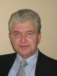 Anatoli a Ischenko合作论文数Department of Chemistry;Moscow State University;Department of Chemistry, Moscow State University6
 David Villeneuve合作论文数Department of Physics, University of Ottawa;National Research Council of Canada;Université du Québec5Igor V Kochikov合作论文数5
 V. V. Petrunin合作论文数4
 V. G. Minogin合作论文数4
 Paul Corkum合作论文数Department of Physics, Faculty of Science, University of Ottawa;Joint Laboratory for Attosecond Science, University of Ottawa4A. P. Cherkun合作论文数3
 Yu. Kudryavtsev合作论文数3A.S. Akhmanov合作论文数2
 Michael Spanner合作论文数Theory and Computation Group, National Research Council of Canada;Theory and Simulation Group, National Research Council of Canada2Peter Dietrich合作论文数2
 Marc Vrakking合作论文数Freie Universität Berlin;Max Born Institute for Nonlinear Optics and Short Pulse Spectroscopy2Georgiy V Girichev合作论文数Ivanovo State University of Chemistry and Technology Department of Physics2
 Denis G. Poydashev合作论文数Institute of Spectroscopy, Russian Academy of Sciences2
 Misha (Mikhail) Ivanov合作论文数Department of Physics, Faculty of Natural Sciences, Imperial College London2А.Л. Малиновский合作论文数1S. Chekalin合作论文数1M. Mehendale合作论文数1O B Serov合作论文数1r j verver合作论文数1MY Ivanov合作论文数1Vs Letokhov合作论文数1v v petrunin合作论文数1Yongfeng Ni合作论文数1
 Leszek Frasinski合作论文数Department of Physics, Faculty of Natural Sciences, Imperial College London1
 Vishal Bhardwaj合作论文数1
 James S. Wright合作论文数Ottawa-Carleton Chemistry Institute, Carleton University1David M. Rayner合作论文数1
 Artemy Irzhak合作论文数Moscow State Institute of Steel and Alloys, Technological University1
 A. A. Lotin合作论文数Institute on Laser and Information Technologies, Russian Academy of Sciences (ILIT RAS),1
 Miguel Cervantes合作论文数Centro de Investigación Biomédica de Michoacán, Instituto Mexicano del Seguro Social (IMSS),1
 George Gibson合作论文数Department of Physics, College of Liberal Arts and Sciences, University of Connecticut1
 Emma Springate合作论文数Rutherford Appleton Laboratory1V. O. Kompanets合作论文数Russian Academy of Sciences Institute of Spectroscopy Troitsk1Harm Muller合作论文数1
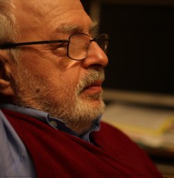 Anatoly Smolovich合作论文数1Tue Haste Andersen合作论文数1V. Petrunin合作论文数1Thomas Vestergaard Andersen合作论文数1Sébastien Zamith合作论文数1DR Matusek合作论文数1Gennady L. Yudin合作论文数1V. Ya. Panchenko合作论文数1
 Lars Bojer Madsen合作论文数Department of Physics and Astronomy, Aarhus University1




