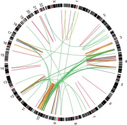
Pediatric tumors undergo a battery of time-sensitive molecular and cytogenetic tests to detect gene fusions and other structural variants. While many labs are beginning to use targeted NGS panels to detect such events, we considered whole transcriptome sequencing (RNA-Seq) to be the ultimate solution as it samples all expressed loci including those only relevant to rare pediatric tumors. Clinical RNA-Seq, although an attractive option, presents several unique challenges including i) How to avoid an informatics bottleneck within a time-sensitive workflow ii) How to minimally burden those reviewing the results and iii) How to standardize and share complex computational analysis methods. Here we present a pilot study of clinical RNA-Seq analyzed in the cloud. Using a highly secure platform, we developed our Rapid RNA-Seq pipeline centered on an extensively validated fusion gene detection algorithm “CICERO.” We initially detected fusion transcripts and structural variants in 78 diverse but well-characterized samples and showed that Rapid RNA-Seq detected all abnormalities found previously. We next ran Rapid RNA-Seq in parallel with standard testing in real time, again showing concordance, but also detecting a large cache of previously undetected events—some of which were targetable. Run times averaged approximately four hours from raw data acquisition to reporting and were not affected by caseload. To aid in clinical review, we developed a rich graphical interface based on our ProteinPaint software (https://pecan.stjude.org/#/home) that allowed pathologists to easily assess the supporting evidence and functional impact of any given event. We make our tools and workflows securely available at www.stjude.cloud.
Graft-versus-host disease (GVHD) is a major cause of morbidity and mortality after allogeneic hematopoietic stem cell transplantation (HSCT). To identify recipient risk factors, a genome-wide study was performed including 481,820 single-nucleotide polymorphisms (SNPs). Two GVHD susceptibility loci (rs17114803 and rs17114808) within the SUFU gene were identified in the discovery cohort (p = 2.85 × 10 −5 ). The incidence of acute GVHD among patients homozygous for CC at SUFU rs17114808 was 69%, which was significantly higher than the 8% rate observed in CT heterozygous patients (p = 0.0002). In an independent validation cohort of 100 patients, 50% of the patients with the CC genotype developed GVHD compared to 8% of the patients with either CT or TT genotype (p = 0.01). In comparison to CC dendritic cells, those from CT expressed higher levels of SUFU mRNA and protein, had lower levels of surface HLA-DR and induced less allogeneic mixed leukocyte response (MLR). Ectopic expression of SUFU in THP-1 derived DCs reduced HLA-DR expression and suppressed MLR, whereas silencing of SUFU enhanced HLA-DR expression and increased MLR. Thus our findings provide novel evidence that recipient SUFU germline polymorphism is associated with acute GVHD and is a novel molecular target for GVHD prevention and treatment.
BackgroundWe sought to improve lumbar spine bone mineral density (LS-BMD) in long-term survivors of childhood acute lymphoblastic leukemia (ALL) using calcium and cholecalciferol supplementation.ProcedureThis double-blind, placebo-controlled trial randomized 275 participants (median age, 17 [9-36.1] years) with age- and gender-specific LS-BMD Z-scores <0 to receive nutritional counseling with supplementation of 1,000mg/day calcium and 800International Unit cholecalciferol or placebo for 2 years. The primary outcome was change in LS-BMD assessed by quantitative computerized tomography (QCT) at 24 months. Linear regression models were employed to identify the baseline risk factors for low LS-BMD and to compare LS-BMD outcomes.ResultsPre-randomization LS-BMD below the mean was associated with male gender (P=0.0024), White race (P=0.0003), lower body mass index (P<0.0001), and cumulative glucocorticoid doses of 5,000mg (P=0.0012). One hundred eighty-eight (68%) participants completed the study; 77% adhered to the intervention. Mean LS-BMD change did not differ between survivors randomized to supplements (0.330.57) or placebo (0.28 +/- 0.56). Participants aged 9-13 years and those 22-35 years had the greatest mean increases in LS-BMD (0.50 +/- 0.66 and 0.37 +/- 0.23, respectively). Vitamin D insufficiency (serum 25[OH]D <30ng/ml) found in 296 (75%), was not associated with LS-BMD outcomes (P=0.78).ConclusionCholecalciferol and calcium supplementation provides no added benefit to nutritional counseling for improving LS-BMD among adolescent and young adult survivors of ALL (93% of whom had LS-BMD Z-scores above the mean at study entry). Pediatr Blood Cancer 2014;61:885-893. (c) 2014 Wiley Periodicals, Inc.
X‐linked lymphoproliferative syndrome (XLP) is caused by mutations in SH2D1A, and is associated with overwhelming infectious mononucleosis, aplastic anemia, hypogammaglobulinemia, and B‐cell lymphomas. However, the frequency of SH2D1A mutations in males who present with B NHL is unknown. Five cases of XLP were diagnosed among 158 males presenting with B NHL (approximately 3.2%). Four of the patients had two episodes of B NHL and one had a single episode of B NHL followed by aggressive infectious mononucleosis. Prospective screening for XLP in males with B‐cell lymphoma at the time of initial diagnosis should be considered. Pediatr Blood Cancer 2013;160:E85–E87. © 2013 Wiley Periodicals, Inc.
To investigate the frequency of isocitrate dehydrogenase 1 (IDH1) and 2 (IDH2) mutations in pediatric acute myeloid leukemia (AML) and acute lymphoid leukemia (ALL), we sequenced these genes in diagnostic samples from 515 patients (227 AMLs and 288 ALLs). Somatic IDH1/IDH2 mutations were rare in ALL (N=1), but were more common in AML, occurring in 3.5% (IDH1 N=3 and IDH2 N=5), with the frequency higher in AMLs with a normal karyotype (9.8%). The identified IDH1 mutations occurred in codon 132 resulting in replacement of arginine with either cysteine (N=3) or histidine (N=1). By contrast, mutations in IDH2 did not affect the homologous residue but instead altered codon 140, resulting in replacement of arginine with either glutamine (N=4) or tryptophan (N=1). Structural modeling of IDH2 suggested that codon 140 mutations disrupt the enzyme's ability to bind its substrate isocitrate. Accordingly, recombinant IDH2 R140Q/W were unable to carry out the decarboxylation of isocitrate to α-ketoglutarate (α-KG), but instead gained the neomorphic activity to reduce α-KG to R(–)-2-hydroxyglutarete (2-HG). Analysis of primary leukemic blasts confirmed high levels of 2-HG in AMLs with IDH1/IDH2 mutations. Interestingly, 3/5 AMLs with IDH2 mutations had FLT3-activating mutations, raising the possibility that these mutations cooperate in leukemogenesis.
Reduced B cell numbers and a mutation in Btk are considered sufficient to make the diagnosis of X-linked agammaglobulinaemia. In the process of conducting family studies, we identified a 58-year-old healthy man with an amino acid substitution, Y418H, in the adenosine-5'-triphosphate binding site of Btk. Immunofluorescence studies showed that this man had 0.85% CD19+ B cells (normal 4-18%) in the peripheral circulation and his monocytes were positive for Btk. He had borderline low serum immunoglobulins but normal titres to tetanus toxoid and multiple pneumococcal serotypes. To determine the functional consequences of the amino acid substitution, a Btk- chicken B cell line, DT40, was transfected with expression vectors producing wild-type Btk or Y418H Btk. The transfected cells were stimulated with anti-IgM and calcium flux and inositol triphosphate (IP3) production were measured. Cells bearing the mutant protein demonstrated consistently a 15-20% decrease in both calcium flux and IP3 production. These findings indicate that even a modest decrease in Btk function can impair B cell proliferation or survival. However, a mutation in Btk and reduced numbers of B cells are not always associated with clinical disease.
Somatic mutations in nucleophosmin (NPM1) occur in approximately 35% of adult acute myeloid leukemia (AML). To assess the frequency of NPM1 mutations in pediatric AML, we sequenced NPM1 in the diagnostic blasts from 93 pediatric AML patients. Six cases harbored NPM1 mutations, with each case lacking common cytogenetic abnormalities. To explore the phenotype of the AMLs with NPM1 mutations, gene expression profiles were obtained using Affymetrix U133A microarrays. NPM1 mutations were associated with increased expression of multiple homeobox genes including HOXA9, A10, B2, B6 and MEIS1. As dysregulated homeobox gene expression is also a feature of MLL-rearranged leukemia, the gene expression signatures of NPM1-mutated and MLL-rearranged leukemias were compared. Significant differences were identified between these leukemia subtypes including the expression of different HOX genes, with NPM1-mutated AML showing higher levels of expression of HOXB2, B3, B6 and D4. These results confirm recent reports of perturbed HOX expression in NPM1-mutated adult AML, and provide the first evidence that the NPM1-mutated signature is distinct from MLL-rearranged AML. These findings suggest that mutated NPM1 leads to dysregulated HOX expression via a different mechanism than MLL rearrangement.
PURPOSE:To improve outcome and study biology of childhood acute lymphoblastic leukemia (ALL) in El Salvador. PATIENTS AND METHODS:Between January 1994 and December 1996, 153 children of El Salvador had newly diagnosed ALL treated in a collaborative program between Hospital Benjamin Bloom and St. Jude Children's Research Hospital (SJCRH). Therapy was based on a modified SJCRH protocol, with uniform remission induction (prednisone, vincristine, L-asparaginase) followed-up by consolidation with teniposide/cytarabine and/or high-dose methotrexate. Continuation treatment was risk-stratified: 123 patients assigned to the high-risk group received weekly rotational drug pairs, and 16 assigned to the standard-risk group received daily 6-mercaptopurine, weekly methotrexate, and monthly pulses of vincristine plus dexamethasone. High risk was defined as: DNA index < 1.16, age 12 months or younger, white blood cell count > or = 50 x 10(9)/L, T-cell immunophenotype, anterior mediastinal mass, central nervous system leukemia at diagnosis, or t(4;11), t(1;19), or t(9;22). Duration of the continuation treatment was 2.5 years in both groups. The median age at diagnosis of all patients was 4.8 (range I d-17 yrs), median leukocyte count was 15 (range 1-766) x 10(9)/L, and sex distribution was equal. RESULTS:Immunophenotypes were early beta-progenitor in 79%, T-cell in 3.9%, and inconclusive in 17% of cases. DNA index was <1.16 in 80.5% and was > or = 1.16 in 19.5% of the 123 known cases. For the analyzes, patients who refused therapy (abandoned treatment) were considered to have treatment failure as of their last follow-up dates. Complete remission was achieved in 126 of 151 (82.4%) patients (11 abandoned therapy during induction). The overall 4-year event-free survival (EFS) rate +/- 1 standard error was 48 +/- 6%. The 4-year EFS rates in patients at high-risk and standard-risk were 46 +/- 7% (n = 121) and 69 +/- 15% (n = 16), respectively (P = 0.20). When patients who refused further treatment are censored, the corresponding 4-year estimates of EFS are 51 +/- 8% and 75 +/- 14%, respectively. CONCLUSIONS:These results suggest that the biology of childhood ALL in El Salvador appears to be similar to that seen in the United States. Risk-directed chemotherapy can successfully be used in developing countries, but risk factors must be carefully determined and applied.
The diagnosis and classification of non-Hodgkin's lymphoma (NHL) has traditionally been made based on morphologic and mununophenotypic criteria. Unfortunately, because of the diverse nature of this group of diseases, rehante solely on these criteria has frequently resulted in misdiagnosis. In addition, investigators have been limited m their ability to define biologically and clinically relevant non-Hodgkin's lymphoma subgroups using this diagnostic approach. The development of modern molecular biological techniques and their use to characterize the genetic abnormalities that are of pathogenic significance in NHL now provides an additional means to identify and to rationally subcategorize these neoplasms. The shortcomings of traditional methods of NHL diagnosis and classification have been especially evident within the morphological subset commonly referred to as the large-cell lymphomas, which comprise approx 25 and 40% of NHL m children and adults, respec tively (1). The marked cytological, immunological, and clinical heterogeneity of this group of tumors suggests that it is comprised of several biologically different neoplasms. However, morphological and immunophenotypic subclassification of the large-cell lymphomas has failed to identify a subset of tumors having a reproducibly different therapeutic response or patient survival rate. Until recently, recurrent genetic abnormalities characteristic of the large-cell lymphomas had not been identified.However, the characterization during the past 2 yr of chromosomal rearrangements mvolvmg the BCL6/LAZ3 zmc finger gene located at 3q27 (which is altered m approx 30% of these tumors [2,3]), and the cloning by our group (4) of the NPM-ALK fusion gene produced by the t(2;5) may now permrt the identrficatton of molecular genetic subtypes of largecell lymphoma that possess unique btological and/or clinical features.
 James R. Downing合作论文数St. Jude Children’s Research Hospital8
 E. M. Azzato合作论文数4J. Nakitandwe合作论文数4
 Scott Newman合作论文数Hutchison-MRC Research Centre and Department of Pathology, University of Cambridge4Jiakun Zhang合作论文数St Jude Childrens Hosp, Dept Dev Neurobiol, Memphis, TN 38105 USA3
 Kim E Nichols合作论文数St Jude Childrens Res Hosp, 332 N Lauderdale St, Memphis, TN 38105 USA3
 Raul C. Ribeiro合作论文数Department of Oncology, Graduate School of Biomedical Sciences, St. Jude Children's Research Hospital3
 Michael Rusch合作论文数Dept Computat Biol, St Jude Childrens Res Hosp3
 Jinghui Zhang (张京慧)合作论文数Central South University2D. Ellison合作论文数2D. W. Ellison合作论文数2
 Michael R. Clay合作论文数Univ Vermont, Coll Med, Dept Pathol, 111 Colchester Ave, Burlington, VT 05401 USA2
 Charles G. Mullighan合作论文数Department of Pathology, St. Jude Children's Research Hospital;Comprehensive Cancer Center, St. Jude Children's Research Hospital2
 Wing Leung合作论文数University of Tennessee;Department of Bone Marrow Transplantation and Cellular Therapy, St. Jude Children’s Research Hospital2
 Ching-Hon Pui (裴正康)合作论文数Leukemia/Lymphoma Division, Department of Oncology, St. Jude Children's Research Hospital2Mary Ellen Conley合作论文数2Vanessa Howard合作论文数2Dale J. Hedges合作论文数Department of Biological Sciences;Biological Computation and Visualization Center;Louisiana State University;Biological Computation and Visualization Center, Louisiana State University2
 Ina Radtke合作论文数2S. Foy合作论文数1E Lovorn合作论文数1H Surprise合作论文数1A Qi合作论文数1Queenie Vong合作论文数1Wm Crist合作论文数1Pl Harrison合作论文数1Jt Sandlund合作论文数1Xd Zhou合作论文数1Fg Behm合作论文数1
 Clay McLeod合作论文数1J. Gu合作论文数1L Carbone合作论文数1J Boyett合作论文数1Ying Li合作论文数1David Miller合作论文数1
 John Lynch合作论文数Dept Commun, Univ Cincinnati1John Easton合作论文数Dept Computat Biol, St Jude Childrens Res Hosp1
 Katherine Clegg Smith合作论文数Department of Health, Behavior and Society, Bloomberg School of Public Health, Johns Hopkins University1
 Kirsten K. Ness合作论文数Human Performance Lab, St. Jude Children's Research Hospital;Department of Epidemiology and Cancer Control, St. Jude Children's Research Hospital;Department of Physical Therapy, College of Health Professions, University of Tennessee Health Science Center;College of Graduate Health Sciences, University of Tennessee Health Science Center1
 Klco Jeffery M合作论文数Dept Pathol & Immunol, Washington Univ1
 Susana C. Raimondi合作论文数Cytogenetics Laboratory, St. Jude Children's Research Hospital1Lemoff Andrew S合作论文数Department of Chemistry, University of California1Marina N合作论文数1
 Mary V. Relling合作论文数Department of Pharmacy and Pharmaceutical Sciences, St. Jude Children's Research Hospital1
 Antillón Klüssmann F合作论文数1
 Tomohiro Kurosaki (黒﨑知博)合作论文数Division of Microbiology and Immunology, Center for Infectious Disease Education and Research, Osaka University;Laboratory for Lymphocyte Differentiation, Center for Integrative Medical Sciences, RIKEN;Laboratory of Lymphocyte Differentiation, WPI Immunology Frontier Research Center, Osaka University1Rencher R合作论文数1Guolian Kang合作论文数1Cai Z合作论文数1




