Purpose: Neoadjuvant chemoradiotherapy (NACRT) followed by esophagectomy is a standard treatment for locally advanced esophageal squamous cell carcinoma (ESCC). Pathological response, assessed using tumor regression grade (TRG), is an established prognostic marker; however, evidence regarding its association with the pattern and timing of recurrence remains limited. This study evaluated recurrence patterns, timing of recurrence, and survival outcomes according to pathological tumor response following NACRT and esophagectomy for ESCC.Methods: We retrospectively analyzed 129 patients with ESCC who underwent NACRT followed by radical esophagectomy between January 2013 and June 2023. Pathological response was assessed according to the College of American Pathologists TRG system and categorized as TRG 0 (complete response), TRG 1 (near-complete response), TRG 2 (partial response), or TRG 3 (poor/no response). Recurrence patterns and timing, disease-free survival, and overall survival were compared across TRG categories.Results: Among 129 patients, 33 (25.6%) developed recurrence. Systemic recurrence was the most common pattern (16/33, 48.5%), followed by combined locoregional and systemic recurrence (9/33, 27.3%) and isolated locoregional recurrence (8/33, 24.2%). Patients with TRG 0 had the lowest proportion of isolated locoregional recurrence (16.7%), with systemic and combined recurrence accounting for 50.0% and 33.3%, respectively. In contrast, TRG 3 patients predominantly developed isolated locoregional recurrence (80.0%), with systemic recurrence occurring in 20.0%; all recurrences in this group occurred within 12 months, suggesting poorer local disease control.Conclusion: Poor pathological response was associated with earlier and predominantly locoregional recurrence, whereas favourable pathological response was associated with a greater proportion of systemic recurrence. These findings suggest that pathological tumor response may help inform postoperative risk stratification and guide individualized surveillance strategies in patients with ESCC.
236 Background: Patients with stage IVA–IVB head and neck squamous cell carcinoma (HNSCC) ineligible for upfront surgery or definitive chemoradiation have poor 5-year survival of 30% to 40%. The prospective NeoLOCUS study showed high response and conversion rates with a regimen combining chemotherapy, low-dose immunotherapy (LD-IO), and oral metronomic therapy (OMT). Here, we report real-world outcomes with the same. Methods: Single-center retrospective study of stage IVA–IVB HNSCC treated with neoadjuvant platinum–taxane chemotherapy plus LD-IO and OMT (erlotinib, methotrexate, celecoxib). Primary endpoint was conversion to radical-intent local therapy. Secondary endpoints were radiologic response, event-free survival (EFS), overall survival (OS), and grade ≥3 adverse events. Results: Forty-three patients were included (Characteristics in Table). After a median 4 (Range 1- 6) neoadjuvant cycles, objective radiologic response was 62.8% (complete response 2.3%; partial response 60.5%, progressive disease 4.7%). Volumetric tumor reduction ≥65% occurred in 54.8%. Conversion to radical-intent therapy was achieved in 69.8%. At median follow-up of 16 months, median EFS was 10.5 months and OS 16.1 months. Thirteen deaths (30.2%) occurred. Median OS was 18.1 months in those receiving radical therapy versus 6.5 months in those who did not. Grade ≥3 adverse events occurred in 14.7%, most commonly anemia and thrombocytopenia (7% each). One grade 4 diarrhea with acute kidney injury required discontinuation. At disease progression, 20.9% received palliative systemic therapy, 2.3% palliative radiotherapy alone and others supportive care. Conclusions: NeoLOCUS regimen demonstrated encouraging response rates and enabled conversion to radical-intent local therapy in about two-thirds of patients with advanced stage IVA–IVB HNSCC, translating into improved survival in this challenging clinical setting. Baseline and treatment characteristics. Characteristic Value Median age, yr (IQR) 54 (45.5–60) Male sex, n (%) 33 (76.7) Stage IVA / IVB, n (%) 25 (58.1) / 18 (41.9) Oral vs non-oral primary, n (%) 28 (65.1) / 15 (34.9) Harmonised ICI dose mg/kg/2wks (IQR) 0.4 (0.2–0.4) Radical-intent local therapy, n (%) 30 (69.8) CTRT + Radical RT, n (%) 24 (55.8) Surgery, n (%) 6 (14.0) Palliative RT / No local therapy 3 (7.0) / 10 (23.3)
Background:Non-surgical management of oral cavity squamous cell carcinoma (OSCC) has poorer outcomes compared to surgery. In borderline resectable tumors, historical neoadjuvant chemotherapy achieves surgical conversion in only about 40%. Combining low-dose immunotherapy and oral metronomic therapy (OMT) with chemotherapy may enhance resection rate and survival. Methods:Between April 2023 and April 2024, patients deemed 'borderline resectable' OSCC based on predefined criteria by a multidisciplinary tumor board were prospectively offered this Phase II single-arm interventional trial setting. Patients received two 21-day cycles of carboplatin, nab-paclitaxel, low-dose nivolumab, and six weeks of erlotinib, methotrexate, celecoxib, with additional cycle(s) if needed. Primary endpoint was R0 resection rate. Secondary endpoints were objective response rate, pathologic response, safety, event-free survival (EFS), and overall survival (OS). Immune biomarkers and Volumetric assessment were exploratory endpoints. The trial was prospectively registered in the Clinical Trial Registry of India (CTRI/2023/04/051617). Findings:Of 34 patients, all except one completed planned neoadjuvant therapy. After 2 cycles, 22 (66·6%) had partial response and 11 had stable disease; none progressed. Twenty six underwent surgery; 25 achieved R0 resection (25/33-75·7% conversion). Four of seven remaining patients received additional cycle(s); three subsequently achieved R0 resection. Overall conversion rate was 90·3% (28/31) excluding 2 patients who refused further treatment. Major pathological response occurred in 12 patients (41·4%), including four with pathological complete response. Grade ≥3 toxicities occurred in 5 of 34 patients (14·7%), with no treatment-related deaths. One patient had grade 4 diarrhea with grade 4 acute kidney injury. Interpretation:The NeoLOCUS regimen offers an affordable, outpatient chemo-immunotherapy approach that improves surgical conversion and pathological response in borderline resectable OSCC. Funding:Fluid Research Grant- Christian Medical College, Vellore, India.
Head and neck cancer (HNC) requires accurate tumor delineation for effective radiotherapy planning. Manual segmentation of tumor regions is time-consuming and subject to considerable inter-observer variability. Although several automated approaches have been proposed, many rely on multimodal imaging such as PET/CT, which is expensive, less accessible in many clinical settings, and increases the burden on patients. In this work, we investigate a CT-only three-dimensional segmentation framework that provides a clinically practical and resource-efficient alternative. CT images of 136 head and neck cancer patients from the publicly available HN1 dataset in The Cancer Imaging Archive (TCIA) were used along with 30 additional cases from a private dataset collected at a tertiary care centre, Christian Medical College (CMC), Vellore, India. A fully automated segmentation model was developed to delineate the primary gross tumor volume (GTV) using the 3D nnU-Net framework. The models were trained using the HN1 dataset and an extended HN1+CMC dataset that included the additional private cases. Performance was evaluated using three-fold cross-validation with standard segmentation metrics including Dice Similarity Coefficient (DSC), Intersection over Union (IoU), and the 95th percentile Hausdorff Distance (HD95). The proposed CT-based model achieved a Global Dice of 0.63 and a Median Dice of 0.60 on the HN1 dataset. When the additional CMC cases were incorporated during training, the performance improved to a Global Dice of 0.65 and a Median Dice of 0.71. These results demonstrate that 3D nnU-Net can effectively segment head and neck tumors from CT images alone. The proposed CT-only approach provides a cost-effective and scalable solution that can support radiotherapy treatment planning and help reduce variability in clinical workflows. ### Competing Interest Statement The authors have declared no competing interest. ### Funding Statement This study did not receive any funding ### Author Declarations I confirm all relevant ethical guidelines have been followed, and any necessary IRB and/or ethics committee approvals have been obtained. Yes The details of the IRB/oversight body that provided approval or exemption for the research described are given below: For this study, we used two datasets: a public dataset, HEAD-NECK-RADIOMICS-HN1, from The Cancer Imaging Archive (TCIA), comprising 137 patients, and a private institutional dataset named HNC, consisting of 30 patients. The private data was collected at Christian Medical College (CMC) Vellore and was ethically approved by the institutional review board. I confirm that all necessary patient/participant consent has been obtained and the appropriate institutional forms have been archived, and that any patient/participant/sample identifiers included were not known to anyone (e.g., hospital staff, patients or participants themselves) outside the research group so cannot be used to identify individuals. Yes I understand that all clinical trials and any other prospective interventional studies must be registered with an ICMJE-approved registry, such as ClinicalTrials.gov. I confirm that any such study reported in the manuscript has been registered and the trial registration ID is provided (note: if posting a prospective study registered retrospectively, please provide a statement in the trial ID field explaining why the study was not registered in advance). Yes I have followed all appropriate research reporting guidelines, such as any relevant EQUATOR Network research reporting checklist(s) and other pertinent material, if applicable. Yes Public data used in this study available at https://www.cancerimagingarchive.net/collection/head-neck-radiomics-hn1/
While AI models are developed in oncology for predicting different clinical outcomes, the focus is often on accuracy and many fail to adequately communicate the degree of certainty in these predictions. To improve clinical decision-making in oncology, this work introduces the idea of uncertainty quantification (UQ) for AI models using an illustrative example. Our goal is to help radiologists and oncologists better understand prediction reliability by integrating UQ. Our illustrative example is a Radiomics Risk Model (RM) for Thymic Epithelial Tumours, developed to provide a basic understanding of the mechanism to evaluate the degree to which individual patient data matches the training set. The study demonstrates the concept of measuring uncertainty in artificial intelligence (AI) models using a simple example of distance measures within the feature space and example cases where uncertainty is addressed with probable causes. The paper highlights specifically where the clinicians may need more information to improve their confidence in their AI-driven assessments for clinical diagnostics.
Abstract Thymic epithelial tumours (TETs) are rare and exhibit varied behaviour and prognosis based on their histological subtype, as classified by the World Health Organization (WHO). These subtypes are further categorized into low–risk and high–risk groups. Low– risk thymomas generally allow for complete surgical resection without adjuvant therapy, while high–risk types often require multimodal treatment due to their aggressive nature. This study aims to evaluate the role of CT radiomics in discriminating between high– and low–risk TETs. Methods This retrospective study included patients treated in a single hospital in India who underwent surgical resection of TETs from 2010 to 2024. Inclusion criteria were confirmed TET diagnosis, had pre–operative CT scans, and had medical and post–operative histopathological confirmation. Conventional CT (semantic) features were manually extracted from radiology reports, while radiomic features were obtained using PyRadiomics. The data was randomly split for training and a hold–out validation set stratified by class. Three classification models were evaluated, each using clinical, semantic, and radiomic features with LASSO regularization. The performance of the models was assessed using Area under the Receiver Operating Curve (AUC), sensitivity, and specificity on the test set. Results Out of 195 enrolled patients, 132 met inclusion criteria and were divided into training (n = 100) and a validation set (n=32). The clinical model included age, presence of Pure Red Cell Aplasia and weight loss, achieving an AUC of 0.69 (95% CI: 0.49–0.87), sensitivity of 0.73 (95% CI: 0.46–1.00), and specificity of 0.53 (95% CI: 0.29–0.76) in the holdout set. The radiomics model included 90th percentile and sphericity as key predictors with AUC of 0.77 (95% CI: 0.56–0.94), sensitivity of 0.82 (95% CI: 0.55–1.00), and specificity of 0.72 (95% CI: 0.52–0.91). The semantic model performed best with AUC of 0.82 (95% CI: 0.62–0.96), sensitivity of 0.82 (95% CI: 0.55–1.00), and specificity of 0.77 (95% CI: 0.57–0.91). Discussion and Conclusion The findings indicate that radiomic features could be valuable in preoperative risk assessment for TETs. Although the conventional CT features–based Semantic models demonstrated superior predictive capability, there is a risk of subjectivity and inter–observer disagreement. ### Competing Interest Statement The authors have declared no competing interest. ### Funding Statement This study was supported by the Internal Fluid Research grant from the institution. ### Author Declarations I confirm all relevant ethical guidelines have been followed, and any necessary IRB and/or ethics committee approvals have been obtained. Yes The details of the IRB/oversight body that provided approval or exemption for the research described are given below: Ethical committee of Christian Medical College has approved this study and has waived the need for informed consent due to the retrospective nature of the study. All images were de-identified for analysis purposes. I confirm that all necessary patient/participant consent has been obtained and the appropriate institutional forms have been archived, and that any patient/participant/sample identifiers included were not known to anyone (e.g., hospital staff, patients or participants themselves) outside the research group so cannot be used to identify individuals. Yes I understand that all clinical trials and any other prospective interventional studies must be registered with an ICMJE-approved registry, such as ClinicalTrials.gov. I confirm that any such study reported in the manuscript has been registered and the trial registration ID is provided (note: if posting a prospective study registered retrospectively, please provide a statement in the trial ID field explaining why the study was not registered in advance). Yes I have followed all appropriate research reporting guidelines, such as any relevant EQUATOR Network research reporting checklist(s) and other pertinent material, if applicable. Yes All data produced in the present study are available upon reasonable request to the authors.
Purpose The treatment of locally advanced unresectable oral cavity cancers (OCCs) is challenging, with limited consensus on optimal management and poor outcomes. Our clinical practice identified a subset of unresectable OCC patients who respond favorably to aggressive alternate treatment with chemotherapy and radiation therapy. We propose a systematic design for optimal selection method that is patient choice-driven while attempting to managing unresectable OCC with radical therapy approach. Methods and Materials This observational pragmatic patient choice-driven cohort study enrolled patients deemed palliative by the multidisciplinary team. Patients were offered a choice between upfront palliation (cohort UPA) or upfront radical (cohort URAD) treatment. After induction chemotherapy, URAD patients were further stratified as responders (R) or nonresponders (N) and offered a choice between radical chemoradiation therapy (responder radical [RRAD] and nonresponders radical) or palliative treatment (responders palliation [RPA] and nonresponders palliation). We compared the overall survival between the cohorts using the University of Washington quality of life version 4 scores. Results A total of 103 patients were screened and 73 enrolled with buccal mucosa 37 (49%) and oral tongue 26 (36%) being major sites; majority 57 (78%) chose URAD and UPA included 16 (22%) patients. After induction chemotherapy, 35 (65%) patients were responders. Of these, 27 (77%) opted to continue radical treatment (RRAD) and 8 (23%) chose palliation (RPA). Among the nonresponders (n = 19), 8 (42%) opted for radical treatment (nonresponders radical) and 11 (58%) chose palliation (nonresponders palliation). Overall, quality of life scores for URAD improved significantly from baseline to postintervention (30-60; P < .05), compared with the UPA (17-25; P = .75) with pain scores being the best in URAD (26-80; P < .05). Following stratification, the RRAD cohort showed median overall survival of 37.9 (95% CI, 18.4-not reached) and RPA was 14.0 (5.0-15.0) months compared with UPA 6.0 (3.0-9.0) months. Conclusions The study assessed the feasibility or futility of managing unresectable OCCs with radical approach instead of palliation. The proposed patient choice-driven stratification protocol showed significantly better quality of life in patients who were optimally selected to undergo aggressive treatment compared with palliative management, with a possible improved survival of at least 9 months.
Locally advanced head and neck squamous cell carcinoma (LAHNSCC) unresectable at presentation has a dismal survival when radical surgery or definitive chemoradiation is not possible. This ambispective cohort study evaluated the addition of low-dose nivolumab to induction chemotherapy (IC) for locally advanced, unresectable or borderline resectable HNSCC. 111 patients with stage III-IVB disease received IC with low-dose nivolumab (< 240 mg or < 3 mg kg-1 biweekly). The median nivolumab dose was 0.51 mg kg-1 biweekly, with a median of 3 doses. Overall response rate per RECIST v1.1 was 75.3% among evaluable patients. 31.6% of oral cavity tumours were rendered resectable, with 32% achieving pathological complete response (pCR). In other sites, the conversion rate to radical chemoradiation was 68.8%. One-year progression-free survival and overall survival were 67% and 83%, respectively, with post-induction radical therapy (p < 0.001) and pCR/radiologic complete response (p < 0.01) correlating with significantly longer PFS. Grade ≥ 3 adverse events occurred in 31.5% of patients. The nivolumab cost was reduced by 88.9% relative to standard dosing. These encouraging conversion rates to definitive therapy, highlight the potential of chemoimmunotherapy as induction therapy in this cohort with a very poor prognosis, with broader implications of cost savings where access to immune checkpoint inhibitors is limited.
Primary mucosal melanomas are rare and aggressive malignancies with limited available information as they are excluded from melanoma trials. They are unique as they exhibit a different molecular profile and mutational burden, when compared to cutaneous melanoma. Activity of commonly used regimens is not well defined, and therefore, there is no consensus on the optimal first-line treatment. The study was conducted at the Christian Medical College, Vellore, India, between January 01, 2012, and January 01, 2022. The hospital information system was used to retrieve data which was then analysed using SPSS software. The primary outcome was progression free survival and overall survival. 50 patients with mucosal melanoma were included. The mean age was 52, with 25 (50%) being males. Anatomically, Anorectal melanomas were 39 (78%), 07 (14%) were of the head and neck and 04 (8%) melanomas were of the genital tract. 19 (38%) had no distant metastasis, while 28 (56%) had distant metastasis. Mutation testing done on 31 patients revealed only 1 (3.2%) patient each with Exon 9 of cKIT, Exon 2 of N-RAS and cKIT. 23 patients in the entire cohort underwent surgery and 10 patients adjuvant radiation. Median OS for 23 patients who had surgery was 39 months (7.7 to 70.2 months). The median overall survival for the entire cohort was 26.6 months (12.8 to 40.3 months). Overall survival in 25 patients with metastasis was 14 months (9.8 to 18.1 months), P value=0.01. Toxicity observed were mainly related to chemotherapy and no Immune related adverse events were noted probably due to small sample size. Details of treatment delivered will be presented. Swimmers plot, indicating survival data will be presented. Surgery can achieve cure in patients with non-metastatic mucosal melanoma. Targetable mutations are uncommon, however molecular testing can help differentiate clear cell sarcoma from mucosal melanoma. Duration of response to checkpoint inhibitor is modest and inferior when compared to cutaneous melanoma. Chemo-immunotherapy or Immunotherapy with TKI combination therapy may achieve better outcomes.
Background:Neoadjuvant chemoradiotherapy (NACRT) using the ChemoRadiotherapy for Oesophageal cancer followed by Surgery Study (CROSS) protocol has improved esophageal cancer outcomes. This study reports the real-world experience of the CROSS regimen for esophageal squamous cell carcinoma (ESCC) regarding its feasibility, safety, and predictors of treatment completion from an Indian tertiary center. Methodology:A retrospective review was conducted for patients with ESCC receiving CROSS (radiation dose: 41.4 Gy) or a modified CROSS (mCROSS; radiation dose: 45 Gy) protocol NACRT between 2015 and 2022. We studied the treatment tolerability, factors predicting NACRT completion, and the effect of completion of its chemotherapy component on the pathological outcomes. Results:Of the109 patients (68.8% males; mean age, 56 ± 9 years; Charlson's comorbidity index [CCI] >2, 19.3%; stage III-IVA, 58%; mean tumor length, 5.5 ± 2.1cm; CROSS, 70.6%; mCROSS, 29.4%), all except 4 completed radiotherapy but only 58 (53.2%) patients completed ≥4 cycles of chemotherapy. Forty-nine patients belonged to the "extended" CROSS trial inclusion criteria. Among the 60 patients who fulfilled the CROSS inclusion criteria, only 51.7% were able to complete ≥4 chemotherapy cycles. The commonest reason for noncompletion of chemotherapy was the occurrence of neutropenia (60.8%). Pretreatment hemoglobin (≥12 vs. <12 g%; odds ratio [OR]: 2.76; 95% confidence interval [CI]: 1.10-6.96; p = 0.031), a low CCI (≤2 vs. >2; OR: 2.98; 95% CI: 1.02-8.73; p = 0.047), and radiation therapy techniques (conformal vs. conventional; OR: 3.29; 95% CI: 1.14-9.50; p = 0.028) were associated with completion of chemotherapy (≥4 cycles). Although there was a trend toward improved R0 resection (95.7 vs. 91.4%), reduced node positivity (17.0 vs. 31.4%), and a high pCR (57.4 vs. 48.6%) in patients completing chemotherapy (≥4 cycles) compared with those not completing chemotherapy (<4 cycles), these differences were statistically nonsignificant. Conclusion:In this study, ESCC patients receiving the CROSS protocol NACRT could complete their radiotherapy component, but a significant proportion exhibited poor chemotherapy tolerance. Neutropenia was a major factor limiting chemotherapy delivery, but anemia, high CCI, and conventional radiation techniques were also associated with noncompletion of chemotherapy. The omission of a few chemotherapy cycles had no significant effect on the pathological response; however, its impact on cancer survival requires further evaluation.
The treatment response to neoadjuvant chemoradiation (nCRT) differs largely in individuals treated for rectal cancer. In this study, we investigated the role of radiomics to predict the pathological response in locally advanced rectal cancers at different treatment time points: (1) before the start of any treatment using baseline T2-weighted MRI (T2W-MR) and (2) at the start of radiation treatment using planning CT. Patients on nCRT followed by surgery between June 2017 to December 2019 were included in the study. Histopathological tumour response grading (TRG) was used for classification, and gross tumour volume was defined by the radiation oncologists. Following resampling, 100 and 103 pyradiomic features were extracted from T2W-MR and planning CT images, respectively. Synthetic minority oversampling technique (SMOTE) was used to address class imbalance. Four machine learning classifiers built clinical, radiomic, and merged models. Model performances were evaluated on a held-out test dataset following 3-fold cross-validation using area under the receiver operator characteristic curves (AUC) with bootstrap 95
Background The WHO 2021 introduced the term pituitary neuroendocrine tumours (PitNETs) for pituitary adenomas and incorporated transcription factors for subtyping, prompting the need for fresh diagnostic methods. Current biomarkers struggle to distinguish between high- and low-risk non-functioning PitNETs. We explored if radiomics can enhance preoperative decision-making. Methods Pre-treatment magnetic resonance (MR) images of patients who underwent surgery between 2015 and 2019 with available WHO 2021 classification were used. The tumours were manually segmented on the T1w, T1-contrast enhanced, and T2w images using 3D Slicer. One hundred Pyradiomic features were extracted from each MR sequence. Models were built to classify (1) somatotroph and gonadotroph PitNETs and (2) high- and low-risk subtypes of non-functioning PitNETs. Feature were selected independently from the MR sequences and multi-sequence (combining data from more than one MR sequence) using Boruta and Pearson correlation. Support vector machine (SVM), logistic regression (LR), random forest (RF), and multi-layer perceptron (MLP) were the classifiers used. Data imbalance was addressed using the Synthetic Minority Oversampling TEchnique (SMOTE). Performance of the models were evaluated using area under the receiver operating curve (AUC), accuracy, sensitivity, and specificity. Results A total of 222 PitNET patients (train, n = 149; test, n = 73) were enrolled in this retrospective study. Multi-sequence-based LR model discriminated best between somatotroph and gonadotroph PitNETs, with a test AUC of 0.84, accuracy of 0.74, specificity of 0.81, and sensitivity of 0.70. Multi-sequence-based MLP model perfomed best for the high- and low-risk non-functioning PitNETs, achieving a test AUC of 0.76, accuracy of 0.67, specificity of 0.72, and sensitivity of 0.66. Conclusions Utilizing pre-treatment MRI and radiomics holds promise for distinguishing high-risk from low-risk non-functioning PitNETs based on the latest WHO classification. This could assist neurosurgeons in making critical decisions regarding surgery or alternative management strategies for PitNETs after further clinical validation.
Radiation recall pneumonitis (RRP) is a type of radiation induced lung injury that develops in a previously irradiated lung field and is triggered by administration of chemotherapeutic or immunomodulating agents. To our knowledge there is only one report of Osimertinib induced RRP. The predominant symptoms include dyspnea and cough which usually resolve after stopping the inciting agent and with glucocorticoids. We describe a 52-year-old lady with lung cancer who developed Osimertinib induced RRP. She had significant dyspnoea and cough despite stopping Osimertinib and treatment with corticosteroids. She was referred to specialist palliative care team for alleviation of symptoms. Her symptoms responded well with non-pharmacological measures and pharmacological agents including opioids and mirtazapine. This is the first report on the effect of supportive care interventions on symptom relief in Osimertinib induced RRP.
Purpose/Objective(s)To assess the Voice Quality of life in non-laryngeal carcinoma who undergo VMAT. Primary objective:1. Subjective assessment of the Voice related QOL in patients undergoing radiation therapy with VMAT technique for non-laryngeal head and neck malignancy using Voice Handicap Index.2. Objective assessment of the Voice related QOL in patients undergoing radiation therapy with VMAT technique for non-laryngeal head and neck malignancy using Video-stroboscopy and voice analysis.Secondary objective: 1. To assess the correlation between Voice related QOL and dose-volume parameters of radiation therapy.Materials/MethodsINCLUSION CRITERIA1. Patients undergoing loco regional radical radiotherapy or chemo radiotherapy with VMAT for non-laryngeal head and neck malignancies.EXCLUSION CRITERIA1. Any tumor primarily involving the larynx or extending from other sub sites.2. Any history of benign laryngeal pathology or surgical interventions in neck or larynx. IRB approval obtained,16 patients with non-laryngeal carcinoma planned for VMAT and at baseline, completion and 3 month follow up three assessments done. 1. Subjective assessment done using Voice handicap index (VHI) 2.Objective assessment done using Video-stroboscopy and voice analysis 3.Dose volume histogram acquired using Treatment planning system (TPS)ResultsMean age of patients is 45 years and most of the patients had oral cavities as a primary. Subjective analysis with VHI showed statistically significant change in the Physical domain at post radiation assessment which became insignificant after 3 months. There is meaningful change in the VHI total score at the end of RT compared to the baseline and after 3 months of radiation therapy, though statistically not significant. Baseline VHI total scores were higher in oral cavity patients. Objective assessments done with video-stroboscopy showed there is a significant change in vocal cord closure patterns at the end of radiation therapy (p=0.02) but became insignificant post 3 months of radiation therapy. Other significant finding found in the objective assessment is vocal edema significantly seen after radiation therapy and 3 months post-radiation therapy. Objective assessments done with Voice analysis couldn't find any significant change in the voice after radiation or post 3 months of radiation therapy. DVH analysis done showed correlation between the laryngeal dose and edema.Conclusion• Volumetric modulated arc therapy (VMAT) minimizes the voice related QOL changes in non-laryngeal cancers. There was strong correlation between higher mean laryngeal dose and laryngeal edema, but it did not affect the Voice related QOL in our study may be due to smaller sample size.
Radiomics involves the extraction of information from medical images that are not visible to the human eye. There is evidence that these features can be used for treatment stratification and outcome prediction. However, there is much discussion about the reproducibility of results between different studies. This paper studies the reproducibility of CT texture features used in radiomics, comparing two feature extraction implementations, namely the MATLAB toolkit and Pyradiomics, when applied to independent datasets of CT scans of patients: (i) the open access RIDER dataset containing a set of repeat CT scans taken 15 min apart for 31 patients (RIDER Scan 1 and Scan 2, respectively) treated for lung cancer; and (ii) the open access HN1 dataset containing 137 patients treated for head and neck cancer. Gross tumor volume (GTV), manually outlined by an experienced observer available on both datasets, was used. The 43 common radiomics features available in MATLAB and Pyradiomics were calculated using two intensity-level quantization methods with and without an intensity threshold. Cases were ranked for each feature for all combinations of quantization parameters, and the Spearman’s rank coefficient, rs, calculated. Reproducibility was defined when a highly correlated feature in the RIDER dataset also correlated highly in the HN1 dataset, and vice versa. A total of 29 out of the 43 reported stable features were found to be highly reproducible between MATLAB and Pyradiomics implementations, having a consistently high correlation in rank ordering for RIDER Scan 1 and RIDER Scan 2 (rs > 0.8). 18/43 reported features were common in the RIDER and HN1 datasets, suggesting they may be agnostic to disease site. Useful radiomics features should be selected based on reproducibility. This study identified a set of features that meet this requirement and validated the methodology for evaluating reproducibility between datasets.
PDF file - 395KB, Figure S1. Pre-surgery plasma ddPCR analysis for PIK3CA mutations. Figure S2. Positive and negative controls for droplet thresholds and counts. Figure S3. Lower Limit of Detection for PIK3CA Exon 9. Supplementary Table S1: Primers used to amplify and sequence FFPE specimens. Supplementary Table S2: Primers and probes used for ddPCR on FFPE samples. Supplementary Table S3: Primers and probes for ddPCR of plasma DNA. Supplementary Table S4: Fractional abundance and 95% confidence intervals of positive ptDNA samples.
Immune checkpoint inhibitors (ICI) have proven efficacy in R/M HNSCC. In LMICs, <5% of patients have access to ICI at approved doses. We assessed the efficacy of low dose ICI in locally advanced/inoperable HNSCC.
Background and purpose: Radiomics models trained with limited single institution data are often not reproducible and generalisable. We developed radiomics models that predict loco-regional recurrence within two years of radiotherapy with private and public datasets and their combinations, to simulate small and multi-institutional studies and study the responsiveness of the models to feature selection, machine learning algorithms, centre-effect harmonization and increased dataset sizes. Materials and methods: 562 patients histologically confirmed and treated for locally advanced head-and-neck cancer (LA-HNC) from two public and two private datasets; one private dataset exclusively reserved for validation. Clinical contours of primary tumours were not recontoured and were used for Pyradiomics based feature extraction. ComBat harmonization was applied, and LASSO-Logistic Regression (LR) and Support Vector Machine (SVM) models were built. 95% confidence interval (CI) of 1000 bootstrapped area-under-the-Receiver-operating-curves (AUC) provided predictive performance. Responsiveness of the models’ performance to the choice of feature selection methods, ComBat harmonization, machine learning classifier, single and pooled data was evaluated. Results: LASSO and SelectKBest selected 14 and 16 features, respectively; three were overlapping. Without ComBat, the LR and SVM models for three institutional data showed AUCs (CI) of 0.513 (0.481–0.559) and 0.632 (0.586–0.665), respectively. Performances following ComBat revealed AUCs of 0.559 (0.536–0.590) and 0.662 (0.606–0.690), respectively. Compared to single cohort AUCs (0.562–0.629), SVM models from pooled data performed significantly better at AUC = 0.680. Conclusions: Multi-institutional retrospective data accentuates the existing variabilities that affect radiomics. Carefully designed prospective, multi-institutional studies and data sharing are necessary for clinically relevant head-and-neck cancer prognostication models.
 Isiah Rajesh合作论文数18
 Hannah Mary Thomas T合作论文数Department of Bioengineering, Christian Medical College18
 Simon Pavamani合作论文数12Aparna Irodi合作论文数Department of Radiology, Christian Medical College and Hospital12
 Manu Mathew合作论文数Christian Med Coll Hosp Prof BV Moses Ctr Evidence Informed Hlth Care H, South Asian Cochrane Network Ctr9
 Anjana Joel合作论文数Department of Medicine Unit I, Christian Medical College9
 Subhashini John合作论文数8S.P. Pavamani合作论文数6Devakumar Devadhas合作论文数6
 Danijela Jelovac合作论文数Department of Otolaryngology-Head and Neck Surgery, School of Medicine, Johns Hopkins University6Julia A. Beaver合作论文数6
 Pedram Argani合作论文数Department of Pathology, School of Medicine, Johns Hopkins University6
 Ben Ho Park合作论文数Division of Hematology Oncology, Department of Medicine, Vanderbilt University Medical Center;Vanderbilt-Ingram Cancer Center, Vanderbilt University Medical Center;Global Cancer Institute6
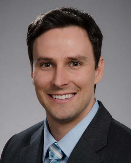 Stephen Bowen合作论文数Department of Radiation Oncology, School of Medicine, University of Washington6
 Antonio C. Wolff合作论文数School of Medicine, Johns Hopkins University6P. Jambunathan合作论文数5P. K. Marimuthu合作论文数5
 Divya Bala Thumaty合作论文数5
 Ajoy Oommen John合作论文数Department of Medical Oncology, Christian Medical College and Hospital5
 Josh Thomas Georgy合作论文数Department of Medical Oncology, Christian Medical College and Hospital5
 Cimino-Mathews Ashley合作论文数Johns Hopkins Univ Hosp5
 Jing Zeng合作论文数Department of Radiation Oncology, University of Washington School of Medicine5
 Vered Stearns合作论文数Breast Center, Weill Cornell Medicine;Johns Hopkins School of Medicine5
 Hubert Vesselle合作论文数Division of Nuclear Medicine, Department of Radiology, School of Medicine, University of Washington;Department of Global Health, University of Washington5
 Paul E. Kinahan合作论文数Department of Bioengineering, College of Engineering and UW Medicine, University of Washington;UW Medical Center, University of Washington5
 B Paul Ravindran合作论文数Department of Radiotherapy, Christian Medical College5r rengan合作论文数4H. Kovilapu合作论文数4Z. Thomas合作论文数4Jill Kessler合作论文数4dustin vandenberg合作论文数4
 Brian G Blair合作论文数4
 Ashish Singh合作论文数4
 Henry Finlay Godson合作论文数Department of Radiation Oncology, Christian Medical College4Michaela J Higgins合作论文数4
 Leonard Wee合作论文数GROW School for Oncology and Developmental Biology, Maastricht University Medical Center+4
 Leslie Cope合作论文数Department of Otolaryngology-Head and Neck Surgery, School of Medicine, Johns Hopkins University4
 Chacko Raju T合作论文数Christian Medical College4
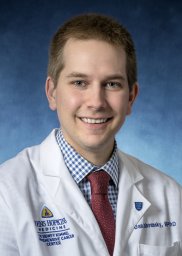 Daniel J. Zabransky合作论文数The Sidney Kimmel Comprehensive Cancer Center, The Johns Hopkins University School of Medicine4Thomas Meera合作论文数4
 Amit Tirkey合作论文数4Toro Patricia Valda合作论文数4Selvamani Backianathan合作论文数4
 Habibi Mehran合作论文数4Hurley Paula J合作论文数4Lange Julie R合作论文数4
 Sunitha Susan Varghese合作论文数4Stacie C. Jeter合作论文数4
 Andre Dekker合作论文数Maastricht University Medical Centre;MAASTRO Clinic;Clinical Data Science Maastricht4




