BACKGROUND:Functional liver imaging has potential to personalize management of Hepatocellular Carcinoma (HCC) by mitigating hepatotoxicity risk. We validated functional liver imaging and dosimetric parameters for risk-stratification in an expanded cohort of patients with HCC. METHODS:We reviewed 109 consecutive patients who underwent Sulfur Colloid (SC)-SPECT/CT scans for radiation therapy (RT) planning and extracted previously reported functional liver imaging metrics. We generated elastic net multivariable Cox models with event-stratified and nested cross-validation folds to predict Overall Survival (OS) and increase in Child-Pugh score ≥ 2 (CP+2). Test-fold patients were risk-stratified, and time-dependent model performance was characterized. ROC analysis generated prognostic cutoffs with confidence intervals to guide functional liver avoidance treatment planning. RESULTS:Cross-validated model concordance was 0.70 (95% CI: 0.67-0.73) for OS and 0.67 (95% CI: 0.63-0.71) for CP+2. Top-ranked OS predictors included tumor volume (HR=1.56, 1.54-1.58), CP-score (HR=1.36, 1.34-1.38), Liver-GTV V20 (HR=1.310, 1.306-1.314), prior liver-directed therapy (HR=0.83, 0.82-0.85), functional liver volume dosimetry (FLV V20) (HR=1.19, 1.14-1.23), and RT-year (HR=0.89, 0.88-0.91). Top-ranked CP+2 predictors were total liver function (TLF) (HR=0.64, 0.63-0.66), Liver-GTV mean dose (HR=1.40, 1.36-1.49), and CP-score (HR=1.19, 1.16-1.23). Test-fold risk groups were defined for each endpoint (log-rank P<0.001). OS model performance stabilized beyond 2 years; CP+2 model stability peaked within 1 year. Optimal strata for 2-yr OS were FLV V20 < 25.8% and Liver-GTV V20 < 25.4%; 1-yr CP+2 strata were TLF < 0.91 and Liver-GTV mean dose < 18.9 Gy. CONCLUSION:Functional liver metrics on SC-SPECT/CT were validated alongside clinical and dosimetric factors within robust outcome models. Testing of personalized RT planning for patients with HCC to preserve liver function is warranted in clinical trials.
Purpose:Although simultaneous integrated boost and protection with proton beam therapy (SIB-PBT) facilitates tumor dose escalation while maintaining organ-at-risk (OAR) dose constraints, clinical outcomes are limited. This study assessed the safety and efficacy of using the SIB-PBT technique in hepatocellular carcinoma (HCC) patients. Methods:We reviewed 47 patients with HCC who underwent SIB-PBT between 2014-2021. The radiation dose ranged from 36-67.5 Gy(RBE) in 15 fractions. SIB-PBT was used for the following reasons: minimize high-dose exposure to organs-at-risk (OARs) (n = 22, 47 %), treat targets with different dose levels (n = 6, 13 %), or both (n = 19, 40 %). Survival, local control, and toxicities were assessed using Kaplan-Meier, Fine-Gray cumulative incidence, and descriptive statistics, respectively. Results:Forty-one patients (87 %) had tumors located ≤2 cm from luminal gastrointestinal (GI) OARs. The median tumor diameter was 9.2 cm (range, 2.0-21.5 cm). The median EQD2 D50%, D95% and D99% of gross tumor volume were 79.8 (range, 51.1-85.9), 66.7 (range, 36.9-84.6) and 50.2 (range, 34.1-83.6) Gy(RBE)10, respectively. Most patients (91 %) received a D0.5 cc of <45 Gy(RBE) to luminal GI OARs. At a median follow-up of 22 months (range, 0.8-77.0 months), the 2-year cumulative incidence of local failure was 12 %. The 2-year progression-free survival and overall survival rates were 12 % (95 % CI 4.7-23.4 %), and 49 % (95 % CI, 33.2-63.2 %), respectively. One patient experienced grade 3 acute nausea/vomiting. No GI bleeding/ulcers or grade 4 + toxicity were observed. CP + 2 occurred in 5 patients. Conclusion:SIB-PBT enables OAR protection along with heterogeneous tumor dose escalation and is a safe and effective treatment for HCC tumors.
8622 Background: Chemotherapy and immune checkpoint inhibitors (chemoICI) for stage IV NSCLC without driver mutations produce variable response patterns and outcomes. Early assessment could establish selective benefit from treatment (de)escalation, consolidation, or adaptation. We evaluated FDG PET imaging and circulating T cell repertoire biomarkers of chemoICI response for clinical decision support. Methods: 35 patients with stage IV NSCLC prospectively enrolled on PET BRIGHT (NCT04151940) from 2019-2023 and received first-line carboplatin-pemetrexed-pembrolizumab. Synchronous FDG PET/CT and peripheral blood draws were performed prior to and after first dose of chemoICI at week 3. Imaging biomarkers included standardized uptake value (SUV) and total lesion glycolysis (TLG) metrics within the primary metabolic tumor volume and across lung lesions. Immunophenotyping included T cell receptor (TCR) sequencing from which TCR diversity metrics were extracted. Spearman correlation of FDG PET and TCR biomarkers with bi-dimensional size reduction after initial chemoICI was estimated. ROC analysis of PET and TCR biomarkers to discriminate chemoICI response status was benchmarked against PD-L1 testing, along with Mann-Whitney testing of groupwise differences. Results: The majority tested negative for PD-L1 (58% TPS<1%). Most patients (63%) had partial response (PR) to initial chemoICI versus 33% with stable disease (SD) and 4% with progressive disease (PD). FDG PET primary tumor peak SUV decreased by a median of 15% [-82%, 63%] at week 3, while TLG decreased by 36% [-55%, 83%]. Higher baseline peak SUV and TLG, along with greater decline after first dose, were correlated with post-chemoICI size reduction (R 0.43-0.64, p=0.001-0.036). PR patients had higher baseline peak SUV (12 vs 6 g/mL, p=0.014) and greater week 3 decline in TLG (39% vs 7%, p=0.032) relative to SD/PD patients. Across lung lesions, PR-patient maximum SUV decreased while SD/PD-patient maximum SUV increased at week 3 (20% vs -19%, p=0.010). FDG PET biomarkers were stronger discriminators of chemoICI response status (AUC 0.75-0.82, p=0.009-0.046) than PD-L1 (AUC 0.60, p=0.42). Higher baseline blood TCR Simpson clonality was positively correlated with size reduction (R 0.70, p=0.004), whereas TCR Pielou evenness was negatively correlated (R -0.68, p=0.006). Higher baseline TCR clonality / lower TCR evenness discriminated chemoICI response status (AUC 0.80-0.84, p=0.028-0.049). TCR diversity changes were uncorrelated with response. Conclusions: FDG PET imaging prior to and after first dose chemoICI, along with baseline circulating T cell repertoire biomarkers, predict future response. Further investigation of these patient- and lesion-level biomarkers to guide personalized (de)escalation, consolidation, or adaptation of regimens for stage IV NSCLC is warranted. Clinical trial information: NCT04151940 .
523 Background: Data on the safety and efficacy of external beam radiation (EBRT) after Yttrium-90 (Y-90) radioembolization for hepatocellular carcinoma (HCC) is limited. We report our experience using EBRT to treat HCC patients who were previously treated with Y-90. Methods: We analyzed 31 HCC patients who received EBRT following Y-90 treatment. Eighteen were treated with photon therapy (40-50 Gy in 5 fractions), and thirteen with proton therapy (42-67.5 Gy in 15 fractions). Twenty-four patients underwent Y-90 segmentectomies, while seven received Y-90 lobar treatment. The median administered Y-90 activity was 44.4 mCi (range 8.3-114.4). Results: Patients had received 1 (n=14), 2 (n=7), or ≥3 (n=10) prior Y-90 treatments. EBRT was administered for various reasons: poor Y-90 response in two patients, technical limitations in 25 patients, and other reasons in four patients. Ten patients had previously received Y-90 within the EBRT area, thirteen outside the EBRT area, and eight in both regions. The median tumor size was 3.8 cm (range 1.6-19.4). Twenty-seven patients had Child-Pugh (CP)-A score, three had CP-B, and one had CP-C baseline liver function. With a median follow-up of 21 months, the 2-year progression-free survival, and overall survival rates were 28%, and 43%, respectively. The 2-year cumulative incidence of local failure was 7%. CP+2 progression was observed in five patients (16%): three had Y-90 delivered outside the EBRT area and two within the EBRT area. Three patients had possible RILD-related deaths. Grade 3+ biliary complications occurred in three patients (10%): one biloma, one liver abscess, and one biliary stricture which resulted in a possible treatment-related death. All three patients had received at least two prior Y-90 treatment overlapping with the area treated with EBRT and had tumors located near the porta hepatis. Conclusions: EBRT for HCC patients previously treated with Y-90 is feasible and offers excellent local control. Hepatic function and biliary toxicities are potential complications and should be weighed against the clinical benefits.
Objective.Vital rules learned from fluorodeoxyglucose positron emission tomography (FDG-PET) radiomics of tumor subregional response can provide clinical decision support for precise treatment adaptation. We combined a rule-based machine learning (ML) model (RuleFit) with a heuristic algorithm (gray wolf optimizer, GWO) for mid-chemoradiation FDG-PET response prediction in patients with locally advanced non-small cell lung cancer.Approach.Tumors subregions were identified using K-means clustering. GWO+RuleFit consists of three main parts: (i) a random forest is constructed based on conventional features or radiomic features extracted from tumor regions or subregions in FDG-PET images, from which the initial rules are generated; (ii) GWO is used for iterative rule selection; (iii) the selected rules are fit to a linear model to make predictions about the target variable. Two target variables were considered: a binary response measure (ΔSUVmean ⩾ 20% decline) for classification and a continuous response measure (ΔSUVmean) for regression. GWO+RuleFit was benchmarked against common ML algorithms and RuleFit, with leave-one-out cross-validated performance evaluated by the area under the receiver operating characteristic curve (AUC) in classification and root-mean-square error (RMSE) in regression.Main results.GWO+RuleFit selected 15 rules from the radiomic feature dataset of 23 patients. For treatment response classification, GWO+RuleFit attained numerically better cross-validated performance than RuleFit across tumor regions and sets of features (AUC: 0.58-0.86 vs. 0.52-0.78,p= 0.170-0.925). GWO+Rulefit also had the best or second-best performance numerically compared to all other algorithms for all conditions. For treatment response regression prediction, GWO+RuleFit (RMSE: 0.162-0.192) performed better numerically for low-dimensional models (p= 0.097-0.614) and significantly better for high-dimensional models across all tumor regions except one (RMSE: 0.189-0.219,p< 0.004).Significance. The GWO+RuleFit selected rules were interpretable, highlighting distinct radiomic phenotypes that modulated treatment response. GWO+Rulefit achieved parsimonious models while maintaining utility for treatment response prediction, which can aid clinical decisions for patient risk stratification, treatment selection, and biologically driven adaptation. Clinical trial: NCT02773238.
Aim: A critical need for hepatocellular carcinoma (HCC) management is understanding how the liver recovers following radiotherapy (RT). We hypothesized that functional liver imaging with 99mTc-sulfur colloid (SC) SPECT/CT provides additional information on liver injury and recovery after RT compared to conventional imaging. Methods: The liver function of patients with HCC was assessed using 99mTc-SC SPECT/CT imaging before and after definitive RT. The anatomical liver volume (ALV) was segmented on CT imaging. Liver function was measured as the total liver function (TLF) encompassing 30% of maximum SC uptake. Changes in ALV and TLF were compared to clinical characteristics. Results: Of 31 patients with evaluable post-RT SC SPECT/CT scans (total of 32), 23 had pre-treatment Child-Pugh (CP)-A and 9 had CP-B/C scores. The median follow-up post-RT was 57 days. The median change in ALV was -1.7% with no significant difference between CP-A and CP-B/C patients (P = 0.26) or between short- (32-99 days) and long-term (271-1120 days) follow-up imaging groups (P = 0.28). The median change in TLF post-RT was -24% and was significantly different between short- and long-term groups (-39% vs. 2%, P = 0.001) and between CP-A and CP-B/C patients (-19% vs. -57%, P = 0.002). TLF significantly decreased following treatment at all radiation dose levels, with the decline correlating with the dose (P < 0.001). Conclusion: Functional imaging provides additional information regarding liver injury and recovery following RT that conventional imaging cannot reveal. Patients with CP-A liver status showed less decline following RT and most had liver function near or above pre-treatment levels.
Abstract Introduction: Cancer survival has increased in part due to treatment advances. However, anthracycline chemotherapy increases cardiovascular (CV) morbidity risk, including atherosclerotic CV disease (ASCVD). Among those receiving anthracyclines, it is yet unclear who is at greatest risk of ASCVD, a major cause of CV mortality. Elucidating pathophysiologic processes involved in the development ASCVD could shed light on strategies to identify those at risk. Methods: Analyses were performed on 279 lymphoma and breast cancer survivors enrolled in the PREVENT study [clinical trial WF-98213) through Wake Forest NCI Community Oncology Research Base (NCORP) and Alliance (A221501)]. CV dysfunction was measured via cardiac MRI (cMRI) at baseline (pre-treatment) and 6-months post-diagnosis to ascertain aortic distensibility and wall thickness in the descending aorta. Biomarkers were measured at baseline and 6-months and were log-transformed to base 2. Multiple linear regression was used to determine associations between biomarker and outcome at 6 months, adjusted for baseline biomarker and cMRI outcome, age, race, sex, body mass index, and smoking history. Results: The mean age (SD) of cancer survivors 49 (12) years; 92% were women. The mean aortic distensibility and mean wall thickness at baseline was 0.002 (0.0014) and 2.99 (0.41), respectively. Table 1 shows the associations between biomarkers and aortic distensibility and wall thickness of the descending aorta. After adjustment for confounders, Arginine, CRP, MPO, and ornithine were associated with 6-month aortic distensibility, and the HDL and SDMA were associated with aortic wall thickness. Conclusions: The findings of this study suggest that biomarkers in oxidative stress and inflammatory pathways may be involved in pathophysiology of ASCVD among cancer survivors receiving anthracyclines. These results require further study in larger cohorts to better define mechanistic pathways involved. Association of Biomarkers with Atherosclerosis-Related Cardiovascular Dysfunction Citation Format: Kerryn W. Reding, Alexi Vasbinder, Nathaniel O'Connell, Biniyam Demissei, Warren Szewczyk, Richard Cheng, Amy Ladd, Alexander Lucas, Juergen Meyer, Stephen Bowen, Fadi Salloum, Ralph D'Agostino, Glenn Lesser, Kathryn Weaver, Bonnie Ky, W. H. Wilson Tang, W. Gregory Hundley. Biomarkers associated with atherosclerosis-related cardiovascular dysfunction in cancer survivors treated with anthracyclines [abstract]. In: Proceedings of the American Association for Cancer Research Annual Meeting 2024; Part 1 (Regular Abstracts); 2024 Apr 5-10; San Diego, CA. Philadelphia (PA): AACR; Cancer Res 2024;84(6_Suppl):Abstract nr 6296.
Background: In the context of a phase II trial of risk-adaptive chemoradiation, we evaluated whether tumor metabolic response could serve as a correlate of treatment sensitivity and toxicity. Methods: Forty-five patients with AJCCv7 stage IIB-IIIB NSCLC enrolled on the FLARE-RT phase II trial (NCT02773238). [18F]fluorodeoxyglucose (FDG) PET-CT images were acquired prior to treatment and after 24 Gy during week 3. Patients with unfavorable on-treatment tumor response received concomitant boosts to 74 Gy total over 30 fractions rather than standard 60 Gy. Metabolic tumor volume and mean standardized uptake value (SUVmean) were calculated semi-automatically. Risk factors of pulmonary toxicity included concurrent chemotherapy regimen, adjuvant anti-PDL1 immunotherapy, and lung dosimetry. Incidence of CTCAE v4 grade 2+ pneumonitis was analyzed using the Fine-Gray method with competing risks of metastasis or death. Peripheral germline DNA microarray sequencing measured pre-defined candidate genes from distinct pathways: 96 DNA repair, 53 immunology, 38 oncology, 27 lung biology.Results: Twenty-four patients received proton therapy, 23 received ICI, 26 received carboplatin-paclitaxel, and 17 pneumonitis events were observed. Pneumonitis risk was significantly higher for patients with COPD (HR 3.78 [1.48, 9.60], p = 0.005), those treated with immunotherapy (HR 2.82 [1.03, 7.71], p = 0.043) but not with carboplatin-paclitaxel (HR 1.98 [0.71, 5.54], p = 0.19). Pneumonitis rates were similar among selected patients receiving 74 Gy radiation vs 60 Gy (p = 0.33), proton therapy vs photon (p = 0.60), or with higher lung dosimetric V20 (p = 0.30). Patients in the upper quartile decrease in SUVmean (>39.7%) were at greater risk for pneumonitis (HR 4.00 [1.54, 10.44], p = 0.005) and remained significant in multivariable analysis (HR 3.34 [1.23, 9.10], p = 0.018). Germline DNA gene alterations in immunology pathways were most frequently associated with pneumonitis.Conclusion: Tumor metabolic response as measured by mean SUV is associated with increased pneu-monitis risk in a clinical trial cohort of NSCLC patients independent of treatment factors. This may be par-tially attributed to patient-specific differences in immunogenicity.& COPY; 2023 Elsevier B.V. All rights reserved. Radiotherapy and Oncology 185 (2023) 1-8
Purpose: Artificial intelligence (AI)-based autocontouring in radiation oncology has potential benefits such as standardization and time savings. However, commercial AI solutions require careful evaluation before clinical integration. We developed a multidimensional evaluation method to test pretrained AI-based automated contouring solutions across a network of clinics. Methods and Materials: Curated data included 121 patient planning computed tomography (CT) scans with a total of 859 clinically approved contours used for treatment from 4 clinics. Regions of interest (ROIs) were generated with 3 commercial AI-based automated contouring software solutions (AI1, AI2, AI3) spanning the following disease sites: brain, head and neck (H&N), thorax, abdomen, and pelvis. Quantitative agreement between AI-generated and clinical contours was measured by Dice similarity coefficient (DSC) and Hausdorff distance (HD). Qualitative assessment was performed by multiple experts scoring blinded AI-contours using a Likert scale. Results: AI1, AI2, and AI3 contours had high quantitative agreement in 27.8%, 32.8%, and 34.1% of cases (DSC >0.9), performing well in pelvis (median DSC = 0.86/0.88/0.91) and thorax (median DSC = 0.91/0.89/0.91). All 3 solutions had low quantitative agreement in 7.4%, 8.8%, and 6.1% of cases (DSC <0.5), performing worse in brain (median DSC = 0.65/0.78/0.75) and H&N (median DSC = 0.76/ 0.80/0.81). Qualitatively, AI1 and AI2 contours were acceptable (rated 1-2) with at most minor edits in 70.7% and 74.6% of ROIs (2906 ratings), higher for abdomen (AI1: 79.2%) and thorax (AI2: 90.2%), and lower for H&N (29.0/35.6%). An end-user survey showed strong user preference for full automation and mixed preferences for accuracy versus total number of structures generated. Conclusions: Our evaluation method provided a comprehensive analysis of both quantitative and qualitative measures of commercially available pretrained AI autocontouring algorithms. The evaluation framework served as a roadmap for clinical integration that aligned with user workflow preference. (c) 2023 American Society for Radiation Oncology. Published by Elsevier Inc. All rights reserved.
AbstractPurposeThe purpose of this study is to investigate the use of a deep learning architecture for automated treatment planning for proton pencil beam scanning (PBS).MethodsA 3‐dimensional (3D) U‐Net model has been implemented in a commercial treatment planning system (TPS) that uses contoured regions of interest (ROI) binary masks as model inputs with a predicted dose distribution as the model output. Predicted dose distributions were converted to deliverable PBS treatment plans using a voxel‐wise robust dose mimicking optimization algorithm. This model was leveraged to generate machine learning (ML) optimized plans for patients receiving proton PBS irradiation of the chest wall. Model training was carried out on a retrospective set of 48 previously‐treated chest wall patient treatment plans. Model evaluation was carried out by generating ML‐optimized plans on a hold‐out set of 12 contoured chest wall patient CT datasets from previously treated patients. Clinical goal criteria and gamma analysis were used to compare dose distributions of the ML‐optimized plans against the clinically approved plans across the test patients.ResultsStatistical analysis of mean clinical goal criteria indicates that compared to the clinical plans, the ML optimization workflow generated robust plans with similar dose to the heart, lungs, and esophagus while achieving superior dosimetric coverage to the PTV chest wall (clinical mean V95 = 97.6% vs. ML mean V95 = 99.1%, p < 0.001) across the 12 test patients.ConclusionsML‐based automated treatment plan optimization using the 3D U‐Net model can generate treatment plans of similar clinical quality compared to human‐driven optimization.
Multiomics data including imaging radiomics and various types of molecular biomarkers have been increasingly investigated for better diagnosis and therapy in the era of precision oncology. Artificial intelligence (AI) including machine learning (ML) and deep learning (DL) techniques combined with the exponential growth of multiomics data may have great potential to revolutionize cancer subtyping, risk stratification, prognostication, prediction and clinical decision-making. In this article, we first present different categories of multiomics data and their roles in diagnosis and therapy. Second, AI-based data fusion methods and modeling methods as well as different validation schemes are illustrated. Third, the applications and examples of multiomics research in oncology are demonstrated. Finally, the challenges regarding the heterogeneity data set, availability of omics data, and validation of the research are discussed. The transition of multiomics research to real clinics still requires consistent efforts in standardizing omics data collection and analysis, building computational infrastructure for data sharing and storing, developing advanced methods to improve data fusion and interpretability, and ultimately, conducting large-scale prospective clinical trials to fill the gap between study findings and clinical benefits.
Boosting Trees are one of the most successful statistical learning approaches that involve sequentially growing an ensemble of simple regression trees ("weak learners"). This paper proposes a gradient Boosted Trees algorithm for Spatial Data (Boost-S) with covariate information. Boost-S integrates the spatial correlation into the classical framework of eXtreme Gradient Boosting. Each tree is constructed by solving a regularized optimization problem, where the objective function takes into account the underlying spatial correlation and involves two penalty terms on tree complexity. A computationally-efficient greedy heuristic algorithm is proposed to obtain an ensemble of trees. The proposed Boost-S is applied to the spatially-correlated FDG-PET (fluorodeoxyglucose-positron emission tomography) imaging data collected from clinical trials of cancer chemoradiotherapy. Our numerical investigations successfully demonstrate the advantages of the proposed Boost-S over existing approaches for this particular application.
ObjectiveCOVID-19 primarily causes pneumonitis but can also cause myocarditis. Injury may be due to a generalised inflammatory immune process or by direct viral infection. Using 18F-fluorodeoxyglucose positron emission tomography/computed tomography (18F-FDG-PET/CT) and cardiac magnetic resonance (CMR) imaging we correlated the metabolic activity/injury between the reticuloendothelial system (bone marrow [BM] and spleen) and myocardial/pulmonary tissue.Methods18F-FDG-PET/CT (n=29, fasted n=27) and CMR (n=23) were performed on hospitalised patients with acute COVID-19. 18F-FDG PET/CT standardised uptake values (SUV) were measured in the spleen, spinal BM, myocardial and pulmonary tissue. Cardiac target-to-background ratio (TBR) was calculated by indexing to blood-pool SUV. Myocarditis was assessed using the sensitive 2018 Lake Louise criteria (LLC), and viral load (by cycle threshold).Results13 patients had myocarditis on CMR (57%), 8 (30%) visually on 18F-FDG-PET/CT. There was no statistical difference comparing LLC positive and negative patients for BM (4.21±0.30, 4.98±0.56, P=0.23), spleen (4.40±0.40, 5.15±0.08, P=0.38) and lung (4.08±0.72, 4.16±0.91, P=0.94) SUV. Lung SUV was significantly associated with BM (r=0.61, P<0.001) and spleen (r=0.48, P<0.05) SUV. Cardiac TBR, T1 and T2 mapping showed no significant association with BM and spleen SUV (P>0.05 for all). Cycle threshold did not correlate with either cardiac TBR and T1 or T2 (p>0.05 for all).ConclusionReticuloendothelial system activation strongly correlated with lung activity, suggesting pulmonary injury is part of a systemic inflammatory process. Cardiac inflammation was not associated with either spleen, BM or viral load, suggesting injury is multifactorial.
Medical imaging provides quantitative and spatial information to evaluate treatment response in the management of patients with non-small cell lung cancer (NSCLC). High throughput extraction of radiomic features on these images can potentially phenotype tumors non-invasively and support risk stratification based on survival outcome prediction. The prognostic value of radiomics from different imaging modalities and time points prior to and during chemoradiation therapy of NSCLC, relative to conventional imaging biomarker or delta radiomics models, remains uncharacterized. We investigated the utility of multitask learning of multi-time point radiomic features, as opposed to single-task learning, for improving survival outcome prediction relative to conventional clinical imaging feature model benchmarks. Survival outcomes were prospectively collected for 45 patients with unresectable NSCLC enrolled on the FLARE-RT phase II trial of risk-adaptive chemoradiation and optional consolidation PD-L1 checkpoint blockade (NCT02773238). FDG-PET, CT, and perfusion SPECT imaging pretreatment and week 3 mid-treatment was performed and 110 IBSI-compliant pyradiomics shape-/intensity-/texture-based features from the metabolic tumor volume were extracted. Outcome modeling consisted of a fused Laplacian sparse group LASSO with component-wise gradient boosting survival regression in a multitask learning framework. Testing performance under stratified 10-fold cross-validation was evaluated for multitask learning radiomics of different imaging modalities and time points. Multitask learning models were benchmarked against conventional clinical imaging and delta radiomics models and evaluated with the concordance index (c-index) and index of prediction accuracy (IPA). FDG-PET radiomics had higher prognostic value for overall survival in test folds (c-index 0.71 [0.67, 0.75]) than CT radiomics (c-index 0.64 [0.60, 0.71]) or perfusion SPECT radiomics (c-index 0.60 [0.57, 0.63]). Multitask learning of pre-/mid-treatment FDG-PET radiomics (c-index 0.71 [0.67, 0.75]) outperformed benchmark clinical imaging (c-index 0.65 [0.59, 0.71]) and FDG-PET delta radiomics (c-index 0.52 [0.48, 0.58]) models. Similarly, the IPA for multitask learning FDG-PET radiomics (30%) was higher than clinical imaging (26%) and delta radiomics (15%) models. Radiomics models performed consistently under different voxel resampling conditions. Multitask learning radiomics for outcome modeling provides a clinical decision support platform that leverages longitudinal imaging information. This framework can reveal the relative importance of different imaging modalities and time points when designing risk-adaptive cancer treatment strategies.
Background Acute COVID‐19–related myocardial, pulmonary, and vascular pathology and how these relate to each other remain unclear. To our knowledge, no studies have used complementary imaging techniques, including molecular imaging, to elucidate this. We used multimodality imaging and biochemical sampling in vivo to identify the pathobiology of acute COVID‐19. Specifically, we investigated the presence of myocardial inflammation and its association with coronary artery disease, systemic vasculitis, and pneumonitis. Methods and Results Consecutive patients presenting with acute COVID‐19 were prospectively recruited during hospital admission in this cross‐sectional study. Imaging involved computed tomography coronary angiography (identified coronary disease), cardiac 2‐deoxy‐2‐[fluorine‐18]fluoro‐D‐glucose positron emission tomography/computed tomography (identified vascular, cardiac, and pulmonary inflammatory cell infiltration), and cardiac magnetic resonance (identified myocardial disease) alongside biomarker sampling. Of 33 patients (median age 51 years, 94% men), 24 (73%) had respiratory symptoms, with the remainder having nonspecific viral symptoms. A total of 9 patients (35%, n=9/25) had cardiac magnetic resonance–defined myocarditis. Of these patients, 53% (n=5/8) had myocardial inflammatory cell infiltration. A total of 2 patients (5%) had elevated troponin levels. Cardiac troponin concentrations were not significantly higher in patients with and without myocarditis (8.4 ng/L [interquartile range, IQR: 4.0–55.3] versus 3.5 ng/L [IQR: 2.5–5.5]; P=0.07) or myocardial cell infiltration (4.4 ng/L [IQR: 3.4–8.3] versus 3.5 ng/L [IQR: 2.8–7.2]; P=0.89). No patients had obstructive coronary artery disease or vasculitis. Pulmonary inflammation and consolidation (percentage of total lung volume) was 17% (IQR: 5%–31%) and 11% (IQR: 7%–18%), respectively. Neither were associated with the presence of myocarditis. Conclusions Myocarditis was present in a third patients with acute COVID‐19, and the majority had inflammatory cell infiltration. Pneumonitis was ubiquitous, but this inflammation was not associated with myocarditis. The mechanism of cardiac pathology is nonischemic and not attributable to a vasculitic process. Registration URL: https://www.isrctn.com; Unique identifier: ISRCTN12154994.
Purpose: We sought to examine the prognostic value of fluorodeoxyglucose-positron emission tomography (PET) imaging during chemoradiation for unresectable non-small cell lung cancer for survival and hypothesized that tumor PET response is correlated with peripheral T-cell function. Methods and Materials: Forty-five patients with American Joint Committee on Cancer version 7 stage IIB-IIIB non-small cell lung cancer enrolled in a phase II trial and received platinum-doublet chemotherapy concurrent with 6 weeks of radiation (NCT02773238). Fluorodeoxyglucose-PET was performed before treatment start and after 24 Gy of radiation (week 3). PET response status was prospectively defined by multifactorial radiologic interpretation. PET responders received 60 Gy in 30 fractions, while nonresponders received concomitant boosts to 74 Gy in 30 fractions. Peripheral blood was drawn synchronously with PET imaging, from which germline DNA sequencing, T-cell receptor sequencing, and plasma cytokine analysis were performed. Results: Median follow-up was 18.8 months, 1-year overall survival (OS) 82%, 1-year progression-free survival 53%, and 1-year locoregional control 88%. Higher midtreatment PET total lesion glycolysis was detrimental to OS (1 year 87% vs 63%, P <.001), progression-free survival (1 year 60% vs 26%, P =.044), and locoregional control (1 year 94% vs 65%, P =.012), even after adjustment for clinical/treatment factors. Twenty-nine of 45 patients (64%) were classified as PET responders based on a priori definition. Higher tumor programmed death-ligand 1 expression was correlated with response on PET (P =.017). Higher T-cell receptor richness and clone distribution slope were associated with improved OS (P =.018-0.035); clone distribution slope was correlated with PET response (P =.031). Conclusions: Midchemoradiation PET imaging is prognostic for survival; PET response may be linked to tumor and peripheral T-cell biomarkers.
Background Patients undergoing chemoradiation and immune checkpoint inhibitor (ICI) therapy for locally advanced non-small cell lung cancer (NSCLC) experience pulmonary toxicity at higher rates than historical reports. Identifying biomarkers beyond conventional clinical factors and radiation dosimetry is especially relevant in the modern cancer immunotherapy era. We investigated the role of novel functional lung radiomics, relative to functional lung dosimetry and clinical characteristics, for pneumonitis risk stratification in locally advanced NSCLC. Methods Patients with locally advanced NSCLC were prospectively enrolled on the FLARE-RT trial (NCT02773238). All received concurrent chemoradiation using functional lung avoidance planning, while approximately half received consolidation durvalumab ICI. Within tumour-subtracted lung regions, 110 radiomics features (size, shape, intensity, texture) were extracted on pre-treatment [ 99m Tc]MAA SPECT/CT perfusion images using fixed-bin-width discretization. The performance of functional lung radiomics for pneumonitis (CTCAE v4 grade 2 or higher) risk stratification was benchmarked against previously reported lung dosimetric parameters and clinical risk factors. Multivariate least absolute shrinkage and selection operator Cox models of time-varying pneumonitis risk were constructed, and prediction performance was evaluated using optimism-adjusted concordance index (c-index) with 95% confidence interval reporting throughout. Results Thirty-nine patients were included in the study and pneumonitis occurred in 16/39 (41%) patients. Among clinical characteristics and anatomic/functional lung dosimetry variables, only the presence of baseline chronic obstructive pulmonary disease (COPD) was significantly associated with the development of pneumonitis (HR 4.59 [1.69–12.49]) and served as the primary prediction benchmark model (c-index 0.69 [0.59–0.80]). Discrimination of time-varying pneumonitis risk was numerically higher when combining COPD with perfused lung radiomics size (c-index 0.77 [0.65–0.88]) or shape feature classes (c-index 0.79 [0.66–0.91]) but did not reach statistical significance compared to benchmark models (p > 0.26). COPD was associated with perfused lung radiomics size features, including patients with larger lung volumes (AUC 0.75 [0.59–0.91]). Perfused lung radiomic texture features were correlated with lung volume (adj R 2 = 0.84–1.00), representing surrogates rather than independent predictors of pneumonitis risk. Conclusions In patients undergoing chemoradiation with functional lung avoidance therapy and optional consolidative immune checkpoint inhibitor therapy for locally advanced NSCLC, the strongest predictor of pneumonitis was the presence of baseline chronic obstructive pulmonary disease. Results from this novel functional lung radiomics exploratory study can inform future validation studies to refine pneumonitis risk models following combinations of radiation and immunotherapy. Our results support functional lung radiomics as surrogates of COPD for non-invasive monitoring during and after treatment. Further study of clinical, dosimetric, and radiomic feature combinations for radiation and immune-mediated pneumonitis risk stratification in a larger patient population is warranted.
 Jing Zeng合作论文数Department of Radiation Oncology, University of Washington School of Medicine28
 Smith Apisarnthanarax合作论文数Experimental Radiation Oncology, M.D. Anderson Cancer Center26
 Paul E. Kinahan合作论文数Department of Bioengineering, College of Engineering and UW Medicine, University of Washington;UW Medical Center, University of Washington26
 Matthew Nyflot合作论文数School of Medicine, University of Washington23
 Hubert Vesselle合作论文数Division of Nuclear Medicine, Department of Radiology, School of Medicine, University of Washington;Department of Global Health, University of Washington22
 Ramesh Rengan合作论文数19
 Daniel S Hippe合作论文数Clinical Research Division, Fred Hutchinson Cancer Center15
 Tobias Chapman合作论文数13Saini Jatinder合作论文数13
 Robert Miyaoka合作论文数Department of Radiology, University of Washington;Imaging Research Laboratory, The University of Washington13Tony Wong合作论文数12
 George A. Sandison合作论文数School of Health Sciences, Purdue University11
 Robert Jeraj合作论文数Department of Medical Physics, School of Medicine and Public Health, University of Wisconsin-Madison9
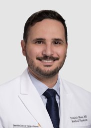 D. Maes合作论文数SCCA Proton Therapy Ctr9Shilpen Patel合作论文数8
 Shouyi Wang合作论文数Department of Industrial and Manufacturing Systems Engineering, University of Texas at Arlington8
 W. Art Chaovalitwongse合作论文数Department of Industrial and Systems Engineering, University of Washington Seattle;Department of Radiology, University of Washington Seattle8
 Hannah Mary Thomas T合作论文数Department of Bioengineering, Christian Medical College7
 Charles Bloch合作论文数Proton Radiation Center, Indiana University7
 Stephanie K Schaub合作论文数Department of Radiation Oncology, University of Washington6
 Juergen Meyer合作论文数Department of Radiation Oncology, University of Wuerzburg6Howard Lee合作论文数6
 Sasidharan Balukrishna合作论文数Department of Radiation Oncology Unit 1, Christian Medical College6
 Soren M. Bentzen合作论文数Division of Biostatistics and Bioinformatics, Department of Epidemiology and Public Health, University of Maryland School of Medicine;Greenebaum Comprehensive Cancer Center, University of Maryland School of Medicine6
 Clemens Grassberger合作论文数Radiation Oncology Division, Fred Hutch Cancer Center;Division of Radiation Oncology, University of Washington School of Medicine;Department of Radiation Oncology, Massachusetts General Hospital, Harvard Medical School6
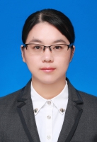 Chunyan Duan (段春艳)合作论文数School of Mechanical Engineering, Tongji University5Rajesh Regmi合作论文数4Rosanna Yeung合作论文数4Santana-Davila Rafael合作论文数4
 George Laramore合作论文数4Liao Jay J合作论文数4
 Kevin Horn合作论文数Huntsman Cancer Institute, University of Utah4
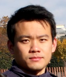 Xiao Liu合作论文数4A. Mcgarry Houghton合作论文数4
 Phawis Thammasorn合作论文数4Lisa J. Forrest合作论文数4
 Paul D Lampe (Paul Lampe)合作论文数Fred Hutchinson Cancer Research Center4Meghan W. Macomber合作论文数3Michael A Deveau合作论文数3Peter Zaki合作论文数3Jie Han合作论文数Dept Ind Mfg & Syst Engn, Univ Texas Arlington3
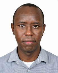 Edward Chege Nganga合作论文数Dept Radiol, Aga Khan Univ Hosp3Samuel Gitau合作论文数Aga Khan Univ Hosp3
 Khalid Makhdomi合作论文数Aga Khan Univ Hosp3Evan D H Gates合作论文数3Angela Fung合作论文数3
 Larry Pierce合作论文数University of Washington3
 William Yuh合作论文数Department of Radiology, School of Medicine, University of Washington3Lee Sylvia M合作论文数3




