Dimethyl-fumarate (DMF) demonstrated efficacy and safety in relapsing–remitting multiple sclerosis (MS) in randomized clinical trials.
Background. The pathological significance and the diagnostic usefulness of intrathecal lc and X free light chain (FLC) synthesis in Multiple Sclerosis (MS) are debated. Methods. Paired cerebrospinal fluid (CSF) and serum specimens from 70 relapsing remitting MS (RAMS), 40 with and 30 without CSF restricted IgG Oligoclonal Band (IgGOB), and 37 from healthy controls (HC) were analyzed. IgG, IgM, kappa FLC and lambda FLC concentrations and indexes were evaluated. All RAMS performed MRI to estimate white and grey matter (WM) pathology. Results. In HC, no intrathecal kappa or lambda FLC synthesis was found, and kappa FLC and lambda FLC Indexes were reciprocally correlated (r = 0.67, p < 0.001). In RAMS, intrathecal kappa FLC or lambda FLC synthesis was demonstrated in respectively 66% and 43% of the cases, the Q(kappa/lambda) ratio was significantly higher compared to HC (17.0 +/- 31.3 vs 0.79 +/- 0.20, p < 0.001) and the correlation between kappa FLC Index and lambda FLC Index was weak (r:0.38, p < 0.05). Intrathecal IgG synthesis was associated with kappa FLC Index (IgG Index: r2 = 0.53, ji = 0.73, p < 0.001; IgG(Loc) : r(2) = 0.37, beta = 0.61, p < 0.001; IgG(IF):r(2) = 0.69, beta = 0.83, p < 0.001), but not with lambda FLC Index, while intrathecal IgM synthesis correlated with XFLC Index (IgM Index: r = 0.41, p < 0.001; IgM(Loc): r = 0.34, p < 0.005; r = 0.45, p < 0.001), but not with kappa FLC Index. 26% of RAMS patients without CSF-restricted IgGOB had increased kappa FLCLoc. Finally, no associations were observed between any CSF and MRI parameters. Conclusions. The demonstration of intrathecal kappa FLC synthesis may further improve the diagnostic usefulness of CSF examination in RAMS. The marked in- creased in Q(kappa/lambda) further suggests a deregulated B-cell activation in MS pathology.
B lymphocytes are thought to play a relevant role in multiple sclerosis (MS) pathology. The in vivo analysis of intrathecally produced B cell-related cytokines may help to clarify the mechanisms of B cell recruitment and immunoglobulin production within the central nervous system (CNS) in MS.
B-cells are thought to play a relevant role in multiple sclerosis (MS) pathology. BAFF (B cell activating factor of the TNF family) is a B-cell survival factor constitutively produced inside the CNS by astrocytes. We studied the intrathecal synthesis of BAFF in MS at clinical onset.Paired serum and cerebrospinal fluid (CSF) specimens from 40 clinically isolated syndromes (CIS) suggestive of MS or early relapse-onset MS (eRRMS) and from 18 healthy controls (HC) were analysed. Patients were classified based on the detection of oligoclonal IgG bands in the CSF (IgGOB+ and IgGOB-). BAFF was detected by highly sensitive ELISA and its ratio (CSF-BAFF/serum-BAFF, Q(BAFF)) and Index (Q(BAFF)/Q(Alb), BAFF-Index) were calculated. IgGOB+ presented lower CSF concentrations of BAFF compared to both HC and IgGOB- (p < 0.05). BAFF Index was significantly lower in IgGOB+ compared to both HC and IgGOB- (p < 0.01). A significant inverse correlation between Q(IgG) and Q(BAFF) (r: -0.4, p < 0.05) and between BAFF index and IgGIF (r: -0.4, p < 0.05) or IgG Index (r: -0.4, p = 0.05) was found in IgGOB+. The decreased CSF levels of BAFF in IgGOB+ at clinical onset suggest the absorption of this factor by intrathecally recruited B cells since the early disease phases. (C) 2016 Elsevier B.V. All rights reserved.
 Paolo Gallo合作论文数24
 Francesca Rinaldi合作论文数Multiple Sclerosis Ctr Veneto Reg CeSMuV, Univ Hosp Padua20M. Puthenparampil合作论文数Univ Padua19Paola Perini合作论文数19L. Federle合作论文数14
 Martino F. Pengo合作论文数6
 Margoni Monica合作论文数6
 Poggiali Davide合作论文数The Multiple Sclerosis Centre of the Veneto Region of Italy (CeSMuV), Padova, Italy.6D. Seppi合作论文数5M. Saiani合作论文数5
 Maria Pia Sormani合作论文数Dipartimento di scienze della salute, University of Genoa5Elisabetta Toffanin合作论文数5Susanna Ruggero合作论文数5V. Poretto合作论文数4S. Franciotta合作论文数3S. Alessio合作论文数3Erica Stropparo合作论文数3
 Massimiliano Calabrese合作论文数Neurol Sect, Univ Verona3Sofia Zywicki合作论文数3Francesca Bovis合作论文数3
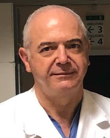 Adolfo Favaretto合作论文数Azienda ULSS 2 Marca Trevigiana3R Cavarretta合作论文数2S. Pravato合作论文数2M. R. Rottoli合作论文数2V. Torri Clerici合作论文数2R. Bergamaschi合作论文数2
 Andrea Lazzarotto合作论文数2A. Riccardi合作论文数2M Ermani合作论文数2Chiara Cazzola合作论文数2Marta Ponzano合作论文数Dept Hlth Sci DISSAL, Univ Genoa2Antonio Zito合作论文数Dept Brain & Behav Sci, Univ Pavia2
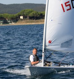 Simone Rossi合作论文数Department of Medicine, Surgery and Neuroscience, University of Siena2Giulia Mallucci合作论文数2
 Antonio Bertolotto合作论文数Neuroscience Institute Cavalieri Ottolenghi2
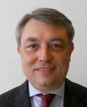 Mario Plebani合作论文数School of Medicine, University of Padova;Department of Laboratory Medicine, University-Hospital of Padova2Marco Rovaris合作论文数2
 Angelo Ghezzi合作论文数Ospedale di Gallarate;Lavoratore autonomo2
 Pietro Annovazzi合作论文数2
 Mauro Zaffaroni合作论文数2Matteo Atzori合作论文数2Paola Cavalla合作论文数Department of Neuroscience;University of Turin2A Fasoli合作论文数1G. Pantano合作论文数1R. Balgera合作论文数1V. Mantero合作论文数1C. Zanetta合作论文数1Lucia Moiola合作论文数1S. La Gioia合作论文数1




