Contrary to numerous assumptions, sonographic examination of the gastrointestinal tract has been performed since the beginning of the 1970 s, making this one of the early clinical indications of diagnostic ultrasound imaging.
Purpose: To investigate if strain elastography could differentiate between metastatic and non-metastatic mesenteric lymph nodes ex-vivo.Materials and Methods: 90 mesenteric lymph nodes were examined shortly after resection from 25 patients including 17 patients with colorectal cancer and 8 patients with Crohn's disease. Ultrasound-based strain elastography was performed with a linear probe. Tissue hardness in lymph nodes was assessed using visual scales and measuring the strain ratio. B-mode characteristics were also recorded. Pathological diagnosis with grading of fibrosis served as the reference standard.Results: 20 lymph nodes were metastatic and 70 lymph nodes were non-metastatic. The strain ratios of metastatic and non-metastatic lymph nodes were significantly different (1.83 vs. 1.42, p = 0.021). The VAS scale (0-100) for tissue hardness gave higher mean values for metastatic than non-metastatic nodes, but the difference was not significant (65.5 vs. 55.0, p = 0.055). There was no difference between lymph nodes in Crohn's and non-metastatic cancer specimens. The metastatic lymph nodes were significantly more fibrotic than the non-metastatic lymph nodes by the ordinal fibrosis score (0-3). In an ROC analysis, quantitative strain imaging was not superior to the measurement of the short-axis diameter of lymph nodes in differentiating metastatic from non-metastatic mesenteric lymph nodes ex-vivo.Conclusion: Strain elastography is correlated to fibrosis in lymph nodes and a significant difference was observed on a group level using the strain ratio. Due to measurement overlap, individual mesenteric lymph nodes could not be identified accurately as metastatic or not in this ex-vivo model by strain imaging alone.
Ultrasonography (US) is a safe and available real-time, high-resolution imaging method, which during the last decades has been increasingly integrated as a clinical tool in gastroenterology. New US applications have emerged with enforced data software and new technical solutions, including strain evaluation, three-dimensional imaging and use of ultrasound contrast agents. Specific gastroenterologic applications have been developed by combining US with other diagnostic or therapeutic methods, such as endoscopy, manometry, puncture needles, diathermy and stents. US provides detailed structural information about visceral organs without hazard to the patients and can play an important clinical role by reducing the need for invasive procedures. This paper presents different aspects of US in gastroenterology, with a special emphasis on the contribution from Nordic scientists in developing clinical applications.
AimStrain elastography is a method for recording tissue hardness. Strain in different areas may be compared using strain ratio (SR). The aims of this study were to validate a previously proposed SR cut-off value of 1.25 for differentiating adenocarcinomas from adenomas and to compare the performance of endorectal ultrasonography (ERUS), strain elastography and MRI in the same patients.MethodA prospective evaluation of 120 consecutive patients with rectal neoplasia, using a predetermined elastography strain ratio cut-off value, was performed to differentiate adenomas from adenocarcinomas. ERUS and MRI were performed according to standard routine at Haukeland University Hospital, defining T0 as adenomas and T1-T4 as adenocarcinomas. Subsequent histopathology was used as the reference standard.ResultsHistopathological evaluation revealed 21 adenomas and 99 adenocarcinomas. Sensitivity, specificity and accuracy (with 95% CI) were as follows: ERUS: 0.96 (0.90-0.99), 0.62 (0.40-0.80) and 0.90 (0.83-0.94); elastography SR: 0.96 (0.90-0.99), 0.86 (0.66-0.96) and 0.94 (0.88-0.97); and MRI: 0.99 (0.94-1.00), 0.07 (0.00-0.31) and 0.87 (0.80-0.93).ConclusionThis study confirms that the elastography SR assessment accurately differentiates sessile adenomas from adenocarcinomas. SR assessment has a superior ability to differentiate adenomas and adenocarcinomas when compared with ERUS and MRI. MRI examination seems unable to recognize adenomas and should be interpreted with care when early-stage rectal neoplasia is suspected.
AIM:Strain elastography is a novel approach to rectal tumour evaluation. The primary aim of this study was to correlate elastography to pT stages of rectal tumours and to assess the ability of the method to differentiate rectal adenomas (pT0) from early rectal cancer (pT1-2). Secondary aims were to compare elastography with endorectal ultrasonography (ERUS) and to propose a combined strain elastography and ERUS staging algorithm.METHOD:In all, 120 consecutive patients with a suspected rectal tumour were examined in this staging study. Patients receiving surgery without neoadjuvant radiotherapy were included (n = 59). All patients were examined with ERUS and elastography. Treatment decisions were made by multidisciplinary team (MDT) assessment, without considering the strain elastography examination.RESULTS:Histopathology identified 21 adenomas, 13 pT1, 9 pT2, 15 pT3 and one pT4. Mean elastography strain ratios were predictive of T stage (P = 0.01). Differentiation of adenomas from early rectal cancer (pT1-2) had sensitivity, specificity and accuracy of 0.82, 0.86 and 0.84 for elastography and 0.82, 0.62 and 0.72 for ERUS. A combined staging algorithm was developed to identify tumours eligible for local resection. Based on MDT evaluation 32% of tumours later identified as pT0 or pT1 were treated with total mesorectal excision, even though a local excision might have sufficed. Combined ERUS and elastography evaluation would have significantly reduced this number to 9% (P = 0.008).CONCLUSION:Elastography may improve the staging of adenomas and early rectal cancer compared with ERUS alone. Combined ERUS and elastography assessment is likely to further improve the selection of patients for local resection.
OBJECTIVES. The aim of this study was to evaluate the diagnostic potential of strain assessment in solid focal pancreatic lesions using real-time elastography in combination with endoscopic ultrasonography (EUS). MATERIAL AND METHODS. Forty-eight solid focal pancreatic lesions in 39 patients were included prospectively over a 3-year period and studied by EUS with real-time elastography (EUS-RTE). Lesions previously described as cystic by CT were not included. Distribution patterns of tissue strain were assessed using strain ratio (SR) measurements, continuous visual analog scale (VAS), and a visual categorical score (VCS), based on color coding of relative strain. Final diagnosis was based on histopathology, fine-needle aspiration cytology, and/or follow-up for ≥6 months. RESULTS. The 48 lesions included 11 adenocarcinomas, 7 malignant neuroendocrine tumors (NETs), 11 benign or indeterminate NETs, 8 focal pancreatic lesions, 2 microcystic adenomas, and 9 other benign lesions. Malignant lesions had significantly higher median SR (7.05 vs. 1.56) and VAS scores (93.0 vs. 63.5) than benign lesions. A receiver operation characteristic curve analysis showed sensitivity of 67% and specificity of 71%, when using SR = 4.4 as a cut-off for malignancy. The highest SR values were found in two benign microcystic adenomas. CONCLUSIONS. EUS-RTE with SR measurements and VAS evaluation demonstrated a significant strain difference between benign and malignant lesions. However, the variation within the entities was substantial and some benign lesions presented with low strain. Benign lesions were generally characterized by a strain similar to reference tissue, whereas malignant lesions were harder. The recorded strain pattern in individual lesions must be interpreted with caution.
Background: To improve management of patients with Crohn's disease (CD), objective measurements of the degree of local inflammation in the gastrointestinal wall are needed. Increased microvessel density and perfusion are typical features of acute inflammation and can be estimated with contrast-enhanced ultrasound (CEUS). The aim of the study was to investigate whether CEUS can provide prognostic information about patients treated medically for an acute exacerbation of CD.Methods: Fourteen patients with CD who received medical treatment for acute exacerbation with systemic steroids or tumor necrosis factor-alpha inhibitors were prospectively recruited. The patients were examined with clinical scoring, blood tests, and CEUS at time 0, 1, 3, and 12 months after initiation of the treatment. Outcome was treatment efficacy or treatment failure defined as change in medical treatment after 1 month or later. The perfusion analysis was performed with a commercially available software program that analyzes the contrast intensity in a selected area, fits the data to a standardized time-intensity curve, and derives several relative perfusion parameters.Results: Six of the 14 patients had treatment failure during the study period. There was a significant difference between the groups for peak contrast enhancement (P = 0.013), rate of wash-in (P = 0.020) and wash-out (P = 0.008), and the area under the time-intensity curve in the wash-in phase (0.013) at the examination 1 month after the start of treatment.Conclusions: Perfusion analysis of the intestinal wall with CEUS 1 month after starting treatment in patients with CD can provide prognostic information regarding treatment efficacy.
The aim of this study was to determine whether there are differences in absolute blood flow between patients with Crohn's disease with inflammation or fibrosis using contrast-enhanced ultrasound. Eighteen patients with fibrotic disease and 19 patients with inflammation were examined. Video sequences of contrast data were analyzed using a pharmacokinetic model to extract the arterial input and tissue residue functions with a custom software, enabling calculation of the absolute values for mean transit time, blood volume and flow. Feasibility of the examination was 89%. The fibrosis group had lower blood volume (0.9 vs. 3.4 mL per 100 mL tissue; p = 0.001) and flow (22.6 vs. 45.3 mL/min per 100 mL tissue; p = 0.003) compared with the inflammation group. There was no significant difference in mean transit time (3.9 vs. 5.5 s). In conclusion, absolute perfusion measurement in the gastrointestinal wall using contrast-enhanced ultrasound is feasible. There seems to be reduced blood volume and blood flow in patients with fibrotic disease.
Purpose: To investigate whether ultrasoundbased strain imaging can discriminate between colorectal adenocarcinomas and stenotic Crohn's lesions in newly resected surgical specimens.Materials and Methods: Resected surgical specimens from 27 patients electively operated for colorectal tumors or stenotic lesions from Crohn's disease were prospectively examined with ultrasonography using a Hitachi HV 900 US scanner with real- time elastography (RTE). Three different methods were applied to assess tissue strain: A four-level categorical visual classification, a continuous visual analog scale (VAS, 0 - 100) and a strain ratio (SR) measurement between the lesion and surrounding reference tissue. The imaged sections were marked and subsequently examined by a pathologist. Results from RTE were evaluated according to diagnosis, degree of fibrosis, inflammatory parameters, tumor stage and grade.Results: 16 sections from Crohn's lesions, 18 sections from adenocarcinomas and 4 sections from adenomas were examined. Both adenocarcinomas and Crohn's lesions were found to be harder than the surrounding tissue, but they could not be discriminated from each other by any of the strain imaging evaluation methods. All adenocarcinomas had significantly higher strain ratios than adenomas. The categorical classification differentiated poorly between Crohn's lesions, adenocarcinomas and adenomas. Categorical evaluation and VAS score showed fair interobserver agreement. SR measurements provided semi- quantitative strain data and added improved information about elasticity properties, despite substantial intra- observer variation.Conclusion: Sonoelastography with SR measurements and visual evaluation of strain differences could not differentiate stenotic Crohn's lesions from adenocarcinomas in resected bowel specimens. A small number of adenomas were found to be significantly softer than adenocarcinomas using the same evaluation methods. The tumor stage or grade did not have a significant impact on the elastography results.
Abstract Purpose: To describe the gastrointestinal (GI) wall thickness and the thickness of individual wall layers in healthy subjects using ultrasound and to determine whether demographic factors, the ultrasound transducer frequency, or a fasting state influences these measurements. Materials and Methods: After overnight fasting, the GI wall thickness and wall layers were measured in several regions with transabdominal, high-frequency ultrasound. 122 healthy subjects aged 23 – 79 were included. All measurements were performed with both 8 and 12-MHz transducers except for the rectum measurement (4 MHz). 23 patients were given a 300 Kcal test meal and re-examined after 30 minutes. Results: Wall thickness measurements of the GI tract with transabdominal ultrasonography are dependent on transducer frequency (p < 0.001), weight (p < 0.001) and age (p < 0.018). The thickness of individual wall layers in the ileum and the sigmoid colon was found to be dependent on both age (p = 0.007) and weight (p < 0.001). The mean wall thickness from the jejunum to the sigmoid colon ranged from 0.9 to 1.2 mm with standard deviations (SD) of 0.3 mm or less. The mean (SD) was 2.9 (0.8) mm in the gastric antrum, 1.6 (0.3) mm in the duodenum, and 2.1 (0.5) mm in the rectum. The gastric antrum was thinner and the ileum and sigmoid colon were thicker after the test meal (p < 0.05). Conclusion: GI wall thickness depends on weight and age. Provided adequate measurement, an abnormal GI wall should be suspected if the thickness exceeds 2 mm except for in the gastric antrum, duodenum and rectum. Reference values for wall thickness can be used regardless of fasting state or probe frequency except for in the gastric antrum.
To investigate if adenocarcinomas, adenomas and Crohn stenoses in resected bowel specimens could be separated by semi-quantification of tissue strain. We investigated surgical specimens from 26 patients, 9 patients with Crohn's disease with stenotic symptoms, 17 patients with colorectal tumours shortly after resection. 39 sections of tumours or stenotic lesions were examined by free-hand ultrasonography. We used Hitachi HV 900 US scanner with Real-Time Elastography and L54M (9-13 MHz) linear probe. Tissue strain in Crohn lesions and tumours was semi-quantified by: strain ratio (SR), visual analog scale (VAS, 0-100) and a four-point categorical visual scale (1-4). The pathological diagnosis was used as reference standard. 15 sections of Crohn stenoses, 17 sections of adenocarcinomas and 4 sections of adenomas were examined. Adenocarcinomas and Crohn stenoses were harder than surrounding tissue, but the entities could not be separated by semi-quantification of strain images: mean strain ratio (2.34 vs. 2.55, p = 0.657), VAS (75,2 vs. 81.6, p = 0.092) and categorical visual scale (2.65 vs. 2.55, p = 0.504). Four adenomas had significantly lower strain compared to adenocarcinomas: mean strain ratio (1.29 vs. 2.34, p < 0.001), VAS (59.8 vs. 75.2, p < 0.001) and by categorical visual scale: (1.50 vs. 2.65, p < 0.001). Free-hand sonoelastography cannot separate Crohn stenoses from adenocarcinomas in resected bowel specimens using strain ratio or visual evaluation. A small number of adenomas were significantly different from adenocarcinomas using the same evaluation methods.
To study the influence of demographic factors on gastrointestinal (GI) wall thickness and individual wall layers in the GI wall using transabdominal ultrasound. The wall thickness in the antrum, jejunum, ileum, colon and rectum and individual wall layers in the ileum and sigmoid colon were examined with transabdominal ultrasound in 122 healthy volunteers of age range 23-79 years. The examination was performed after approximately 8 hours fasting using 8 and 12 MHz linear ultrasound transducers. Gender, age, weight, height and tobacco use of the subjects were recorded. Multiple linear regression applying a mixed model was used for analysis. In the ileum, the mucosa and submucosa were 0.4 ± 0.1mm (mean±SD) each and the proper muscle was 0.4 ± 02mm. In the sigmoid colon, the corresponding values were 0.3 ± 0.1mm and 0.5 ± 0.1mm, respectively. There was an overall increase in GI wall thickness with decreasing transducer frequency (p < 0.001) and with increasing weight (p = 0.046) in the duodenum and sigmoid colon. Increasing age was related to increased thickness of the GI wall in the ileum and sigmoid colon (p < 0.001), and in the same locations, weight (p < 0.001) and age (p = 0.002) were associated with thickening of all wall layers. Increasing weight is associated with wall thickening in the duodenum and sigmoid colon and increasing age with wall thickening in both the ileum and sigmoid colon. This seems to be due to a general thickening of all wall layers.
Purpose: Real-time elastography (RTE) is an ultrasound-based method for the visualization of relative strain distribution in soft tissues. Strain ratio is a semi-quantitative measurement of strain differences between two user-defined areas in an elastogram. The aim of this study was to evaluate the impact of the size and location of a reference area when measuring the strain ratio of focal lesions in a tissue-mimicking phantom and in normal liver tissue. We also investigated whether the strain ratio was affected by changing the scanner parameter: elasticity dynamic range (E-dyn).Materials and Methods: Two investigators individually collected data by scanning 4 spherical inclusions with different elasticity in a phantom in which the elastic modulus was known in both the lesions and the background. Subsequently, a liver scan was performed in-vivo using the same scanning protocol. Five different setups with changes in reference area position or size were tested. All eight levels of the scanner setting E-dyn were recorded for each setup and the strain ratio was measured in 3 different representative elastograms for each recording situation.Results: The four inclusions had significantly different mean strain ratio levels (p < 0.01) when compared to the surrounding material. Changing the position of the reference area to a deeper position influenced the strain ratio measurements significantly for all phantom lesions and in the liver. Changing the size of the reference area, while keeping the center depth unchanged, did not influence the mean strain ratio levels significantly. The strain ratio was independent of the E-dyn parameter setting. The intra- and interobserver reliability was high when measuring the strain ratio with a free-hand technique.Conclusion: Strain ratio provides reproducible measurements of inclusions representing different elastic contrasts using a free-hand technique in vitro. Changes in the distance of the reference areas to the ultrasound probe, representing the stress source, seem to have a significant impact on strain ratio measurements.
Aim Real-time elastography visualizes tissue compliance using an ultrasound platform. Elastography has been used, particularly in the breast, to characterize indeterminate lesions on B-mode imaging as either benign or malignant. The primary aim of this study was to assess the feasibility of routine endorectal elastography to evaluate rectal neoplasia. The secondary aim was to correlate elastography data with histopathological end-points. Method Sixty-nine patients referred to the outpatient clinic of the Department of Colorectal Surgery at Haukeland University Hospital for the evaluation of rectal tumours were included in this prospective cohort study. All patients underwent digital rectal examination, rigid rectoscopy with biopsy, endorectal ultrasonography and endorectal elastography. In each case a strain ratio was calculated, comparing the tumour tissue with adjacent reference tissue that appeared normal on ultrasound scanning. Results Histopathologically there were 23 adenomas and 45 adenocarcinomas. One patient died before surgical treatment. Adequate elastography images were obtained in 66/69 (96%) patients. Optimal discrimination of malignant and benign lesions was obtained using a strain ratio cut-off value of 1.25 (sensitivity, 0.93; specificity, 0.96; and accuracy, 0.94). Conclusion Endorectal elastography can be performed as an integral part of the clinical evaluation of rectal tumours and has good patient compliance. The method is a promising modality for the discrimination between adenocarcinoma and adenoma of the rectum.
To investigate if sonoelastography could differentiate between metastatic and non-metastatic lymph nodes and to semi-quantify fibrosis in lymph nodes. We investigated 98 mesenterial lymph nodes from 26 patients representing 18 patients with colorectal cancer and 8 Crohn patients shortly after resection. We used Hitachi HV 900 US scanner with Real-Time Elastography and L54M (9-13 MHz) linear probe. Tissue hardness in lymph nodes was semi-quantified by: strain ratio, visual analog scale (VAS) and a four-point categorical visual scale. The pathological diagnosis was used as reference standard and a four-point semi-quantification of fibrosis was done for each lymph node. 22 lymph nodes were metastatic and 76 lymph nodes were non-metastatic (30 from Crohn specimens and 46 from cancer specimens). Metastatic and non-metastatic lymph nodes were different for median strain ratio (1.94 vs. 1.36, p = 0.037) and fibrosis score (1.55 vs. 0.03, p < 0.001). Semi-quantification of strain by mean strain ratio (2.05 vs. 1.43, p = 0.073), VAS (64.0 vs. 55.1, p = 0.111) and categorical classification (2.05 vs. 1.64, p = 0.154) were not significantly different for metastatic and non-metastatic lymph nodes (p>0.05). The metastatic lymph nodes were more fibrotic than the non-metastatic lymph nodes (1.55 vs. 0.03, p < 0.001). Semi-quantification of strain images of mesenterial lymph nodes in resected tissue could separate metastatic from non-metastatic lymph nodes using median SR. Metastatic lymph nodes were significantly more fibrotic than the non-metastatic lymph nodes.
BackgroundFunctional chest pain is commonly reproduced by bag distension in the esophageal body. It is unknown whether such pain is primarily associated with mechanical stress and strain (force-deformation) or with changes in mucosal perfusion.MethodsFourteen patients (6M, 8F, average age 55.9 years) underwent ramp bag distension before and after injection of 20 mg butylscopolamine bromide (BS) using a novel bag catheter incorporating endosonography and laser Doppler perfusion monitoring. Healthy subjects served as controls. Mucosal perfusion was evaluated and stress and strain were computed and related to the sensation.Key ResultsThe symptom score increased with bag volume (P < 0.001). Volume as a function of pressure was higher in patients than in controls (P < 0.001), both before and during BS. The stress-strain relationship was exponential and indicated a stiffer esophageal wall in patients especially before BS (P < 0.01). The stress-strain curves indicate increased muscle tone in the functional chest pain patients. The perfusion decreased with increasing symptom score from visual analog scale 1-7 during BS. The decrease was on average 18.9% in patients and 19.7% in controls (P = ns). Multiple regression analysis from distensions during BS showed that the discomfort/pain sensations depended on stress and strain (P < 0.001) and with stress as the largest contributor. Perfusion did not contribute.Conclusions & InferencesPain evoked by bag distension in patients with functional chest pain is stress-dependent rather than dependent on mucosal perfusion. Furthermore, the esophagus of the patients was characterized by more pronounced muscle tone during the distensions.
 Odd Helge Gilja合作论文数Department of Clinical Medicine, Faculty of Medicine, University of Bergen;National Centre for Ultrasound in Gastroenterology, Haukeland University Hospital62
 Trygve Hausken合作论文数Department of Medicine, Haukeland University Hospital;University of Bergen52
 Lars Birger Nesje合作论文数29B. Børkje合作论文数16
 Hans Gregersen合作论文数California Medical Innovations Institute16E Schrumpf合作论文数13A Berstad合作论文数13Knut Matre合作论文数12
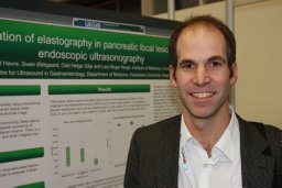 Roald Flesland Havre合作论文数University of Bergen, Haukeland University Hospital12
 Arnold Berstad合作论文数Institute of Medicine, University of Bergen;Lovisenberg Diakonale Hospital11
 Ole Didrik Laerum合作论文数The DCCC Brain Tumor Center10
 Sabine Leh合作论文数Institute of Medicine, University of Bergen10
 Paul Fockens合作论文数Waikato Hospital, Health New Zealand;Amsterdam UMC;Vrije Universiteit Amsterdam;Faculty of Medicine, University of Amsterdam;PROCOLO - Center for Colon Examination9D Martens合作论文数8S.-O. Molin合作论文数8
 Thomas Rösch合作论文数Zentrum für Radiologie und Endoskopie, Universitätsklinikum Hamburg-Eppendorf;Klinik und Poliklinik für Interdisziplinäre Endoskopie, Universitätsklinikum Hamburg-Eppendorf8K Vetvik合作论文数7
 Gunnar Baatrup合作论文数Department of Surgery, Haukeland University Hospital7
 Kim Nylund合作论文数Institute of Medicine, University of Bergen7DW SKAGEN合作论文数7
 Dag Arne Lihaug Hoff合作论文数7J. E. R. Waage合作论文数6Fe Silverstein合作论文数5
 Amitabh Chak合作论文数Department of Medicine, School of Medicine, Case Western Reserve University;Digestive Health Research Institute, School of Medicine, Case Western Reserve University5Michael B Kimmey合作论文数5Michael B. Kimmey合作论文数5
 Roy W. Martin合作论文数5Giancarlo Caletti合作论文数the Department of Gastroenterology, University of Bologna5WR Lees合作论文数4
 Peter Vilmann合作论文数Department of Clinical Medicine, University of Copenhagen;Dept. of Surgery, Gastrointestinal Unit, Herlev Hospital;Department of Endoscopy, Copenhagen University Herlev Hospital4Thomas J. Savides合作论文数Indianapolis4J. G. Hatlebakk合作论文数4Thomas Roesch合作论文数4
 Kenneth J. Chang合作论文数Division of Gastroenterology and Executive, School of Medicine, University of California4
 Robert Bjerknes合作论文数The Gade Institute, University of Bergen4TL TIO合作论文数4
 Jan Gunnar Hatlebakk合作论文数Department of Clinical Medicine, Faculty of Medicine, University of Bergen4
 Jacques Deviere合作论文数Université Libre de Bruxelles;Department of Gastroenterology, Erasme University Hospital4A Hatfield合作论文数3N Frank合作论文数3
 Josef Menzel合作论文数3
 Stephen McClave合作论文数Department of Gastroenterology, School of Medicine, University of Louisville3Andreas Heimdal合作论文数University Hospital of Trondheim, Norwegian University of Science and Technology3Nils Thune合作论文数3Ch Nattermann合作论文数3Arne Skarstein合作论文数3




