Diabetic foot ulcers (DFUs) are the leading cause of amputations in people with diabetes, mainly due to poor wound healing. This study evaluated DFU trial designs and analysed observational data from the two prospective Diabetic Foot Consortium (DFC) studies to estimate longitudinal healing and amputation rates, assess the effects of wound surface area and duration on healing, and inform sample size considerations for future trials. We analysed data from Open Wound Master Protocol (MP, n = 419) and c-Myc Biomarker (n = 140). The primary outcome was complete wound healing. Time-to-event analysis estimated healing and amputation rates, with amputations classified as non-healed. Logistic regression assessed the prediction of healing based on baseline wound characteristics, with model performance evaluated using AUC (area under the curve). Sample size calculations were performed to achieve 80% power. In the MP study, healing rates were 26% by week 12% and 49% by week 32; amputation rates were 4% and 10%, respectively. The c-Myc study showed 38% healing and 2.5% amputation by week 12. Wound area and duration significantly predicted healing (AUC ≥ 0.70 by 24 weeks). For smaller treatment effects, predicted healing rates varied by up to 40% between small and large wounds, impacting sample size. Contemporary DFU trial designs and healing times remained largely unchanged over two decades. While shorter trials (e.g., 12 weeks) theoretically require smaller sample sizes due to reduced outcome variance, they risk failing to capture the full treatment effect. Trial design must account for baseline wound characteristics, which should inform eligibility criteria and follow-up.
Chronic, non-healing wounds are sustained by Staphylococcus aureus biofilms, yet how biofilm reprograms the immune cells tasked with resolving injury has remained unclear. We show that biofilm-derived soluble cues, not bacterial contact, generate a previously undescribed biofilm-associated macrophage (BAM) subset marked by high Macrophage receptor with collagenous structure (MARCO) and suppressed MERTK, which recognizes apoptotic cells but fails to engulf them. Using isogenic S. aureus USA300 variants of graded biofilm capacity, only high biofilm-conditioned medium elicited this state in human macrophages; MARCO neutralization restored corpse clearance. Mechanistically, biofilm factors drove phospho-C/EBPβ into the nucleus to repress MERTK, severing sensing from engulfment. Single-cell and Xenium spatial transcriptomics, with cytometry by time of flight (CyTOF) and PhenoCycler, localized BAMs to biofilm-proximal niches as a hybrid CD64+CD163+ state outside M1/M2 categories, populating 60%-80% of infected chronic wounds. In vivo, myeloid-restricted MARCO overexpression by tissue nanotransfection recapitulated impaired efferocytosis and persistent inflammation, nominating MARCOhiMERTKlo macrophages as a druggable checkpoint in biofilm-associated disease.
Barrier function of human skin maintains hydration by preventing excessive transepidermal water loss (TEWL), blocks the entry of pathogens and allergens, regulates thermal and chemical exchange with the environment, and sustains immune equilibrium. A wound arises when this barrier is breached, resulting in a loss of structural integrity and barrier function. An invisible wound represents a focal loss of human skin barrier function without any visible cut, ulcer, or defect. Despite appearing intact, the skin in these regions is functionally compromised. Current U.S. Food and Drug Administration (FDA) criteria define complete wound closure as full re-epithelialization without drainage or dressing needs, confirmed during two assessments at least 14 days apart. Wounds may satisfy this structural requirement for wound closure yet fail to restore barrier function. Recent studies demonstrate that wounds closed meeting FDA closure criteria but exhibiting elevated TEWL at the wound site (i.e., invisible wound) are more likely to recur, highlighting the clinical importance of achieving functional wound closure. Invisible wounds occur across the lifespan and arise from a wide range of everyday and high-risk exposures. They can result from age-related skin thinning, sunburn, and the routine use of cosmetic chemicals on vulnerable or sensitized skin. They may also develop after minor trauma that leaves no laceration, from mechanical stress imposed by prosthetic use, and from barotrauma or other subclinical mechanical forces commonly encountered by warfighters. Although outwardly undetectable, these silent disruptions weaken the skin's protective barrier, undermine systemic health, and contribute to chronic morbidity.
Wound healing emerges from a tightly orchestrated bioelectric landscape shaped by ion gradients, membrane potentials, and redox dynamics physical cues that direct cell migration, immune activation, and epithelial organization long before biochemical gradients take form. Recent advances reveal that electrical signals constitute a master regulatory layer: Transient receptor potential (TRP)-channel mediated ion flux governs early wound polarity; endogenous transepithelial potential collapse triggers rapid electric fields that guide keratinocyte and fibroblast migration; and connexin-dependent gap-junction coupling coordinates tissue-level responses across multicellular sheets. Electroceutical strategies exploit these principles by recalibrating electrical and electrochemical environments rather than targeting single molecules. This shift enables simultaneous modulation of ion-channel gating, cytoskeletal dynamics, growth-factor signaling, and immunometabolic programs reshaping whole-tissue behavior in ways unattainable with classical pharmacology. Key breakthroughs demonstrate that controlled electrical stimulation can reprogram human macrophages toward reparative phenotypes, enhance keratinocyte electrotaxis even under diabetic conditions, accelerate fibroblast-driven matrix assembly, and amplify endothelial angiogenic responses. Microbial communities respond in the opposite direction. Biofilms, long considered antibiotic-impervious, depend on exquisitely tuned membrane potential, proton motive force, and redox stratification for cohesion and persistence. Low-intensity electrical cues disrupt this energetics, collapsing efflux pump function, silencing quorum systems, loosening EPS architecture, and destabilizing metabolic heterogeneity effects impossible to escape through single gene mutation. Overall, these discoveries frame electroceuticals as system-level disruptors of microbial order and restorers of host coordination. With the emergence of AI-enabled, closed-loop bioelectronic dressings capable of sensing and responding to wound physiology in real time, electricity is poised to become a foundational operating principle for next-generation regenerative and anti-infective therapy.
The COVID-19 pandemic highlighted critical limitations in conventional pulse oximetry, especially in diverse populations. This study evaluates the performance of noninvasive devices for assessing tissue oxygenation in a cohort of 20 healthy individuals, with a focus on device sensitivity, physiological and demographic variables. Tissue oxygenation was measured using devices: spatial frequency domain imaging (SFDI), transcutaneous oxygen measurement (TCOM), wearable photoplethysmography (WD), and pulse oximetry (PO) during baseline, ischemia, and reperfusion phases. Comparative analyses were performed across devices, sex and Fitzpatrick skin types. Among the four devices, SFDI uniquely detected significant differences in tissue oxygen saturation (StO2), highlighting its sensitivity to tissue heterogeneity. PO and WD data showed moderate correlation (r = 0.44-0.59); SFDI and TCOM demonstrated fair correlation (r = 0.23-0.36). Although sex-based differences were minimal, the SFDI revealed significantly different recovery metrics. Notably, SFDI provided deeper insights into hemoglobin dynamics (HbO2, dHb, HbP1, HbP2), which was not captured by point-measurement devices. Results demonstrated that SFDI offers superior spatial/spectral resolution for mapping tissue oxygenation, particularly in detecting skin-type variations. However, device-specific limitations such as motion artifacts and melanin interference necessitate further optimization. This work supports the development of more inclusive and accurate non-invasive monitoring tools for clinical use.Trial registration: ClinicalTrials.gov ID NCT05784103.
Diabetic wounds are complicated by underlying peripheral vasculopathy. Reliance on vascular endothelial growth factor (VEGF) therapy to improve perfusion makes logical sense, yet clinical study outcomes on rescuing diabetic wound vascularization have yielded disappointing results. Our previous work has identified that low endothelial phospholipase Cy2 (PLCy2) expression hinders the therapeutic effect of VEGF on the diabetic ischemic limb. In this work, guided by single- cell RNA sequencing of human wound edge, we test the efficacy of gene-targeted therapeutic demethylation intending to improve VEGF-mediated neovascularization. PLCy2 expression was diminished in all five identified diabetic wound-edge endothelial subclusters encompassing arterial, venous, and capillary cells. Such low expression was associated with hyper- methylated PLCy2 promoter. PLCy2 promoter was also hyper- methylated at murine diabetic ischemic wound edge. To specifically demethylate endothelial PLCy2 promoter during VEGF therapy, a CRISPR-dCas9-based demethylation cocktail was delivered to the ischemic wound edge using tissue nanotransfection (TNT) technology. Demethylation-based upregulation of PLCy2 during VEGF therapy improved wound tissue blood flow with an increased abundance of von Willebrand factor (vWF)+/PLCy2+ vascular tissue elements by activating p44/ p42-mitogen-activated protein kinase (MAPK) / hypoxiainducible factor [HIF]-1a pathway. Taken together, TNT- based delivery of plasmids to demethylate the PLCy2 gene promoter activity led to significant improvements in VEGF therapy for cutaneous diabetic wounds, resulting in better perfusion and accelerated wound closure.
Diabetic wounds are complicated by underlying peripheral vasculopathy. Reliance on vascular endothelial growth factor (VEGF) therapy to improve perfusion makes logical sense, yet clinical study outcomes on rescuing diabetic wound vascularization have yielded disappointing results. Our previous work has identified that low endothelial phospholipase Cγ2 (PLCγ2) expression hinders the therapeutic effect of VEGF on the diabetic ischemic limb. In this work, guided by single-cell RNA sequencing of human wound edge, we test the efficacy of gene-targeted therapeutic demethylation intending to improve VEGF-mediated neovascularization. PLCγ2 expression was diminished in all five identified diabetic wound-edge endothelial subclusters encompassing arterial, venous, and capillary cells. Such low expression was associated with hypermethylated PLCγ2 promoter. PLCγ2 promoter was also hypermethylated at murine diabetic ischemic wound edge. To specifically demethylate endothelial PLCγ2 promoter during VEGF therapy, a CRISPR-dCas9-based demethylation cocktail was delivered to the ischemic wound edge using tissue nanotransfection (TNT) technology. Demethylation-based upregulation of PLCγ2 during VEGF therapy improved wound tissue blood flow with an increased abundance of von Willebrand factor (vWF)+/PLCγ2+ vascular tissue elements by activating p44/p42-mitogen-activated protein kinase (MAPK) → hypoxia-inducible factor [HIF]-1α pathway. Taken together, TNT-based delivery of plasmids to demethylate the PLCγ2 gene promoter activity led to significant improvements in VEGF therapy for cutaneous diabetic wounds, resulting in better perfusion and accelerated wound closure.
Coronavirus with intact infectivity attached to PPE surfaces pose significant threat to the spread of COVID-19. We tested the hypothesis that an electroceutical fabric, generating weak potential difference of 0.5V, disrupts the infectivity of coronavirus upon contact by destabilizing the electrokinetic properties of the virion. Respiratory coronavirus particles (105) were placed in direct contact with the fabric for 1 or 5 minutes. Viral particles (2.5-4x10⁴) were recovered from the fabric. Following one minute of contact, zeta potential of the coronavirus was significantly lowered indicating destabilization of its electrokinetic properties. Size-distribution plot showed appearance of aggregation of the virus. Testing of the cytopathic effects of the virus showed eradication of infectivity as quantitatively assessed by PI-calcein and MTT cell viability tests. This work provides the rationale to consider the studied electroceutical fabric, or other materials with comparable property, as material of choice for the development of PPE in the fight against COVID-19.
Recent advancements in cell and tissue biology have fundamentally changed our understanding of cellular behavior, revealing that both stem and nonstem cells exhibit remarkable plasticity and adaptability. This discovery has paved the way for revolutionary medical drug therapies that leverage cell and tissue reprogramming to repair or regenerate damaged tissues, offering new hope for conditions that were once considered irreversible. Tissue reprogramming involves the activation of specific molecular pathways to convert the function of residual tissue to compensate for the loss of tissue function to aging, trauma, or disease processes. By targeting these pathways, emerging drugs can promote regenerative processes, enabling the restoration of tissue function lost due to aging, injury, or disease. These therapies have shown promising results in preclinical studies addressing a wide range of diseases. Unlike traditional treatments, which focus primarily on managing symptoms, tissue reprogramming therapies offer a dynamic approach that can fundamentally alter cellular states, leading to functional recovery. This review explores the current state of cell and tissue reprogramming, highlighting its potential applications in regenerative medicine and the challenges that must be addressed for successful clinical translation. As our understanding of cellular plasticity continues to evolve, these innovative therapies stand at the forefront of a new era in medicine, with the potential to transform treatment paradigms and significantly improve patient outcomes across a wide range of conditions. Significance Statement Breakthrough technologies have transformed our understanding of cell and tissue biology, uncovering that cells and tissues possess remarkable adaptability and fluidity in their roles. This revelation has opened up exciting possibilities in regenerative medicine, where emerging drug therapies aim to harness and reprogram cells to repair or regenerate damaged tissues. An emerging class of medical drugs will activate the body’s natural regenerative abilities, offering the potential to restore tissue function lost due to aging, injury, or disease.
Collagen, the most abundant structural protein in the human body, plays a vital role in wound healing, tissue repair, and skin integrity. Collagen-based products-ranging from wound dressings, skin substitutes, dental and orthopedic scaffolds, to topical cosmetics and oral supplements-have proliferated rapidly across healthcare and consumer markets. Medical applications leverage collagen's biocompatibility, biodegradability, and scaffold-forming properties to manage chronic wounds, burns, and bone defects, while emerging technologies such as recombinant collagen and phage-integrated dressings target future innovations. Topical collagen improves skin hydration but is unlikely to replace endogenous collagen; injectable fillers offer temporary cosmetic enhancement with some potential risks. Oral collagen supplements, although marketed for skin, joint, and hair health, primarily serve as incomplete proteins and require cautious interpretation, as rigorous clinical evidence supporting transformative outcomes remains limited. Specific formulations such as undenatured type II collagen show promise for inflammatory joint conditions by promoting immune tolerance. In wound care, collagen-based scaffolds enhance healing by supporting fibroblast proliferation, reducing inflammation, and modulating moisture balance, while novel crosslinked matrices and living skin equivalents push regenerative medicine boundaries. Not all collagen-based products are the same. As the global collagen market surges toward $18.7 billion by 2030, users must distinguish between marketing claims and evidence-based benefits of specific preparations. Proper product selection should be guided by clinical context, molecular source (animal, marine, recombinant), and intended use with awareness of underlying scientific evidence critical to therapeutic success. Continued innovation, rigorous validation, mechanism of action studies and rigorous clinical testing are essential to fully realize collagen's therapeutic potential across medicine and wellness.
OBJECTIVE:The National Institute of Diabetes and Digestive and Kidney Diseases Diabetic Foot Consortium tested the hypothesis that compromised restoration of the skin barrier function of closed diabetic foot ulcers (DFUs), as measured by high transepidermal water loss (TEWL), is associated with an increased risk of DFU recurrence. RESEARCH DESIGN AND METHODS:This was a multicenter noninterventional study measuring TEWL in 418 adult participants with diabetes and a recently healed DFU. TEWL was measured at the center of the closed wound and at an anatomically similar reference area on the contralateral foot within 2 weeks of wound closure (visit 1); measurements were repeated at a wound closure confirmation visit 2 weeks later (visit 2). Participants were observed for up to 16 weeks to assess for wound recurrence. Participant self-reported and clinician assessments of DFU wound recurrence were recorded. RESULTS:DFU recurrence by week 16 occurred in 21.5% of participants. Mean TEWL at the center of the healed DFU at visit 1 was higher for those with recurrence compared with those without (P = 0.006). Among participants with high TEWL (>30.05 g · m-2 · h-1), 35% reported wound recurrence by 16 weeks versus 17% of those with low TEWL. The odds ratio for recurrence for participants with high TEWL was 2.66 (P < 0.001). Self-reported wound recurrence was highly concordant with clinician assessment of wound recurrence. CONCLUSIONS:Compromised wound healing mechanisms culminating in wound closure associated with defective skin barrier function is associated with increased risk of DFU recurrence. Measurement of TEWL has value as a predictor of functional wound healing and could affect clinical practice, leading to better outcomes.
Objective: Wound closure is skin reepithelialization confirmed at two consecutive clinical visits 2 weeks apart. Our objective was to identify participant characteristics, including transepidermal water loss (TEWL), associated with complete wound closure of diabetic foot ulcers (DFUs) and reopening of a DFU within 2 weeks after initial closure in the National Institute of Diabetes and Digestive and Kidney Diseases-sponsored Diabetic Foot Consortium TEWL prospective observational cohort study of wound recurrence. At the site of wound closure, TEWL measures restoration of skin barrier function and functional wound closure. Approach: Four hundred and sixty-six eligible participants had physician-assessed wound closure at baseline. Of which, 418 (90%) had confirmed closure 2 weeks later and remained in the study, whereas 29 had their DFU reopen 2 weeks later and were not eligible for follow-up (i.e., screen failures). We compared baseline characteristics of 418 enrolled and 29 screen fail individuals using Wilcoxon rank sum and Fisher's exact tests p value for continuous and categorical outcomes, respectively. Results: There were no statistically significant differences in demographics, including age, sex, race, education, employment status, social support, or dressing change requirements between groups. The failure to maintain closure group had longer median duration of index DFU before initial closure (25.8 vs. 14 weeks, p = 0.003), higher frequency of prior total contact casting use (37% vs. 14%, p = 0.003), and a higher median initial TEWL measurement at the healed ulcer midpoint (27.1 vs. 21.0 g/m2/h, p = 0.006). Innovation: TEWL measurement at the site of wound closure can assess functional capacity of the skin in conjunction with current standards of wound closure end point in DFU and has significant potential to add quantitative measurement to assist in clinical assessment of healing wounds. Conclusion: Individuals with DFU who did not maintain wound closure had higher TEWL values at baseline, longer DFU wound duration, and more prior off-loading use. These findings are clinically relevant as a higher TEWL measurement demonstrates incomplete functional wound closure, supporting the use of TEWL to identify a healed DFU.
Uncontrolled fibrosis via excess deposition of extracellular matrix (ECM) is a hallmark of hypertrophic scars and keloids. A decellularized ECM biomaterial from porcine small intestinal submucosa (SIS; Biodesign or BioD, Cook Biotech, Inc.) is widely used in clinical applications for tissue repair. The objective of the current study was to test the effects of BioD scaffolds, as compared with collagen constructs, on normal human skin (nFB) and keloid fibroblasts (kFBs). Immortalized human dermal fibroblasts (hFBs) and human keloid fibroblasts (hKFs) were utilized for all experiments. Cells were cultured either on BioD membranes or on collagen gel (used as a control). To investigate pro-fibrotic signaling pathways, real-time quantitative PCR (qPCR), ELISA, and gene knockdown studies were conducted on cultured cells. ECM gene expression array revealed that BioD significantly attenuated (p < 0.05) the expression of thrombospondin-1 and fibronectin-1, two drivers of fibrosis in nFB as well as kFB. BioD-repressed thrombospondin-1 and fibronectin-1 gene expression manifested as significant downregulation (n = 5–6; p < 0.05) of both proteins in nFB and kFB. The levels of latent transforming-growth factor (LAP-TGFβ-1) were markedly reduced (n = 5; p < 0.05) in both nFB and kFB cultured on BioD, but not the other constructs. Knockdown of FN1 using siRNA significantly attenuated (n = 5, p < 0.05) pro-fibrotic responses, including expression of Col1A1 and the levels of LAP-TGFβ-1 in nFB, suggesting that downregulation of FN1 by BioD is one of the primary underlying mechanisms of attenuated pro-fibrotic responses in keloid fibroblasts. This study reports that a decellularized ECM scaffold may significantly attenuate pro-fibrotic responses in both normal and keloid fibroblasts via TSP1 and FN1-dependent mechanisms.
Significance: Skin lipids are essential for various skin functions including maintaining barrier integrity, regulating hydration, and providing protection against microbes and inflammatory irritants. Along with skin health, the role of lipids in the etiology of macroangiopathic diseases, such as atherosclerosis of arteries, is well recognized. Recent Advances: In diabetes, lipid dysregulation is evident and may contribute to the diverse complications of the disease. Diabetic vasculopathy primarily reflects the dysfunction and deterioration of existing blood vessels, as their preservation is key in preventing the progression of vascular disease and reducing the need for compensatory angiogenesis. In the peripheral diabetic skin of the limbs, diabetic vasculopathy runs alongside peripheral neuropathy. Although a causative link between the two is plausible, direct evidence in support of such claim is scanty. Critical Issues: Diabetic skin is known to be compromised in many ways, including weakened barrier functionality and diabetes-induced alterations in the extracellular matrix, likely stemming from chronic inflammation, which may directly affect vascular integrity and nerve health. Both, in the compromised skin and within wounds, microbial pathogens and their enzymes may metabolize host lipids, driving inflammatory reactions and exacerbating the pathogenesis of diabetic vasculopathy and related neuropathy. Future Directions: This review focuses on lipid mediators such as sphingolipids, resolvins, oxidized low-density lipoproteins and their specific downstream signaling pathways to obtain a comprehensive understanding of diabetic complications relevant to wound healing. Through lipid-based strategies, this review hopes to inspire the development and utilization of individualized, precision-based approaches to manage diabetic vasculopathy and neuropathy.
 Chandan K. Sen合作论文数McGowan Institute for Regenerative Medicine, University of Pittsburgh School of Medicine;Mary Ann Liebert, Inc.;Bioengineering MDPI;JoVE;Genes and Nutrition;DNA & Cell Biology376
 Savita Khanna合作论文数Davis Heart and Lung Research Institute, The Ohio State University Medical Center214
 Subhadip Ghatak合作论文数Centre for Liver Research, Department of Gastroenterology, Institute of Post Graduate Medical Education and Research, Kolkata 700020. West Bengal, India63
 Gayle Gordillo合作论文数512 Davis Heart and Lung Research Institute, Ohio State University62Amitava Das合作论文数55
 Lester Packer合作论文数Department of Pharmacology and Pharmaceutical Sciences, School of Pharmacy, Health Science Center, University of Southern California;University of California, Berkeley50
 Surya Gnyawali合作论文数42
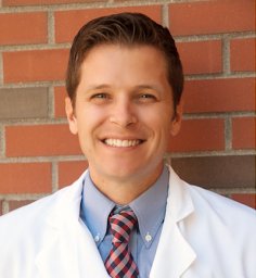 Cameron Rink合作论文数Davis Heart and Lung Research Institute, The Ohio State University Medical Center37
 Singh Kanhaiya合作论文数Department of Molecular &Human Genetics, Banaras Hindu University;Department of Molecular & Human Genetics, Banaras Hindu University36
 Shomita S Mathew-Steiner合作论文数Comprehensive Wound Center, Indiana University School of Medicine30
 Nandini Ghosh合作论文数Indiana University Health Comprehensive Wound Center, Indiana University School of Medicine27
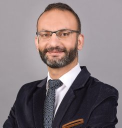 Mohamed El Masry合作论文数Indiana University Health Comprehensive Wound Center, Indiana University School of Medicine25
 Mithun Sinha合作论文数Structural Genomics Section, Saha Institute of Nuclear Physics24
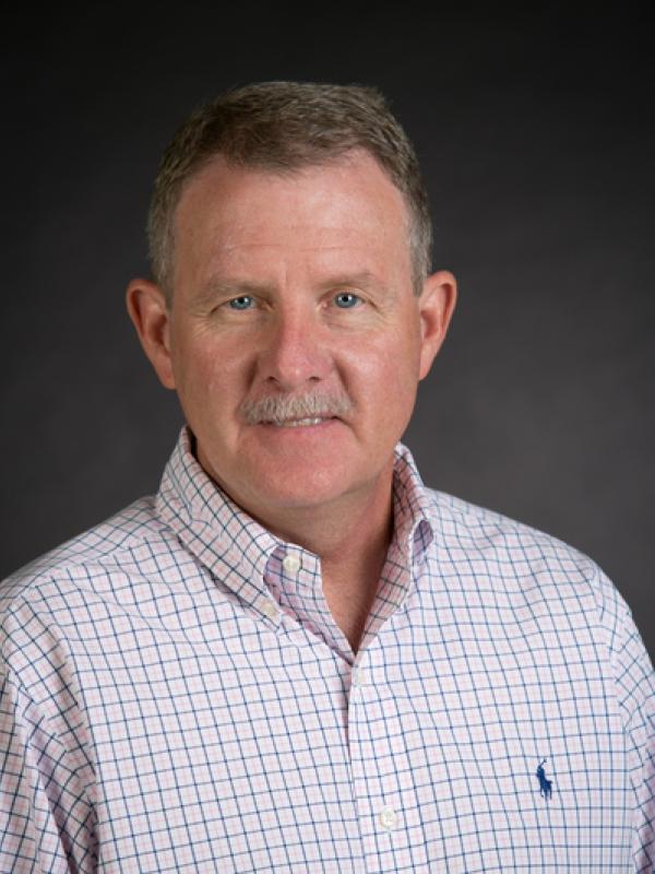 Daniel Wozniak合作论文数Department of Microbiology, College of Arts and Sciences, The Ohio State University22
 Piya Das Ghatak合作论文数Davis Heart &, Comprehensive Wound Center;Lung Research Institute, The Ohio State University Wexner Medical Center;Davis Heart & Lung Research Institute, The Ohio State University21
 Sabyasachi Biswas合作论文数21
 Kasturi Ganesh合作论文数Hist, Univ Calicut20
 Debasis Bagchi合作论文数Department of Pharmacological and Pharmaceutical Sciences, College of Pharmacy, University of Houston19
 Pradipta Banerjee合作论文数School of Bio Science and Technology, Vellore Institute of Technology University16
 Mustafa Atalay合作论文数Institute of Biomedicine, School of Medicine, University of Eastern Finland15Manashi Bagchi合作论文数14Chaffee Scott合作论文数Wexner Medical Center, The Ohio State University14
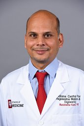 Manishekhar Kumar合作论文数Indiana University Health Comprehensive Wound Center, Indiana University School of Medicine13
 Heather Powell合作论文数Department of Materials Science and Engineering, College of Engineering, The Ohio State University;Department of Biomedical Engineering, The Ohio State University13
 Jaideep Banerjee合作论文数Departments of Surgery and12
 Anu Sharma合作论文数12
 Jay Zweier合作论文数Department of Internal Medicine, College of Medicine, The Ohio State University;Division of Cardiovascular Medicine, College of Medicine, The Ohio State University12Valerie Bergdall合作论文数12
 Durba Pal合作论文数Department of Biomedical Engineering, Indian Institute of Technology Ropar10
 Dickerson Ryan合作论文数Davis Heart and Lung Research Institute, The Ohio State University Medical Center10Sujit Kumar Mohanty合作论文数Cincinnati Children's Hospital Medical Center and the Department of Pediatrics, University of Cincinnati College of Medicine10
 Elgharably Haytham合作论文数Department of Surgery, Davis Heart &Lung Research Institute, The Ohio State University;Davis Heart & Lung Research Institute, The Ohio State University10Chan Yuk Cheung合作论文数10
 Oren Tirosh合作论文数Department of Molecular and Cell Biology, University of California10Kobuchi H合作论文数9David E. Laaksonen合作论文数9
 Soma Datta合作论文数School of Life Sciences, Visva-Bharati University9E. Routier合作论文数8Ayan Biswas合作论文数School of Medicine, University of Alabama at Birmingham8
 Motaz Abas合作论文数Sch Med, Ross Univ8
 Sedat Kacar合作论文数Indiana Univ Hlth Comprehens Wound Ctr, Indiana Univ Sch Med8
 Periannan Kuppusamy合作论文数Department of Radiology, Geisel School of Medicine, Dartmouth College8
 Srivastava Rajneesh合作论文数8
 Yashika Rustagi合作论文数Department of Biotechnology, Jaypee Institute of Information Technology8
 Narasimham L Parinandi合作论文数College of Medicine, The Ohio State University8SEN C合作论文数7S. Dixith合作论文数7Jared Radtke合作论文数7




