Supplementary Figure S8. Myeloid cell transcriptional diversity in response to mutant-selective and broad spectrum RAS inhibitors.
Supplementary Figure S15. Clinical implications of mucinous lineage differentiation in human KRAS-mutant NSCLC cohorts
Supplementary Figure S3. Acute effects of active RAS inhibition in the KP2 and KL2 KrasG12C-mutant lung adenocarcinoma models
Supplementary Figure S10. MAPK pathway modulation in immune and stromal cell subsets in response to active RAS inhibition.
Supplementary Figure S4. Acute inhibition of active RAS in KRASG12C-mutant lung adenocarcinoma.
Supplementary Figure S2. Models and mechanisms of acquired resistance to RAS inhibitors
Supplementary Figure S1. Activity of RAS inhibitors in immune-competent pre-clinical models of KRASG12C-mutated NSCLC.
Supplementary Figure S11. Characterization of tumor-cell transcriptional heterogeneity following RAS inhibition.
Supplementary Table S1. Comparisons of anti-tumor activity of distinct RAS inhibition strategies.
Supplementary Figure S12. Evolution and regulatory networks of RASGTP inhibitor-tolerant persister cells.
Abstract Primary resistance to targeted therapies, immunotherapies, and gene therapies in NSCLC continues to be a significant challenge. TUSC2 tumor suppressor gene therapy has shown promising anti-tumor efficacy by overcoming resistance to targeted therapy and enhancing checkpoint blockade immunotherapy, including in a mutant KRAS/LKB1-driven immunotherapy-resistant NSCLC model. TUSC2 protein expression is downregulated or absent in over 80% of NSCLC and 100% of SCLC cases., TUSC2 mediates cancer cell death through several mechanisms: inhibiting MAPK and mTOR signaling pathways, arresting cell growth, inducing programmed cell death, and activating immune responses. We established models primarily resistant to TUSC2 gene therapy to find biomarkers indicative of TUSC2 gene therapy resistance in NSCLC patient-derived xenografts (PDXs), PDX-derived organoids (PDXOs), and cell lines. A panel of 10 NSCLC cell lines screened for TUSC2 sensitivity showed resistance in 50% of the cell lines, as assessed by annexin V staining and colony formation assays. We evaluated TUSC2 sensitivity in 12 NSCLC PDXOs using ATP-based viability assays in 3D culture following TUSC2 or empty vector transfection. While some PDXOs were highly responsive to TUSC2 within 72 hours post-transfection, 50% of PDXOs exhibited primary resistance. We developed TC314AR (Acquired Resistance) PDX tumors and xenograft models (A549, H1299, H23AR) in NSG mice and treated them with TUSC2 gene therapy. 20-30% of tumors in every model showed resistance, with no significant reduction in size compared to the control tumors after treatment. Protein expression profiling using reverse-phase protein array (RPPA) analysis of 500 proteins showed distinct expression signatures, with several candidate biomarkers significantly altered in resistant cell lines and PDXOs. RPPA analysis of residual tumors from both the xenograft and PDX models revealed significant but model-specific alterations in protein expression between responders and non-responders. Comparative analyses across the three models showed low expression of TROP2 and high expression of PTEN as potential biomarkers of primary resistance. Overexpression of TROP2 in H1299 and H460 cells increased TUSC2-induced apoptosis. These findings suggest that TROP2 and PTEN may serve as biomarkers to predict TUSC2 response and guide therapeutic strategies in NSCLC. Citation Format: Ismail M. Meraz, Renduo Song, Shuhong Wu, Yi Xu, Meng Feng, Lihui Gao, Chenghui Ren, Qi Wang, Jun Li, Mourad Majidi, Jing Wang, Mark Berger, Jack A. Roth. TROP2 and PTEN are biomarkers of primary resistance to TUSC2 gene therapy in non-small cell lung cancer (NSCLC) [abstract]. In: Proceedings of the American Association for Cancer Research Annual Meeting 2026; Part 1 (Regular Abstracts); 2026 Apr 17-22; San Diego, CA. Philadelphia (PA): AACR; Cancer Res 2026;86(7 Suppl):Abstract nr 391.
Supplementary Figure S14. Mucinous tumor cells exhibit higher baseline expression of phospho-ERK1/2 and relative refractoriness to MAPK suppression following treatment with RMC-7977.
Background:Thoracic SMARCA4-deficient undifferentiated tumor (SMARCA4-UT) is a rare, highly virulent neoplasm lacking a standardized therapeutic protocol. Because current evidence is scarce, systemic management guidelines are not well-defined. We aimed to address this lack of data by synthesizing individual patient-level data from case reports and retrospective series. Our goal was to characterize treatment patterns and survival, focusing on combination strategies using immune checkpoint inhibitors (ICIs). Methods:A systematic literature search was performed in PubMed, Embase, and Web of Science from database inception to November 30, 2024, in accordance with PRISMA guidelines. Individual patient data were extracted from eligible published case reports and case series and pooled for unified survival analysis. Aggregated survival data from retrospective cohort studies were summarized for cross-study comparison. Overall survival (OS) was analyzed using the Kaplan-Meier method, and treatment-specific outcomes were visualized using swimmer plots and forest plots. Results:A total of 32 studies comprising 239 patients with thoracic SMARCA4-UT were included, consisting of 26 patients with individual-level data and 8 retrospective cohort studies. Patients were mostly male heavy smokers, and most cohort studies consisted of advanced-stage disease. In the pooled individual patient dataset, 46.2% of patients received first-line chemotherapy plus immunotherapy (Chemo-IO). The median OS (mOS) of pooled cases was 15.0 months, exceeding that reported in most retrospective cohorts treated predominantly with chemotherapy or immunotherapy monotherapy (mOS range, 4.0-7.7 months). Long-term survival was observed almost exclusively in patients receiving Chemo-IO or multimodal treatment. Clinical benefit from Chemo-IO was also observed in patients with negative PD-L1 expression. Conclusions:Our analysis suggests that combination therapy based on ICIs correlates with better survival in thoracic SMARCA4-UT, regardless of PD-L1 expression. Data support using immunotherapy-based regimens early as a first-line treatment for this aggressive disease. These real-world findings offer guidance for clinical decision-making while prospective trials are unavailable.
Supplementary Figure S6. Histological heterogeneity of lung tumors in the KrasG12C/+;Lkb1-/- GEM model.
Abstract Sotorasib (AMG510) and adagrasib (MRTX849) have shown significant efficacy in KRASG12C mutant NSCLC, but acquired resistance occurs within 6–12 months. While some resistance arises from new mutations, over half of the resistant cases lack identifiable genomic alterations. We hypothesize that resistance is driven by signaling network rewiring, creating new therapeutic vulnerabilities. To investigate acquired resistance (AR) mechanisms, multiple AR models, including cell lines (H23AR & H358AR), PDXs (TC303AR & TC314AR), CDXs (H358AR CDX), and PDXOs (PDXO303AR & PDXO314AR) were developed. H23AR and H358AR cells displayed >600-fold and 200-fold and PDXO303AR and PDXO314AR, exhibited >300-fold and >100-fold resistance to sotorasib, respectively compared to their parental counterparts, however, no additional mutations in KRAS or other potential genetic alterations were identified. The AR cells and PDXOs also showed comparable resistance to adagrasib. Proteomic and phosphoproteomic analyses in TC303AR & TC314AR PDXs identified distinct protein signatures associated with KRAS reactivation, mTORC1 signaling upregulation, and PI3K/AKT/mTOR pathway activation. PI3K protein levels were significantly elevated in AR PDXs, H23AR, and H358AR cells. Pharmacological inhibition of PI3K with copanlisib or genetic knockout via CRISPR-Cas9 restored sotorasib sensitivity, suppressed colony formation, and inhibited downstream effectors, including p-AKT, p-mTOR, p-S6, p70S6K, p-GSK3β, and p-PRAS40 in AR cells. copanlisib also sensitized both acquired and primary resistant PDXOs and synergized with sotorasib in restoring drug sensitivity. We found that p4E-BP1 was significantly upregulated in H23AR and H358AR cells, and copanlisib suppressed its expression. The level of p4E-BP1 expression was correlated with Sotorasib sensitivity in PI3K knockout clones, where the most sensitive clone displayed reduced or no p4E-BP1 expression. CRISPR-Cas9-mediated knockout of 4E-BP1, either alone or in combination with PI3K knockout, dramatically restored sotorasib sensitivity to levels comparable to parental cells. Suppression of 4E-BP1 hyperphosphorylation required dual inhibition of mTORC1 and mTORC2, and treatment with AZD8055 or sapanisertib (mTORC1/2 dual inhibitors) significantly dephosphorylated 4E-BP1 and restored sotorasib sensitivity in resistant cells and PDXOs. In contrast, everolimus (a mTORC1-selective inhibitor) did not restore sotorasib sensitivity. In PDX, CDX, and xenograft models in vivo, the combination of sotorasib with either copanlisib or sapanisertib resulted in robust, synergistic, and durable tumor regression at well-tolerated doses. These findings showed the critical role of PI3K/mTOR signaling as a bypass mechanism of resistance to KRASG12C inhibitors. We conclude that mTORC1/2 mediated inhibition of p4E-BP1 and combination strategies targeting this pathway effectively overcomes acquired resistance to KRASG12C inhibitors in NSCLC.
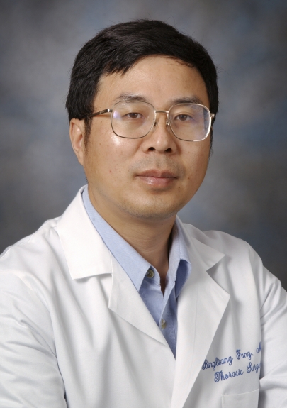 Bingliang Fang合作论文数Thoracic and Cardiovascular Surgery, MD Anderson Cancer Center59
 Jack A. Roth合作论文数Department of Thoracic and Cardiovascular Surgery, Division of Surgery, The University of Texas MD Anderson Cancer Center43
 John V. Heymach合作论文数Department of Thoracic-Head & Neck Med Onc, Division of Cancer Medicine, The University of Texas MD Anderson Cancer Center;Graduate School of Biomedical Sciences, The University of Texas MD Anderson Cancer Center31
 li Wang合作论文数27
 Stephen G. Swisher合作论文数Department of Thoracic and Cardiovascular Surgery, Division of Surgery, The University of Texas MD Anderson Cancer Center;Lung Cancer20
 Don L. Gibbons合作论文数Department of Thoracic-Head & Neck Med Onc, Division of Cancer Medicine, The University of Texas MD Anderson Cancer Center20
 Maria E. Salvatierra合作论文数17Rashi Raghulan合作论文数17Mariela A. Moreno Ayala合作论文数17Ximo Pechuan-Jorge合作论文数17Heladio P. Ibarguen合作论文数17Nataliya Tovbis Shifrin合作论文数17Haniel A. Araújo合作论文数17Mallika Singh合作论文数17Richard Lee合作论文数17Teng Zhou合作论文数17Xin Hu合作论文数Univ Texas MD Anderson Canc Ctr, Dept Genom Med, Houston, TX 77030 USA17
 Harshit Shah合作论文数Department of Pharmaceutical Sciences, North Dakota State University17
 Do Minh Truong合作论文数17Negrao Marcelo V合作论文数Heart Institute (InCor),, University of São Paulo Medical School17
 Frank Rojas合作论文数17
 Luisa M Solis Soto合作论文数Division of Pathology-Lab Medicine, Department of Translational Molecular Pathology, University of Texas M.D. Anderson Cancer Center17
 Jacqueline A Smith合作论文数Division of Genetics and Genomics, Roslin Institute17
 Elsa Quintana合作论文数Facultad de Medicina, Universidad de Valencia17Ferdinandos Skoulidis合作论文数17
 David S Hong合作论文数Department of Investigational Cancer Therapeutics, Division of Cancer Medicine, The University of Texas MD Anderson Cancer Center17Jingjing Jiang合作论文数16Ran Zhang合作论文数14
 Fuminori Teraishi合作论文数14
 Jianjun Zhang (张建军)合作论文数College of Stomatology, Shanghai Jiao Tong University13Lidong Zhang合作论文数13Xiaoshan Zhang合作论文数11Xiaoli Wei合作论文数9John W Davis合作论文数9
 Hongbo Zhu (朱洪波)合作论文数Sir Run Run Shaw Hospital, Zhejiang University School of Medicine9Ismail M Meraz合作论文数8
 Jinsong Liu合作论文数Department of Pathology/Laboratory Medicine, The University of Texas MD Anderson Cancer Center;Graduate School of Biomedical Sciences, The University of Texas MD Anderson Cancer Center8
 John Minna合作论文数Hamon Center for Therapeutic Oncology Research, The University of Texas Southwestern Medical Center;Department of Internal Medicine, The University of Texas Southwestern Medical Center;Department of Pharmacology, The University of Texas Southwestern Medical Center8
 Wayne L. Hofstetter合作论文数Department of Thoracic and Cardiovascular Surgery, Division of Surgery, The University of Texas MD Anderson Cancer Center8
 Wei Guo合作论文数Department of Biology, School of Arts & Sciences, University of Pennsylvania7Pataer Apar合作论文数7Meng Qing H合作论文数7Cao Mengru合作论文数7Dai Bingbing合作论文数6
 Luhua Wang(Michael)合作论文数The University of Texas MD Anderson Cancer6Xingxiang Pu合作论文数6
 Reza J. Mehran合作论文数Department of Thoracic and Cardiovascular Surgery, Division of Surgery, The University of Texas MD Anderson Cancer Center6Chenghui Ren合作论文数5




