Alterations in the BCOR gene, including internal tandem duplications (ITDs) of exon 15 have emerged as important oncogenic changes that define several diagnostic entities. In pediatric cancers, BCOR ITDs have recurrently been described in clear cell sarcoma of kidney (CCSK), primitive myxoid mesenchymal tumor of infancy (PMMTI), and central nervous system high-grade neuroepithelial tumor with BCOR ITD in exon 15 (HGNET-BCOR ITDex15). In adults, BCOR ITDs are also reported in endometrial and other sarcomas. The utility of multiplex targeted RNA sequencing for the identification of BCOR ITD in pediatric cancers was investigated. All available archival cases of CCSK, PMMTI, and HGNET-BCOR ITDex15 were collected. Each case underwent anchored multiplex PCR library preparation with a custom-designed panel, with BCOR targeted for both fusions and ITDs. BCOR ITD was detected in all cases across three histologic subtypes using the RNA panel, with no other fusions identified in any of the cases. All BCOR ITDs occurred in the final exon, within 16 codons from the stop sequence. Multiplex targeted RNA sequencing from formalin-fixed, paraffin-embedded tissue is successful at identifying BCOR internal tandem duplications. This analysis supports the use of anchored multiplex PCR targeted RNA next-generation sequencing panels for identification of BCOR ITDs in pediatric tumors. The use of post-analytic algorithms to improve the detection of BCOR ITD using DNA panels was also explored.
We set out to identify the origins of the Árpád Dynasty based on genome sequencing of DNA derived from the skeletal remains of Hungarian King Béla III (1172–1196) and eight additional individuals (six males, two females) originally interred at the Royal Basilica of Székesfehérvár. Y-chromosome analysis established that two individuals, Béla III and HU52 assign to haplogroups R-Z2125 whose distribution centres near South Central Asia with subsidiary expansions in the regions of modern Iran, the Volga Ural region and the Caucasus. Out of a cohort of 4340 individuals from these geographic areas, we acquired whole-genome data from 208 individuals derived for the R-Z2123 haplogroup. From these data we have established that the closest living kin of the Árpád Dynasty are R-SUR51 derived modern day Bashkirs predominantly from the Burzyansky and Abzelilovsky districts of Bashkortostan in the Russian Federation. Our analysis also reveals the existence of SNPs defining a novel Árpád Dynasty specific haplogroup R-ARP. Framed within the context of a high resolution R-Z2123 phylogeny, the ancestry of the first Hungarian royal dynasty traces to the region centering near Northern Afghanistan about 4500 years ago and identifies the Bashkirs as their closest kin, with a separation date between the two populations at the beginning of the first millennium CE.
Cranial fasciitis is a benign myofibroproliferative lesion of the scalp and underlying bones typically occurring in the pediatric population. Histologically, it is characterized by loose fascicles of stellate cells in a fibromyxoid background, findings similar to those described in the closely related variant nodular fasciitis. Previously characterized as a reactive process, the identification of USP6 translocations in over 90% of nodular fasciitis cases prompted their reclassification as a clonal neoplastic process. Unlike nodular fasciitis, the molecular underpinnings of cranial fasciitis are less clear. While a subset of cranial fasciitis has been associated with Wnt/β-catenin pathway dysregulation, recent case reports suggest that this entity may also harbor USP6 fusions, a finding we sought to further investigate. We identified fifteen archival cases of cranial fasciitis, five females and ten males ranging in age from 3 months to 9 years (median 11 months), composed of formalin-fixed paraffin-embedded and fresh frozen tissues (11 and 4 cases respectively). Samples were evaluated on an RNA-based targeted sequencing panel targeting genes recurrently rearranged in neoplasia, including USP6. Five of fifteen cases (33%) were positive for USP6 rearrangements predicted to result in the fusion of the entire USP6 coding region to the promoter of the 5′ partner, (three of which were novel): two SERPINH1-USP6 (novel) and one each of COL3A1-USP6 (novel), SPARC-USP6, and MYH9-USP6. These results demonstrate the recurrent nature of USP6 rearrangements in cranial fasciitis, and highlight the success of targeted RNA sequencing in identifying known and novel fusion partners. The identification of USP6 promoter-swapping rearrangements is helpful in understanding the underlying biology of cranial fasciitis, and reinforces its biologic relationship to nodular fasciitis. Targeted RNA sequencing is a helpful tool in diagnosing this pseudosarcomatous lesion.
Abstract Background: Molecular characterization of tumor and/or host has the potential to advance the management of pediatric cancer and high risk hematologic disease, but the clinical utility of integrating genomic profiling into standard clinical practice has been limited. The PIPseq Program at Columbia University has instituted prospective CLIA-compliant genomic sequencing for newly diagnosed, high risk, relapsed or refractory pediatric cancer patients and patients referred for bone marrow transplantation. Methods: Families are consented for clinical cancer whole-exome sequencing (cWES) or constitutional whole-exome sequencing (WES) with opt out options for return of results, exclusion of results from medical records, receipt of American College of Medical Genetics (ACMG) recommended secondary germline variants, and data/ sample use in research. Molecular characterization utilizes next generation cWES, WES, RNAseq (transcriptome), or targeted sequencing of select cancer genes. Clinical cancer reports include: known tumor type-specific actionable somatic mutations (Tier 1); somatic mutations actionable in other tumor types, in targetable pathways, or in well-established cancer genes (Tier 2); other somatic mutations in cancer genes (Tier 3); and somatic variants of uncertain significance (VUS; Tier 4). Reports for cWES testing also note translocations, significantly over expressed genes, segmental copy number variation, and germline variants. Institutional Review Board approval was obtained to conduct a retrospective review of results to date. Five categories were developed to assess clinical utility and describe significance: 1) diagnostic, 2) prognostic, 3) potentially actionable target, 4) other critical role in decision making, and 5) implications for health maintenance and genetic counseling. Results: Since January 2014, adequate tissue samples were available for 47 patients, including 31 (66%) with solid tumors and 16 (34%) with hematologic conditions. Testing included cWES (n=8), cWES with transcriptome (n=15), transcriptome only (n=1), targeted somatic panel (n=8), constitutional WES only (n=6), and multiple sequencing platforms (n=7). Normal tissue was obtained from buccal swab (n=8), blood (n=18), and unaffected tissue (n=1). Three families opted out of receiving secondary findings. Genomic aberrations were reported in 41/47 patients. Of the 127 cancer alterations found, 70 (55%) were in 15 patients with hematologic disease (median 2, range 1-11) and 57 (45%), were in 26 patients with solid tumors (median 1, range 1-6). Among the hematologic cases, alterations of known or potential clinical relevance were categorized as Tier 1 (n=0), Tier 2 (n=27), Tier 3 (n=2) mutation, or translocation (n=4); whereas in solid tumors these were categorized as Tier 1 (n=1), Tier 2 (n=14), Tier 3 (n=3) mutation, or translocation (n=9). Twenty-four Tier 4 somatic VUS were identified in hematologic specimens and 26 in solid tumor specimens. Genomic interrogation informed diagnosis in 10 patients (3 previously unknown); provided new prognostic information in 4; identified potentially actionable targets in 15; influenced clinical decision making regarding bone marrow transplant in 2; and revealed cancer or other disease predisposition in 7. Secondary germline ACMG findings in BRCA1 and PMS2 were found. Germline APC mutation was confirmed in one patient and germline VUS in SDHC was seen in another. Novel germline findings were also observed in RUNX1, MLL2 and DICER1. Overall, the PIPseq platform provided clinically impactful results in 30/47 cases (64%). Conclusions: Utilizing a CLIA-compliant prospective WES-based platform, more than half of selected patients derived clinically impactful information. The potential clinical utility of genomic sequencing in pediatric hematology-oncology has likely been underestimated. This abstract is also presented as Poster 50. Citation Format: Julia L. Glade Bender, Jennifer A. Oberg, Maria Luisa Sulis, Filamon Dela Cruz, Anthony N. Sireci, Susan J. Hsiao, Darrell J. Yamashiro, Carrie Koval, Wendy K. Chung, Stephen G. Emerson, Rebecca Zylber, Samantha Cano, Danielle P. Denney, Stuart Andrews, Peter L. Nagy, Mahesh M. Mansukhani, Andrew L. Kung. Precision in Pediatric Sequencing (PIPseq): Clinical implementation of genomic sequencing into pediatric hematology-oncology practice. [abstract]. In: Proceedings of the AACR Precision Medicine Series: Integrating Clinical Genomics and Cancer Therapy; Jun 13-16, 2015; Salt Lake City, UT. Philadelphia (PA): AACR; Clin Cancer Res 2016;22(1_Suppl):Abstract nr PR01.
 Tamara Restrepo合作论文数2Al-Ibraheemi Alyaa合作论文数2
 Sekhar Duraisamy合作论文数2
 Peter Nagy合作论文数Nagy Lab, Department of Plant Pathology, College of Agriculture, Food and Environment, University of Kentucky2
 Church Alanna J合作论文数2Marian H. Harris合作论文数2Nicholas Rouse合作论文数1Stuart J. Andrews合作论文数1R. Seth Pinches合作论文数1György Szabados合作论文数1Carrie Koval合作论文数1Rebecca J. Zylber合作论文数1Natalia Ekomasova合作论文数1Harrison K Tsai合作论文数1Elza Khusnutdinova合作论文数1Lilya Gabidullina合作论文数1Joanna Plunkitt合作论文数1Karan Kapuria合作论文数1Janos Jeney合作论文数1Sergey Litvinov合作论文数1Goran Runfeldt合作论文数Mill Mito Project1Murat Dzhaubermezov合作论文数1Danielle Pendrick Denney合作论文数1Susan J Hsiao合作论文数Columbia Univ, Med Ctr, Dept Pathol & Cell Biol, New York, NY USA1Huijie Chen合作论文数1
 Endre Neparaczki合作论文数Department of Archaeogenetics, Institute of Hungarian Research1Jacques Chiaroni合作论文数1
 Katherine A. Janeway合作论文数Dana-Farber Cancer Institute;Harvard Medical School1
 Doron Behar合作论文数Estonian Biocentre, Institute of Genomics, University of Tartu1
 Julia Glade Bender合作论文数1
 Julie Di Cristofaro合作论文数1Wasman Jay合作论文数1
 Sanda Alexandrescu合作论文数1Sireci Anthony N合作论文数1
 Vera A. Paulson合作论文数Department of Laboratory Medicine & Pathology, University of Washington1Stojanov Ivan James合作论文数1
 Jennifer Oberg合作论文数1Stephen G. Emerson合作论文数Departments of Medicine|Pathology , and Pediatrics|University of Pennsylvania School of Medicine1
 Andrew Kung合作论文数Department of Pediatrics, Memorial Sloan Kettering Cancer Center1Kásler Miklós合作论文数1
 Wendy Chung合作论文数Department of Pediatrics, Columbia University;Division of Genetics and Genomics, Boston Children’s Hospital;Harvard Medical School1
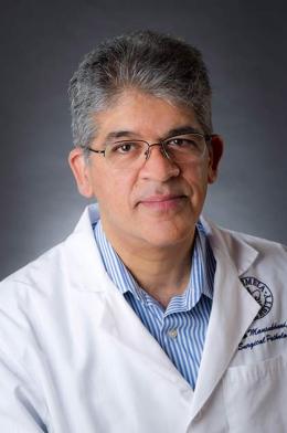 Mahesh M. Mansukhani合作论文数Department of Pathology and Cell Biology, Columbia University;Laboratory of Personalized Genomic Medicine, Columbia University;Department of Pathology and Cell Biology, Columbia University Irving Medical Center1Deborah Chute合作论文数1Judit Olasz合作论文数1Darrell J. Yamashiro合作论文数1Zoltán Szentirmay合作论文数1Maria Luisa Sulis合作论文数1Va Lip合作论文数1
 Zoltan Maroti合作论文数Department of Pediatrics, University of Szeged1




