Heart failure affects over 64 million individuals globally, contributing to elevated mortality rates and substantial healthcare costs. This study investigates the potential of retinal optical coherence tomography features combined with routine clinical variables as biomarkers for the detection of heart failure, exploring a potential avenue for improved risk assessment and screening support using explainable machine-learning tools. A comprehensive dataset of normal and heart failure patients' demographic and medical records including retinal measurements from both eyes was used. Among the machine learning models employed, the Extreme Gradient Boosting model demonstrated the best performance, achieving an accuracy of 73.31%, a precision of 71.81%, and an area under the receiver operating characteristic curve of 0.837. Explainability analyses further revealed that macular thickness metrics, particularly in the inner temporal subfield, inner nasal subfield, and outer superior subfields of the left eye, along with key clinical indicators such as age, body mass index, and glycated hemoglobin, were the most influential predictors of heart failure status. Local explanation methods also provided patient-level reasoning consistent with overall cohort patterns. To our knowledge, this is the first study to use an integrated, explainable approach incorporating bilateral retinal optical coherence tomography measurements with routine clinical indicators for heart failure detection, providing an interpretable and accessible alternative to black-box models while helping address the cost, invasiveness, and limited accessibility of existing heart failure diagnostic tools.
Dysfunction in the autonomic nervous system function during sleep is common in type 2 diabetes mellitus and is associated with increased risk of diabetic related complications. While heart rate variability is a standard non-invasive measure of autonomic function, the utility of symbolic heart rate transition motifs to characterize sleep-specific autonomic dysfunction in diabetes has not been explored. Here, we analysed 5-min electrocardiogram segments during daytime and sleep from 35 male participants ((51 ± 17) years old) with type 2 diabetes mellitus. Frequency-domain heart rate variability metrics and 27 symbolic motifs based on 3-beat heart rate transitions were computed. Logistic regression models adjusted for age and body mass index were used to test associations with diabetic complications. The high frequency power of heart rate variability was significantly higher during sleep than daytime (p = 0.012), reflecting enhanced parasympathetic activity. Several symbolic motifs showed differential prevalence between sleep and day. In particular, motif [1, 1, -1] was significantly more prevalent during sleep and was independently associated with diabetic complications (β = -1.8, p = 0.023). Motif [1,1,-1] along with [-1, 1, 1], showed strong positive correlations with high frequency power during day and sleep [Day: [1,1,-1]: r = 0.70 (p = 7.0e-06), [-1,1,1]: r = 0.78 (p = 4.5e-07)] [Sleep: [1,1,-1]: r = 0.77 (p = 5.7e-07), [-1,1,1]: r = 0.80 (p = 3.1e-07)], suggesting a link to vagal tone. The study's findings provide a novel, non-invasive approach to sleep-based autonomic monitoring in type 2 diabetes mellitus by showing that the symbolic heart rate motifs during sleep reflect unique autonomic patterns associated with diabetic complications. These results demonstrate the diagnostic utility of motif-based HR analysis for diabetes and sleep medicine.
Chronic Kidney Disease (CKD) is a progressive condition that requires accurate diagnosis and staging for effective clinical management. Conventional CKD diagnosis relies on estimated Glomerular Filtration Rate (eGFR), a measure of kidney function derived from serum biomarkers such as serum creatinine (SCr) and cystatin C (SCysC). However, eGFR calculations may be inaccurate when applied to diverse patient populations. This study proposes a machine learning (ML) system that integrates regression-based eGFR estimation, metaheuristic optimization using the Grey Wolf Optimizer (GWO), and multi-class classification with various ML models to enhance CKD staging and classification. The model estimates eGFR using three established CKD Epidemiology Collaboration (CKD-EPI) equations incorporating SCr, SCysC, and their combined values. Regression models assess predictive performance, specifically Linear Regression (LR) and Support Vector Regression (SVR). SVR demonstrates superior performance compared to LR for CKD-EPISCr-SCysC achieved a root mean squared error (RMSE) of 3.03, a mean absolute percentage error (MAPE) of 2.97%, and a coefficient of determination (R2) score of 0.97. The application of GWO for hyperparameter tuning has resulted in a 37.3% reduction in root mean square error (RMSE), a 37.4% drop in mean absolute percentage error (MAPE), and a 2.06% improvement in R2 to improve the precision of prediction. Once the model fine-tunes the eGFR estimations, it feeds them into various algorithms for CKD stage classification, including Support Vector Machine (SVM), Decision Tree (DT), Random Forest (RF), and Extreme Gradient Boosting (XGBoost). Among these, XGBoost achieves the highest classification accuracy of 97.76%, along with an F1-score of 97.45%, demonstrating its effectiveness in CKD staging. Shapley Additive Explanations (SHAP) provide global and local feature importance insights, enhancing clinical decision-making and model transparency. Future research will validate the model using more extensive and more diverse datasets. Additionally, it will incorporate extra clinical parameters, including biomarkers and genetic data, to enhance the precision of CKD risk prediction. This research enhances AI-driven nephrology by providing a scalable, interpretable, and highly accurate solution for diagnosing and managing CKD.
This paper introduces a novel methodology for estimating diagnostic information loss in fetal phonocardiogram (fPCG) signals during transmission. fPCG, a non-invasive technique for monitoring fetal heart sounds, is crucial for prenatal health evaluation. However, the clinical information in the fPCG signal can be distorted during transmission, particularly over wireless channels, posing a significant challenge in telehealth care monitoring. To address this issue, we propose a new approach for assessing the quality of received fPCG signals. Our method involves compressing, encoding, and transmitting the fPCG signals using an Ultra-Wide Band (UWB) transceiver system. The effectiveness of this approach is evaluated using fPCG signals from a public database, and various compression and transmission frameworks are tested. Performance quality measures are derived by comparing the original and reconstructed signals, demonstrating our method’s ability to estimate the extent of clinical information loss accurately. Experimental results demonstrate that our method provides valuable insights into improving the reliability and accuracy of remote fetal monitoring systems. This research opens new avenues for enhancing the quality of transmitted biomedical signals in telemedicine applications and suggests further studies to validate this approach in real-world clinical settings.
Assessing fetal health traditionally involves techniques like echocardiography, which require skilled professionals and specialized equipment, making them unsuitable for low-resource settings. An emerging alternative is Phonocardiography (PCG), which offers affordability but suffers from challenges related to accuracy and complexity. To address these limitations, we propose a deep learning model, Fetal Heart Sounds U-NetR (FHSU-NETR), capable of extracting both fetal and maternal heart rates directly from raw PCG signals. FHSU-NETR is designed for practical implementation in various healthcare environments, enhancing accessibility and reliability of fetal monitoring. Due to its enhanced capacity to simulate remote interactions and capture global context, the suggested pipeline utilizes the self-attention mechanism of the transformer. Validated with data from 20 normal subjects, including a case of fetal tachycardia arrhythmia, FHSU-NETR demonstrated exceptional performance. It accurately identified most of the fetal heartbeat locations with a low mean difference in fetal heart rate estimation (-2.55±10.25 bpm) across the entire dataset, and successfully detected the arrhythmia case. Similarly, FHSU-NETR showed a low mean difference in maternal heart rate estimation (-1.15±5.76 bpm) compared to the ground-truth maternal ECG. The model's exceptional ability to identify arrhythmia cases within the dataset underscores its potential for real-world application and generalization. By leveraging the capabilities of deep learning, our proposed model holds promise to reduce the reliance on medical experts for the interpretation of extensive PCG recordings, thereby enhancing efficiency in clinical settings.
Over 64 million people worldwide are affected by heart failure (HF), a condition that significantly raises mortality and medical expenses. In this study, we explore the potential of retinal optical coherence tomography (OCT) features as non-invasive biomarkers for the classification of heart failure subtypes: left ventricular heart failure (LVHF), congestive heart failure (CHF), and unspecified heart failure (UHF). By analyzing retinal measurements from the left eye, right eye, and both eyes, we aim to investigate the relationship between ocular indicators and heart failure using machine learning (ML) techniques. We conducted nine classification experiments to compare normal individuals against LVHF, CHF, and UHF patients, using retinal OCT features from each eye separately and in combination. Our analysis revealed that retinal thickness metrics, particularly ISOS-RPE and macular thickness in various regions, were significantly reduced in heart failure patients. Logistic regression, CatBoost, and XGBoost models demonstrated robust performance, with notable accuracy and area under the curve (AUC) scores, especially in classifying CHF and UHF. Feature importance analysis highlighted key retinal parameters, such as inner segment-outer segment to retinal pigment epithelium (ISOS-RPE) and inner nuclear layer to the external limiting membrane (INL-ELM) thickness, as crucial indicators for heart failure detection. The integration of explainable artificial intelligence further enhanced model interpretability, shedding light on the biological mechanisms linking retinal changes to heart failure pathology. Our findings suggest that retinal OCT features, particularly when derived from both eyes, have significant potential as non-invasive tools for early detection and classification of heart failure. These insights may aid in developing wearable, portable diagnostic systems, providing scalable solutions for personalized healthcare, and improving clinical outcomes for heart failure patients.
Congenital heart diseases (CHDs), caused by structural abnormalities in the heart and blood vessels, pose a significant public health concern and contribute significantly to the socioeconomic burden, particularly in pediatric populations. Phonocardiograms (PCGs), as a non-invasive and cost-effective diagnostic modality, capture vital acoustic signals that reflect the mechanical activity of the heart and can reveal pathological patterns associated with various CHD types. This study investigates the minimum signal duration required for accurate automatic classification of heart sounds and evaluates signal quality using the root mean square of successive differences (RMSSD) and the zero-crossing rate (ZCR). Mel-frequency cepstral coefficients (MFCCs) are extracted as features and fed into a transformer-based residual one-dimensional convolutional neural network (1D-CNN) for classification. Experimental results show that a threshold of 0.4 for RMSSD and ZCR yields optimal classification performance, with a minimum signal length of 5 seconds required for reliable results. Shorter segments (3 seconds) lack sufficient diagnostic information, while longer segments (15 seconds) may introduce additional noise. The proposed model achieves a maximum classification accuracy of 93.69% with 5-second signals.
Phonocardiography is a widely used procedure for understanding the functioning of human heart and diagnosing heart diseases. However, the Phonocardiogram (PCG) signals obtained during this process are susceptible to noise, so its imperative to eliminate such noise to ensure accurate heart disease diagnosis. Various algorithms are available for denoising biomedical signals, and this study aims to compare the effective- ness of two distinct algorithmic approaches: Discrete Wavelet Transform (DWT) based methods and adaptive-based methods for denoising PCG signals. The DWT-based approach involves breaking down the PCG signal into different frequency sub- bands using filtering and down-sampling techniques, enabling noise reduction within specific frequency ranges. On the other hand, adaptive-based algorithms employ techniques such as Least Mean Square (LMS) and Recursive Least Squares (RLS) to estimate and remove noise from the original signal. To assess the performance of these denoising methods, various performance metrics are utilized, including Mean Square Error (MSE), Signal-to-Noise Ratio (SNR), Peak Signal-to-Noise Ratio (PSNR), Mean Absolute Error (MAE), and Percentage Root Mean Square Difference (PRD). The results of this study indicate that different methods of DWT and adaptive-based algorithms effectively reduce noise in PCG signals. However, DWT exhibits superior performance in high-noise environments, while adaptive- based algorithms excel when dealing with signals characterized by higher noise power. These findings emphasize the importance of selecting a denoising algorithm based on the specific noise characteristics of the analysis environment and application.
Chronic kidney disease (CKD) is a major worldwide health problem, affecting a large proportion of the world’s population and leading to higher morbidity and death rates. The early stages of CKD sometimes present without visible symptoms, causing patients to be unaware. Early detection and treatments are critical in reducing complications and improving the overall quality of life for people afflicted. In this work, we investigate the use of an explainable artificial intelligence (XAI)-based strategy, leveraging clinical characteristics, to predict CKD. This study collected clinical data from 491 patients, comprising 56 with CKD and 435 without CKD, encompassing clinical, laboratory, and demographic variables. To develop the predictive model, five machine learning (ML) methods, namely logistic regression (LR), random forest (RF), decision tree (DT), Naïve Bayes (NB), and extreme gradient boosting (XGBoost), were employed. The optimal model was selected based on accuracy and area under the curve (AUC). Additionally, the SHAP (SHapley Additive exPlanations) and LIME (Local Interpretable Model-agnostic Explanations) algorithms were utilized to demonstrate the influence of the features on the optimal model. Among the five models developed, the XGBoost model achieved the best performance with an AUC of 0.9689 and an accuracy of 93.29%. The analysis of feature importance revealed that creatinine, glycosylated hemoglobin type A1C (HgbA1C), and age were the three most influential features in the XGBoost model. The SHAP force analysis further illustrated the model’s visualization of individualized CKD predictions. For further insights into individual predictions, we also utilized the LIME algorithm. This study presents an interpretable ML-based approach for the early prediction of CKD. The SHAP and LIME methods enhance the interpretability of ML models and help clinicians better understand the rationale behind the predicted outcomes more effectively.
In this letter, the Fourier–Bessel domain adaptive wavelet transform (FBDAWT) is proposed for the automated detection of anxiety stages using the single-channel wearable electrocardiogram (ECG) sensor signal. The modes or components are evaluated using the FBDAWT of the ECG signal. The increment entropy and energy features are computed from each mode of ECG data. The cross gradient boosting (XGBoost) model is employed for the normal versus light anxiety versus moderate anxiety versus severe-anxiety-based detection task using the FBDAWT domain ECG signal features. The wearable-sensor-based ECG signals from a publicly available database are used to assess the performance of the proposed approach. The results show that the XGBoost model has obtained the accuracy, F1-score, and Kappa score values of 92.27%, 92.13%, and 0.89, respectively. We have compared the performance of the proposed FBDAWT domain approach with existing methods for anxiety detection using physiological signals.
Chronic kidney disease (CKD) remains one of the most prominent global causes of mortality worldwide, necessitating accurate prediction models for early detection and prevention. In recent years, machine learning (ML) techniques have exhibited promising outcomes across various medical applications. This study introduces a novel ML-driven nomogram approach for early identification of individuals at risk for developing CKD stages 3–5. This retrospective study employed a comprehensive dataset comprised of clinical and laboratory variables from a large cohort of diagnosed CKD patients. Advanced ML algorithms, including feature selection and regression models, were applied to build a predictive model. Among 467 participants, 11.56% developed CKD stages 3–5 over a 9-year follow-up. Several factors, such as age, gender, medical history, and laboratory results, independently exhibited significant associations with CKD (p < 0.05) and were utilized to create a risk function. The Linear regression (LR)-based model achieved an impressive R-score (coefficient of determination) of 0.954079, while the support vector machine (SVM) achieved a slightly lower value. An LR-based nomogram was developed to facilitate the process of risk identification and management. The ML-driven nomogram demonstrated superior performance when compared to traditional prediction models, showcasing its potential as a valuable clinical tool for the early detection and prevention of CKD. Further studies should focus on refining the model and validating its performance in diverse populations.
Assessing fetal well-being using conventional tools requires skilled clinicians for interpretation and can be susceptible to noise interference, especially during lengthy recordings or when maternal effects contaminate the signals. In this study, we present a novel transformer-based deep learning model called fetal heart sounds U-Net Transformer (FHSU-NETR) for automated extraction of fetal heart activity from raw phonocardiography (PCG) signals. The model was trained using a realistic synthetic dataset and validated on data recorded from 20 healthy mothers at the pregnancy outpatient clinic of Tohoku University Hospital, Japan. The model successfully extracted fetal PCG signals; achieving a heart rate mean difference of −1.5 bpm compared to the ground-truth calculated from fetal electrocardiogram (ECG). By leveraging deep learning, FHSU-NETR would facilitates timely interpretation of lengthy PCG recordings while reducing the heavy reliance on medical experts; thereby enhancing the efficiency in clinical practice.
Breast cancer is the most prevalent cancer among women, with a high mortality rate. The early detection of breast cancer using medical imaging techniques helps reduce the number of deaths caused by this disease. Thermogram imaging is safer and less expensive than mammography for diagnosing breast cancer. The automated analysis of thermogram images using artificial intelligence (AI) methods is an interesting approach to detect breast cancer. This article proposes a novel multiscale analysis domain interpretable deep learning (MSADIDL) approach for automatically detecting breast cancer using thermogram images. The 2D empirical wavelet transform (2DEWT) with fixed boundary points (FBPs) is employed for the multiscale analysis of thermogram images and evaluation of modes or subbands. All the modes of the thermogram images are used as the input to the MSADIDL model for the automated detection of breast cancer. The MSADIDL architecture comprises seven individual deep neural networks (DNNs) connected in parallel. The outputs of the individual DNNs are concatenated and then used as the input to the dense layers, after which the output layer evaluates the probability score for the automated categorization of normal versus cancerous classes. A publicly available thermogram imaging dataset is utilized to evaluate the performance of the proposed MSADIDL approach. The results show that the proposed MSADIDL approach has obtained an accuracy value of 99.54% for both fivefold cross-validation (CV) and hold-out validation cases using all seven modes of thermogram images. The MSADIDL model has achieved an accuracy higher than all of the transfer learning-based breast cancer detection techniques using thermogram images. The suggested MSADIDL model has shown higher accuracy when compared with different existing methods to detect breast cancer using thermogram images.
Fetal well-being assessment using conventional tools requires skilled clinicians for interpretation and may be heavily affected by noise if recorded for lengthy durations or by the contaminated maternal effects. In this paper, we propose for the first time a deep learning-based model, fetal heart sounds U-Net (FHSU-NET), for automated extraction of fetal heart activity illustrated as sound waves in raw phonocardiography (PCG). A total of 20 healthy mothers were included in this study to train and validate FHSU-NET following a leave-one-subject-out (LOSO) cross-validation scheme. The model successfully extracted fetal PCG with a median root mean square error (RMSE) of 0.702 [IQR: 0.695-0.706] relative to ground-truth. The median error in heart rate estimated using the ground-truth and FHSU-NET was 18.507 [IQR: 11.996-23.215] with a correlation of 0.642 (p-value = 0.002) and Bland-altman mean difference of 5.18. The proposed model paves the way towards implementing deep learning in clinical settings to decrease the high dependency on medical experts when interpreting lengthy PCG.
The phonocardiogram (PCG) signal deciphers the mechanical activity of the heart, and it consists of the fundamental heart sounds (FHSs) (S1 and S2), murmurs, and other associated sounds (S3 and S4). Detection of FHS activity (FHSA) is vital for the automated analysis of PCG signals to diagnose various heart valve diseases. This article proposes a time–frequency-domain (TFD) deep neural network (DNN) approach for automated FHSA detection using PCG signals. The modified Gaussian window-based Stockwell transform (MGWST) is used to obtain the time–frequency representation (TFR) of PCG signals. The Shannon–Teager–Kaiser energy (STKE), smoothing, and thresholding techniques are then employed to evaluate the segmented heart sound components. The TFD Shannon entropy (TFDSE) features are computed from the segmented heart sound components of the PCG signal. The DNN developed based on the stacked autoencoders (SAEs) is used for the automated identification of FHSA components. The performance of the proposed approach is evaluated using two publicly available standard databases (Database 1: Michigan heart sound and murmur database and Database 2: PhysioNet Computing in Cardiology Challenge 2016). The results demonstrate that the proposed approach has achieved the accuracy, sensitivity, specificity, and precision values of 99.55%, 99.93%, 99.26%, and 99.02% for Database 1 and 95.43%, 97.92%, 98.32%, and 97.60% for Database 2, respectively. It is shown that the proposed FHSA detection approach has obtained better accuracy than existing methods.
The damage to the heart valves causes heart valve disorders (HVDs). The detection of HVDs is crucial in a clinical study as these diseases may cause congestive heart failure, hypertrophy, and stroke. The phonocardiogram (PCG) signal reveals information regarding the mechanical activity of the heart. The early detection of HVDs using PCG signal is vital to minimize the chances of cardiac arrest and other cardiac complications. This article proposes the time–frequency-domain deep learning (TFDDL) framework for automatic detection of HVDs using PCG signals. The time–frequency (TF)-domain representations of PCG signals are evaluated using both time-domain polynomial chirplet transform (TDPCT) and frequency-domain polynomial chirplet transform (FDPCT). The deep convolutional neural network (CNN) model is used to detect four types of HVDs using the TF images of PCG signals obtained using both the TDPCT and FDPCT methods. The proposed TFDDL approach is evaluated using PCG signals from public databases. For the detection of HVDs using TDPCT- and FDPCT-based TF images of PCG signals, the suggested approach has achieved overall accuracy values of 99% and 99.48%, respectively. For the classification of normal and abnormal heart sound classes, the proposed TFDDL approach has obtained an accuracy of 85.16% using PCG signals from the Physionet challenge 2016 database. The proposed TFDDL framework is compared with TF-domain transfer learning models such as residual network (ResNet-50) and visual geometry group (VGGNet-16). The overall accuracy values obtained using VGGNet-16 and ResNet-50 are less than the proposed deep CNN model for the detection of HVDs. The proposed TFDDL model can be validated in real-time using heart sound signals recorded from different subjects for automated identification of HVDs.
Phonocardiogram (PCG) signals are contaminated with various noise signals, which hinders the accurate diagnostic interpretation of the signal. Discrete wavelet transform (DWT) is a well-known technique used to remove noise from PCG signals and improve signal quality. The performance of DWT-based denoising depends upon several parameters involved in the process, such as mother wavelets used for decomposition, the number of decomposition levels (DLs), thresholding technique used and the threshold estimation rule followed. In this work, an investigative study is carried out to select the optimal parameter values which give the best denoising performance. The metrics such as mean-square error (MSE), normalized-mean-square error (NMSE), root-mean-square error (RMSE), percentage root-mean-square difference (PRD) and signal-to-noise ratio (SNR) are used to evaluate the performance of the denoising in this study. The results obtained show that the fifth-order Coiflet wavelet is best suited for denoising PCG signals when applied with the soft thresholding (ST) function and rigrsure threshold selection rule. Also, the optimum number of DLs resulting in better performance is level 6. SNR value obtained from the studies shows the efficacy of the parameters selected for denoising. The denoised PCG signals provide accurate information to determine various kinds of heart valve-related disorders (HVDs).
In this article, a novel time-frequency (TF) analysis approach based on the Fourier-Bessel series expansion (FBSE) domain discrete Stockwell transform (DST) is proposed. The Toeplitz matrix is formulated using the FBSE coefficients of the non-stationary signal. The Gaussian matrix is multiplied by the Toeplitz matrix to obtain the windowed Fourier-Bessel Toeplitz (WFBT) matrix. The FBSE-DST time-frequency representation (TFR) is evaluated based on inverse FBSE of WFBT matrix followed by Bessel function root to frequency transformation. The synthetic signals such as multicomponent damped sinusoidal, multicomponent based amplitude modulation (AM), multicomponent based frequency modulation (FM) signals, and real-time signals such as the electroencephalogram (EEG) signals are used to assess the performance of the proposed FBSE-DST approach. The Renyi-entropy measure is computed in each signal case to compare the performance of FBSE-DST and DST techniques. The results of the proposed FBSE-DST technique provide the Renyi entropy values of 15.90, 18.28, and 18.67, respectively, for the TFRs of multicomponent damped sinusoidal, multicomponent AM, multicomponent FM signals. The Renyi entropy values obtained using FBSE-DST are lower than that obtained by the DST approach for the TF analysis of different synthetic signals.
 Rajesh Kumar Tripathy合作论文数19R. N. Ponnalagu合作论文数12
 Ahsan H. Khandoker合作论文数Department of Electrical & Electronic Engineering, The Universityof Melbourne9
 Rajendra Acharya合作论文数School of Mathematics, Physics and Computing, University of Southern Queensland;Kumamoto University7
 Ram Bilas Pachori合作论文数Department of Electrical Engineering, Indian Institute of Technology Indore5P. Bachan合作论文数4
 P B Natarajan合作论文数3
 Jay Karhade合作论文数Dept Elect & Elect Engn, BITS Pilani3Namareq Widatalla合作论文数3
 Dinesh Kumar Dash合作论文数Dept Elect & Commun Engn, Parala Maharaja Engn Coll3
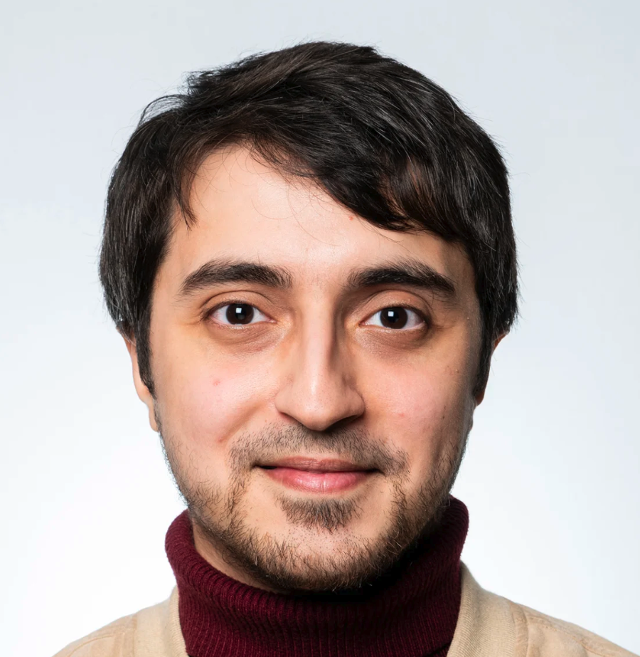 Mohanad Alkhodari合作论文数Department of Biomedical Engineering, Khalifa University;Institute of Biomedical Engineering, University of Oxford3
 Murad Almadani合作论文数2
 Sachin Trankatwar合作论文数Department of Electrical and Electronics Engineering, Birla Institute of Technology and Science2
 Shaswati Dash合作论文数Dept EEE, BITS Pilani2
 Pranjali Gajbhiye合作论文数Dept Elect & Elect Engn, BITS Pilani2
 Jishan Mehedi合作论文数Advanced Digital and Embedded Systems Lab;Department of Electronics & Telecommunication Engineering;Jadavpur University;Advanced Digital and Embedded Systems Lab, Jadavpur University2
 Ganapati Panda合作论文数School of Electrical Sciences, Indian Institute of Technology Bhubaneswar;National Institute of Technology Meghalaya2
 Nurul Hasan合作论文数1
 Mohammad Abdullah Al Mamun合作论文数1
 R. Karthik合作论文数1
 Shelesh Saraswat合作论文数1M.Naga Sravani合作论文数1Ashirwad Ray合作论文数1Abhishek Varshney合作论文数1
 Md Hassanuzzaman合作论文数1Anurag Gade合作论文数1
 Tejas Radhakrishnan合作论文数Department of Electrical and Electronics Engineering, Birla Institute of Technology and Science - Pilani1
 Umesh Chandra Samal合作论文数KIIT University1
 Manish Sharma合作论文数Pharmacology Department, Regional Research Laboratory, Jammu Tawi, India.1
 Prusty, S.B.合作论文数Dept. of Electron. & Commun. Eng., Nat. Inst. of Technol.;c1
 Mario R Arrieta Paternina合作论文数1
 Sibasankar Padhy合作论文数Dept. of Electron. & Electr. Eng., Indian Inst. of Technol. Guwahati;c;Dept. of Electron. & Electr. Eng., Indian Inst. of Technol. Guwahati1
 Alejandro Zamoramendez合作论文数CINVESTAV, IPN - Unidad Guadalajara;c;CINVESTAV, IPN - Unidad Guadalajara1
 Leontios Hadjileontiadis合作论文数Department of Biomedical Engineering, Khalifa University of Science and Technology/Department of Electrical Engineering and Computer Science, Khalifa University of Science and Technology/State Conservatory of Thessaloniki1
 Khawza I. Ahmed合作论文数Department of Electrical Engineering;Arizona State University;Department of Electrical Engineering, Arizona State University1
 Ganesh Naik合作论文数Fac Engn & Informat Technol, Univ Technol Sydney1
 Siamak Yousefi合作论文数Department of Ophthalmology, College of Medicine, University of Tennessee Health Science Center;Department of Genetics, Genomics, and Informatics, University of Tennessee Health Science Center1
 R. Mostafa合作论文数Mobile and Portable Radio Research Group, Virginia Polytechnic Institute and State University1




