Background:Heterozygous c.283+1G>A and c.283G>A variants in the THRB gene, encoding for thyroid hormone receptor (TR)β1 and β2, lead to autosomal dominant macular dystrophy (ADMD). We report the detailed clinical characterization of two first-degree relatives with ADMD, heterozygous for THRB c.283+1G>A, and an unrelated ADMD patient with a novel variant, c.283G>C. The genomic and molecular consequences of both variants were studied. Methods:gDNA and mRNA were obtained from leukocytes. Clinical characterization included biochemistry, bone density and body composition, ECG, echocardiography, ultrasound, audiometry and color-vision. In vitro assays investigated TR function and DNA binding. Results:The patients manifested no resistance to thyroid hormone beta (RTHβ) and had normal FT4 and TSH. Detailed studies in two patients showed no goiter, tachycardia, hypercholesterinemia or hepatic steatosis. Hearing was not impaired. Both had impaired color vision and reduced bone density. RT-PCR from all three patients revealed skipping of exon 4 exclusive to TRβ1, producing a deletion of 87 amino acids in the N-terminal domain (TRβ1ΔNTD). In vitro, DNA-binding affinity of TRβ1ΔNTD to DR4-TRE with or without RXRα was comparable to TRβ1WT. Surprisingly, TRβ1ΔNTD was transcriptionally twice more active than TRβ1WT with a similar EC50 for T3, demonstrating gain-of-function of TRβ1ΔNTD. THRA expression in leukocytes was increased by 3-fold compared to unrelated controls and different from RTHβ patients. Conclusion:These THRB splice site variants produce TRβ1 exon 4 skipping, resulting in a gain-of-function mutant, TRβ1ΔNTD. This explains the dominant ADMD phenotype devoid of RTHβ and suggests a TRβ1 gain-of-function syndrome.
Background: Heterozygosity of genetically encoded misfolded mutant thyroglobulin (TG, the thyroid hormone protein precursor) occurs with a frequency estimated at 1-in-217 people worldwide, resulting in subclinical hypothyroidism that largely escapes medical detection. However, patients carrying biallelic TG mutation, when untreated, are frankly hypothyroid unless and until they develop a massive goiter. To date, TG is the only proven endogenous precursor protein for thyroid hormone synthesis in vertebrates. Our objective was to understand how homozygous expression of mutant TG protein that cannot undergo secretion to the iodination site in the thyroid follicle lumen can be compatible with patients or animal models ultimately achieving normal/near-normal circulating T4 and T3 levels. Notably, in both monoallelic and biallelic disease, dead thyrocytes have been reported in the lumen of thyroid follicles. Methods: We recently engineered mice with homozygous Tg-KO , making it possible to study circulating T4 and T3 in the complete absence of TG protein expression. We also employed immunoblotting methods to test for the presence of thyroidal T4-containing and T3-containing protein. Results: We found that, concomitant with goiter growth, animals completely lacking TG (like patients or animals with biallelic TG defect) are able to eventually normalize circulating T4 levels when their goiter has sufficiently enlarged, while maintaining serum T3 at ∼two-thirds normal levels throughout life. Very little T3-containing protein is synthesized in the thyroid glands of Tg-KO mice; however, T4-containing protein is synthesized on the ghosts of dead thyrocytes, with T3 generated from circulating T4 deiodination. Albeit inefficient, thyroidal T4-containing protein content is not appreciably different in the presence of homozygous mutant TG or in the complete absence of TG. Conclusions: Our data suggest that in biallelic TG mutation, thyroid stimulating hormone (TSH)-driven iodination underlies inefficient T4 formation derived from the iodoproteome of dead thyrocytes. In the presence of homozygous mutant TG or in the complete absence of TG, normalizing circulating T4 requires massive goiter growth to generate sufficient cells to sustain this inefficient hormonogenesis mechanism.
Thyroid hormones are produced in the thyroid gland and metabolized in the peripheral tissues. The major pathway of thyroid hormone metabolism is the removal of an iodine atom from the phenolic or tyrosyl ring by deiodinating enzymes, the so-called deiodinases. Three distinct types of deiodinase have been identified, namely type 1 (D1), type 2 (D2) and type 3 (D3), which differ in main function and expression profile. Measuring the activities of D1, D2 and D3 is an indispensable tool in research on thyroid hormone metabolism. At present, only a limited number of research laboratories worldwide measure deiodinase activity using a variety of assays and protocols. Unlike diagnostic labs, research labs rarely participate in external quality control due to limited availability for most analytes or enzymes. However, implementing a quality assurance program for deiodinase assays is crucial to ensure consistent enzyme activity across laboratories. The present study provides the results of a method comparison between five established laboratories with experience in measuring deiodinase activity. The results showed that there are considerable differences in deiodinase activity levels determined by participating laboratories, which could be partially explained by differences in techniques and protocols. We therefore concluded that in most cases, absolute deiodinase activities can only be compared within the same laboratory. External quality control only adds value when all the laboratories use the same technique with their own optimized protocols. The use of internal controls is recommended to ensure that the correct enzymatic activity is being measured over time in the same laboratory.
CONTEXT:Congenital hypothyroidism (CH) is a leading cause of preventable intellectual disability worldwide if left untreated. Thyroid peroxidase (TPO) is a key enzyme that uses hydrogen peroxide from the DUOX/DUOXA system to oxidize iodide for thyroid hormone synthesis. OBJECTIVE:This work aimed to identify the pathogenic TPO variants responsible for CH. METHODS:Variants identified by whole-exome sequencing were analyzed using in silico tools and structural modeling for pathogenicity. TPO function was assessed through in vitro studies on intracellular trafficking, enzymatic activity, and interaction with DUOX/DUOXA proteins. RESULTS:Six TPO variants were identified: p.G395D, p.V618M, p.M706V, and p.T725P in family 1, and p.R648G and p.G771R in families 2 and 3, respectively. Affected individuals in family 1 exhibited compound heterozygous or homozygous variants for the 4 variants. In silico analyses showed incomplete concordance in predicting pathogenicity. In vitro studies confirmed p.G395D as the primary pathogenic variant in family 1, and p.R648G and p.G771R in families 2 and 3. Notably, p.V618M, p.M706V, and p.T725P did not impair TPO function, either individually or in combination, suggesting that these regions are not critical for enzymatic activity. Further functional analyses revealed that p.G771R is essential for proper membrane insertion, whereas p.R648G is necessary for enzymatic activity. CONCLUSION:When multiple TPO variants occur within the same family, a combination of in silico and in vitro analyses can help identify the variant responsible for the phenotype. In silico methods, however, cannot predict the different mechanisms of impairment, such as enzyme activity vs cellular localization, where the protein's topology is essential for normal function.
Patterning of mammalian endoderm into lung and thyroid lineages depends upon a correct early expression of a homeobox domain-containing transcription factor, Nkx2-1. However, the gene networks distinguishing the differentiation of those lineages remain largely unknown. In this work, by using mouse stem cell lines, scRNA-seq, and transcriptomic and chromatin accessibility profiling, we show that Foxe1 knockout impairs Nkx2-1+ cell differentiation and maturation into thyroid follicular-like cells. Concomitantly, a subset of Foxe1 null/Nkx2-1+ cells follows a lung epithelial differentiation program and form lung-like organoids harboring cells transcriptionally similar to mouse fetal lung types. Chromatin analyses reveal that, while accessibility at the Pax8 locus is reduced, loci associated with lung programs are in an open configuration, indicating that lung fate can be adopted without additional chromatin remodeling. These findings demonstrate that Foxe1 loss destabilizes thyroid commitment but also creates a permissive state in which Nkx2-1+ foregut progenitors can adopt an alternative lung fate. Our study illustrates how the interplay between transcription factors and chromatin context governs lineage decisions in vitro and provides a platform to investigate mechanisms underlying organ specification and plasticity.
It is now recognized that patient and animal models expressing genetically encoded misfolded mutant thyroglobulin (TG, the protein precursor for thyroid hormone synthesis) exhibit dramatic swelling of the endoplasmic reticulum (ER), with ER stress and cell death in thyrocytes - seen both in homozygotes (with severe hypothyroidism) and heterozygotes (with subclinical hypothyroidism). The thyrocyte death phenotype is exacerbated upon thyroidal stimulation (by thyrotropin [TSH]), as cell death is inhibited upon treatment with exogenous thyroxine. TSH stimulation might contribute to cytotoxicity by promoting ER stress or by an independent mechanism. Here we've engineered KO mice completely lacking Tg expression. Like other animals/patients with mutant TG, these animals rapidly developed severe goitrous hypothyroidism; however, thyroidal ER stress was exceedingly low - lower even than that seen in WT mice. Nevertheless, mice lacking TG exhibited abundant thyroid cell death, which depended upon renegade thyroidal iodination; cell death was completely suppressed in a genetic model lacking effective iodination or in Tg-KO mice treated with propylthiouracil (iodination inhibitor) or iodide deficiency. Thyrocytes in culture were killed not in the presence of H2O2 alone, but rather upon peroxidase-mediated iodination, with cell death blocked by propylthiouracil. Thus, in the thyroid gland bearing Tg mutation(s), TSH-stimulated iodination activity triggers thyroid cell death.
Disclosure: C.E. Citterio: None. B. Morales-Rodriguez: None. X. Liao: None. C. Vu: None. R. Nguyen: None. J. Tsai: None. J. Le: None. I. Metawea: None. M. Liu: None. D.P. Olson: None. S. Refetoff: None. P. Arvan: None. Thyroid hormones (T4 and T3) are indispensable for sustaining vertebrate life, and their deficiency gives rise to a wide range of symptoms characteristic of hypothyroidism, affecting 5-10% of the world’s population. T3 and T4 may make distinct contributions to the physiology of different organ systems, and a subset of hypothyroid patients treated with T4 do not fully normalize their symptoms despite achieving normal TSH levels. The precursor for thyroid hormone synthesis is thyroglobulin (Tg), consisting of upstream regions I-II-III (responsible for synthesis of most T4) and the C-terminal CholinEsterase-Like (ChEL) domain (responsible for synthesis of most T3, which can also be generated extrathyroidally by T4 deiodination). Genetically-engineered mice with a thyroid gland designed for disproportionate T3 generation from the T3-forming ChEL domain of Tg, named ChEL-KI, are capable of thyroidal T3 synthesis but largely incompetent for T4 synthesis such that T4-to-T3 conversion contributes little. We have examined the physiology of a thyroidally-derived T3-centric hormonal environment. Compared to cog/cog mice with conventional hypothyroidism (low serum T4and T3), body size was greater in ChEL-KI mice; although these animals with profound T4 deficiency, despite normal circulating T3 levels, did exhibit a marked elevation of serum TSH and developed a large goiter. ChEL-KI mice exhibited normal expression of the hepatic markers of thyroid hormone action ME1 and D1, indicating that these markers do not require normal circulating levels of T4 but are supported by circulating levels of T3. In contrast with the liver, the CNS is thought to rely substantially on local T3 production from T4 via D2-mediated 5’-deiodination. We examined additional CNS functions that have been reported to be linked to phenotypes in hypothyroid rodents and humans. Both ChEL-KI and cog/cog mice exhibited similar behavioral abnormalities such as impaired motor activity and locomotion, as well as increased anxiety-like behavior compared to euthyroid controls. Thus, we conclude that normal circulating T3 cannot efficiently replace the role of T4 in supporting these behaviors that require local generation of T3 in the CNS. This work highlights tissue-specific differences in T3 versus T4 action, reflecting key considerations in hypothyroid patients receiving thyroid hormone replacement therapy. Presentation: Saturday, July 12, 2025
Pendred syndrome (PDS) is an autosomal recessive disease caused by variants in SLC26A4 manifesting thyroid dyshormonogenesis. Patients typically present with goiter and sensorineural hearing loss (SNHL). The prevalence of PDS in non-African populations is estimated to be between 7.5 and 10 per 100,000, while its occurrence in African populations has not been reported with molecular analysis. This study, conducted at a university research center in Miami, USA and Khartoum, Sudan, to investigate PDS in Sudanese families with congenital hypothyroidism (CH). It involved 32 Sudanese families with children diagnosed with CH between 2016 and 2023. Patients underwent clinical evaluation, thyroid function tests, and genetic sequencing. Two disease-causing SLC26A4 variants were identified in two consanguineous families with first-cousin parents. One homozygous nonsense variant causing premature termination, p.Trp482*, previously reported as part of a compound heterozygous defect together with p.Gly102Arg, while the other homozygous defect was a previously reported missense variant, p.Thr410Met. In 32 families (72 individuals) whole exome sequencing data revealed 56.3 This report extends our understanding of the severity of the phenotypes caused by deleterious bi-allelic variants in SLC26A4. Recurrent SLC26A4 variants observed in our cohort likely reflect high consanguinity rather than a founder effect. SLC26A4 screening could be a part of the molecular testing for children presenting with congenital or early-onset SNHL in Sudan.
Thyroid hormones (thyroxine, T4, and triiodothyronine, T3) are indispensable for sustaining vertebrate life, and their deficiency gives rise to a wide range of symptoms characteristic of hypothyroidism, affecting 5–10% of the world’s population. The precursor for thyroid hormone synthesis is thyroglobulin (Tg), a large iodoglycoprotein consisting of upstream regions I-II-III (responsible for synthesis of most T4) and the C-terminal CholinEsterase-Like (ChEL) domain (responsible for synthesis of most T3, which can also be generated extrathyroidally by T4 deiodination). Using CRISPR/Cas9-mediated mutagenesis, we engineered a knock-in of secretory ChEL into the endogenous TG locus. Secretory ChEL acquires Golgi-type glycans and is properly delivered to the thyroid follicle lumen, where T3 is first formed. Homozygous knock-in mice are capable of thyroidal T3 synthesis but largely incompetent for T4 synthesis such that T4-to-T3 conversion contributes little. Instead, T3 production is regulated thyroidally by thyrotropin (TSH). Compared to cog/cog mice with conventional hypothyroidism (low serum T4 and T3), the body size of ChEL-knock-in mice is larger; although, these animals with profound T4 deficiency did exhibit a marked elevation of serum TSH and a large goiter, despite normal circulating T3 levels. ChEL knock-in mice exhibited a normal expression of hepatic markers of thyroid hormone action but impaired locomotor activities and increased anxiety-like behavior, highlighting tissue-specific differences in T3 versus T4 action, reflecting key considerations in patients receiving thyroid hormone replacement therapy.
Intracellular trafficking of secretory and membrane proteins from the endoplasmic reticulum (ER) to the cell surface, via the secretory pathway, is crucial to the differentiated function of epithelial tissues. In the thyroid gland, a prerequisite for such trafficking is proper protein folding in the ER, assisted by an array of ER molecular chaperones. One of the most abundant of these chaperones, Glucose-Regulated-Protein-170 (GRP170, encoded by Hyou1), is a noncanonical hsp70-like family member. Thyroid follicular epithelial cells abundantly express GRP170, but the role of this abundant ER chaperone in thyrocytes remains unknown. Here, we have examined the effect of inducible Pax8-specific (thyroid and kidney) deficiency of GRP170 in mice, in parallel with siRNA-treated PCCL3 (rat) thyrocytes for knockdown of GRP170. Thyrocyte-specific loss of GRP170 in vivo triggers primary hypothyroidism with a deficient thyroidal response to Thyroid-Stimulating Hormone (TSH). In addition, knockdown of GRP170 in PCCL3 thyrocytes inhibits the folding and forward trafficking of TSH receptors to the cell surface. Taken together, our findings suggest that GRP170 contributes to the conformational maturation of TSH receptors and thyroid gland responsiveness to TSH, which is required for proper regulation of thyroid hormone synthesis.
Objective: To evaluate the combined administration of propylthiouracil (PTU) and levothyroxine (LT4) in managing monocarboxylate transporter 8 (MCT8) deficiency and identify optimal therapeutic dosages. Methods: This multicenter case series involved 12 male patients with MCT8 deficiency whose parents/guardians consented to PTU and LT4 treatment. Data were collected from January 2008 to June 24, 2024. The study focused on treatment safety and outcomes, analyzing baseline and last encounter biochemical, metabolic, and anthropometric parameters. Statistical analyses included Wilcoxon signed ranks tests and generalized estimated equations to assess effects on thyroid and metabolic markers, and receiver operating characteristics curves to predict optimal dose. Results: Patients showed a significant reduction in serum total triiodothyronine (TT3) concentration and TT3/TT4 ratio, with increased serum TT4 and free T4 (fT4) concentrations. The use of PTU effectively reduced TT3 concentration by 25% at an average dose of 6.8 mg/kg/day, while LT4 increased fT4 concentration by 40% from baseline at an average dose of 4.3 µg/kg/day. Thyrotropin concentration was undetectable on treatment. No statistical differences were observed in metabolic and physical parameters between baseline and last encounter overall for the group, but six of eight patients for whom these data were available had an increase in weight (z-score). There were no adverse effects on liver function or granulocyte numbers noted throughout the period of observation. Conclusion: Combined treatment with PTU and LT4 normalized serum T3, fT4, and TT4 in patients with MCT8 deficiency. Individualized dose adjustments were crucial for achieving therapeutic goals, indicating the need for personalized treatment plans.
Abstract Disclosure: H. Grasberger: None. A. Dumitrescu: None. X. Liao: None. E. Swanson: None. R.E. Weiss: None. P. Srichomkwun: None. T. Pappa: None. J. Chen: None. T. Yoshimura: None. P. Hoffmann: None. M. Franca: None. K. Onigata: None. S. Costagliola: None. J. Ranchalis: None. M.R. Vollger: None. A.B. Stergachis: None. J.X. Chong: None. M.J. Bamshad: None. G. Smits: None. G. Vassart: None. S. Refetoff: None. We previously identified a novel form of dominantly inherited resistance to TSH (RTSH), without TSH or TSH receptor mutations, that was linked to a locus on chromosome 15q (Grasberger et al., Hum. Genet. 2005). At birth, affected individuals present with nongoitrous euthyroid hyperthyrotropinemia that persists into adulthood and is frequently associated with high serum thyroglobulin levels not correlated with the degree of TSH elevation. Using whole genome sequencing we now report that noncoding mutations in an intergenic (TTTG)4 short tandem repeat (STR) underlie this form of RTSH in all 82 affected subjects from 12 unrelated families of different ethnic origins. Affected were heterozygous for the deletion of one repeat unit (10 families), or a single nucleotide change within the STR (two families). The STR maps to a primate-specific retrotransposon (AluSx1) with weak thyroid-specific enhancer-like chromatin signature. Paradoxically, some affected individuals appear prone to manifest proliferative thyroid disease since three of the affected subjects in our cohort required thyroidectomy for large nodular goiters in middle age. To investigate the consequence of these noncoding mutations we performed single-molecule chromatin fiber sequencing (Fiber-seq) and RNA-seq on thyroid specimens from subjects representing the two different mutations, and control thyroid tissues (healthy and nontoxic multinodular goiter) without STR mutation. Studies revealed that the mutant STRs stabilize transcription factor (TF) occupancy over the STR site, altering the spacing of adjacent FOXE1 binding elements. Increased TF occupancy at the mutated STR site actuates a complete enhancer cluster that selectively upregulates a bicistronic micro-RNA locus 35 kb downstream. The miRNA primary transcript is specifically overexpressed from the mutant STR allele leading to marked overexpression of the mature miRNA products, MIR7-5P and MIR1179. This was not observed in primary skin fibroblasts from RTSH subjects, consistent with thyroid specific regulation of the enhancer and lack of an extrathyroidal phenotype. An imbalance in signaling pathways targeted by MIR7-5P provides a working model for this novel cause of RTSH. These findings broaden our current knowledge of genetic defects altering pituitary-thyroid feedback regulation and highlight the function of this locus as primate-specific modulator of thyroid function. Presentation: 6/1/2024
Impaired sensitivity to thyroid hormones encompasses disorders with defective transport of hormones into cells, reduced hormone metabolism, and resistance to hormone action. Mediated by heritable single-gene defects, these rare conditions exhibit different patterns of discordant thyroid function associated with multisystem phenotypes. In this context, challenges include ruling out other causes of biochemical discordance, making a diagnosis using clinical features together with the identification of pathogenic variants in causal genes, and managing these rare disorders with a limited evidence base. For each condition, the present guidelines aim to inform clinical practice by summarizing key clinical features and useful investigations, criteria for molecular genetic diagnosis, and pathways for management and therapy. Specific, key recommendations were developed by combining the best research evidence available with the knowledge and clinical experience of panel members, to achieve a consensus.
Thyrotropin (TSH) is the master regulator of thyroid gland growth and function. Resistance to TSH (RTSH) describes conditions with reduced sensitivity to TSH. Dominantly inherited RTSH has been linked to a locus on chromosome 15q, but its genetic basis has remained elusive. Here we show that non-coding mutations in a (TTTG)(4) short tandem repeat (STR) underlie dominantly inherited RTSH in all 82 affected participants from 12 unrelated families. The STR is contained in a primate-specific Alu retrotransposon with thyroid-specific cis-regulatory chromatin features. Fiber-seq and RNA-seq studies revealed that the mutant STR activates a thyroid-specific enhancer cluster, leading to haplotype-specific upregulation of the bicistronic MIR7-2/MIR1179 locus 35 kb downstream and overexpression of its microRNA products in the participants' thyrocytes. An imbalance in signaling pathways targeted by these micro-RNAs provides a working model for this cause of RTSH. This finding broadens our current knowledge of genetic defects altering pituitary-thyroid feedback regulation.
Patients with mutations in the thyroid hormone (TH) cell transporter monocarboxylate transporter 8 (MCT8) gene develop severe neuropsychomotor retardation known as Allan-Herndon-Dudley syndrome (AHDS). It is assumed that this is caused by a reduction in TH signaling in the developing brain during both intrauterine and postnatal developmental stages, and treatment remains understandably challenging. Given species differences in brain TH transporters and the limitations of studies in mice, we generated cerebral organoids (COs) using human induced pluripotent stem cells (iPSCs) from MCT8-deficient patients. MCT8-deficient COs exhibited (i) altered early neurodevelopment, resulting in smaller neural rosettes with thinner cortical units, (ii) impaired triiodothyronine (T3) transport in developing neural cells, as assessed through deiodinase-3-mediated T3 catabolism, (iii) reduced expression of genes involved in cerebral cortex development, and (iv) reduced T3 inducibility of TH-regulated genes. In contrast, the TH analogs 3,5-diiodothyropropionic acid and 3,3',5-triiodothyroacetic acid triggered normal responses (induction/repression of T3-responsive genes) in MCT8-deficient COs, constituting proof of concept that lack of T3 transport underlies the pathophysiology of AHDS and demonstrating the clinical potential for TH analogs to be used in treating patients with AHDS. MCT8-deficient COs represent a species-specific relevant preclinical model that can be utilized to screen drugs with potential benefits as personalized therapeutics for patients with AHDS.
To evaluate the combined administration of propylthiouracil (PTU) and levothyroxine (LT4) in managing MCT8 deficiency and identify optimal therapeutic dosages.
Abstract Disclosure: C.E. Citterio: None. B. Morales-Rodriguez: None. P. Lee: None. R. Bhavana: None. M. Liu: None. D.P. Olson: None. X. Liao: None. S. Refetoff: None. P. Arvan: None. Vertebrate life is unsustainable without thyroid hormone. The only known precursor for thyroid hormone synthesis is thyroglobulin (Tg), composed of upstream regions I-II-III and a C-terminal cholinesterase-like (ChEL) domain. Tg protein, regulated by TSH, is secreted to the lumen of thyroid follicles, where thyroxine (T4) is formed primarily at the N-terminus, and triiodothyronine (T3) is formed primarily in the ChEL domain. Previous in vitro studies indicated that the isolated ChEL domain (following a signal peptide) is competent for dimerization, secretion, iodination, and T3 generation, but little to no T4 generation. However, the in vivo biological relevance of the C-terminal ChEL domain of Tg (in the absence of the remainder of the Tg molecule) is entirely unknown. Here, we used CRISPR/Cas9-mediated mutagenesis to create a large edition in the endogenous mouse Tg gene including all 5’ upstream regulatory elements yet encoding only the Tg-ChEL domain (Flag-tagged, immediately downstream of the endogenous signal peptide, and followed by a stop codon and strong polyadenylation signal). After confirming the mutagenesis, we bred the mice to homozygosity (and heterozygosity). We provide T4 supplementation to pregnant homozygotes and females with suckling pups but find that once the animals have weaned, the homozygous mutant mice live to adulthood without the need for thyroid hormone supplementation. We have begun initial characterization of these mice. At ∼3 months, serum T4 levels in homozygous mutant mice are below the lower limit of our assay (<1.7 µg/dL). In the setting of circulating T4 deficiency, the homozygous mice develop a goiter accompanied by high serum TSH levels (∼17,000 mU/L). Histological examination reveals significantly smaller thyroid follicles in homozygous mutant mice compared with the follicles of heterozygous controls (that exhibit a significantly lower TSH level, ∼2,000 mU/L). In homozygous mutant mice provided with T4 supplementation, immunofluorescence staining detects Flag-ChEL primarily within the thyroid follicular lumen, and biochemical analysis of thyroid lysates from these mice (by immunoblotting with anti-Flag antibodies) confirms the presence of complex (Golgi/post-Golgi modified) N-glycans, indicating that Flag-ChEL is competent for intracellular trafficking through the secretory pathway in thyrocytes in vivo. Additionally, by immunoblotting of thyroid lysates from the homozygous mice with anti-T3 mAb, we detect a T3-containing protein at the expected molecular weight of Flag-ChEL. This new mouse model establishes that in vivo the C-terminal ChEL domain of Tg is exported through the thyrocyte secretory pathway to the follicular lumen, where thyroid hormonogenesis takes place, and the mice make sufficient thyroid hormone to sustain life. Interestingly, the animals exhibit significant T4 deficiency, resulting in high TSH, and goiter. Presentation: 6/2/2024
 Alexandra M. Dumitrescu合作论文数Dept. of Human Genetics, University of Chicago Chicago98Xiao-Hui Liao合作论文数84Roy E Weiss合作论文数77RE Weiss合作论文数47Gilbert Vassart合作论文数37
 Helmut Grasberger合作论文数Department of Medicine;Klinikum Innenstadt;Department of Medicine, Klinikum Innenstadt33Joachim Pohlenz合作论文数31
 LC Moeller合作论文数Klinik für Endokrinologie, Diabetologie und Stoffwechsel, Universität Duisburg-Essen26H Seo合作论文数24Yoshiharu Murata合作论文数24Hisao Seo合作论文数20
 Onno E. Janßen合作论文数Department of Medicine, Endokrinologikum Hamburg Betriebsgesellschaft mbH;Asklepios Medical School20Jeffrey A. Knauf合作论文数20
 James A. Fagin合作论文数Division of Subspecialty Medicine, Memorial Sloan Kettering Cancer Center19
 Yoshitaka Hayashi合作论文数Environm Med Res Inst, Nagoya Univ18
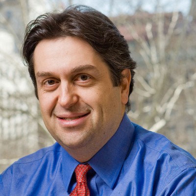 Ronald A. Ghossein合作论文数Memorial Sloan Kettering Cancer Center18Victor S Fang合作论文数17Leslie J. Degroot合作论文数17H BROCAS合作论文数17Caterina Di Cosmo合作论文数16T Sunthornthepvarakul合作论文数15Vatche Tchekmedyian合作论文数15Prasanna P Tamarapu合作论文数15Vincent P Reuter合作论文数15E VANCAUTER合作论文数15
 Elisa de Stanchina合作论文数Sloan Kettering Institute, Memorial Sloan Kettering Cancer Center15
 Mååg Jesper L V合作论文数15
 Saqcena Mahesh合作论文数From the Department of Biological Sciences, Hunter College of the City University of New York15Tiedje Vera合作论文数Klinik für Endokrinologie und Stoffwechselerkrankungen, Universitätsklinikum Essen.15
 Krishnamoorthy Gnana Prakasam合作论文数Dipartimento di Medicina Molecolare e Biotecnologie Mediche, University of Naples Federico II15
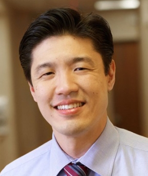 Alan L. Ho合作论文数David H. Koch Center for Cancer Care, Memorial Sloan Kettering Cancer Center;Division of Solid Tumor Oncology, Department of Medicine, Memorial Sloan Kettering Cancer Center15
 Ping Chi合作论文数Human Oncology and Pathogenesis Program, Memorial Sloan-Kettering Cancer Center/Department of Medicine, Memorial Sloan-Kettering Cancer Center/Department of Medicine, Weill Cornell Medical College15
 Luis Javier Leandro García合作论文数Spanish National Cancer Research Centre15Sabine Costagliola合作论文数15
 Richard Koche合作论文数Ctr Epigenet Res, Mem Sloan Kettering Canc Ctr15Georges Copinschi合作论文数14Nh Scherberg合作论文数13
 Juan Bernal合作论文数13Ferrara Alfonso Massimiliano合作论文数13
 Enio Martino合作论文数Department of Endocrinology and Metabolism, University of Pisa13Jacques Samarut合作论文数Université Lyon 1, Université de Lyon13Theodora Pappa合作论文数11
 Sirimon Reutrakul合作论文数11Luca Persani合作论文数11Joao Anselmo合作论文数11
 Daanniieell Desir合作论文数11David Sarne合作论文数10




