Background: There is little evidence of the biomechanical performance of medial collateral ligament (MCL) reconstructions for restoring stability to the MCL-deficient knee regarding valgus, external rotation (ER), and anteromedial rotatory instability (AMRI). Hypothesis: A short isometric reconstruction will better restore stability than a longer superficial MCL (sMCL) reconstruction, and an additional deep MCL (dMCL) graft will better control ER and AMRI than single-strand reconstructions. Study design: Controlled laboratory study. Methods: Nine cadaveric human knees were tested in a kinematics rig that allowed tibial loading while the knee was flexed-extended 0 degrees to 100 degrees. Optical markers were placed on the femur and tibia and displacements were measured using a stereo camera system. The knee was tested intact, and then after MCL (sMCL + dMCL) transection, and loaded in anterior tibial translation (ATT), ER, varus-valgus, and combined ATT + ER (AMRI loading). Five different isometric MCL reconstructions were tested: isolated long sMCL, a short construct, each with and without dMCL addition, and isolated dMCL reconstruction, using an 8 mm-wide synthetic graft. Results: MCL deficiency caused an increase in ER of 4 degrees at 0 degrees of flexion (P = .271) up to 14 degrees at 100 degrees of flexion (P = .002), and valgus laxity increased by 5 degrees to 8 degrees between 0 degrees and 100 degrees of flexion (P < .024 at 0 degrees-90 degrees). ATT did not increase significantly in isolated MCL deficiency (P > .999). All 5 reconstructions restored native stability across the arc of flexion apart from the isolated long sMCL, which demonstrated residual ER instability (P <= .047 vs other reconstructions). Conclusion: All tested techniques apart from the isolated long sMCL graft are satisfactory in the context of restoring the valgus, ER, and AMRI stability to the MCL-deficient knee in a cadaveric model.
Purpose: This work developed a novel preclinical test of total knee replacements (TKRs) in order to explain TKR instability linked to patient dissatisfaction. It was hypothesized that stability tests on the isolated moving prostheses would provide novel comparative data on the stability and kinematics among TKR designs. Methods: Three TKR designs, DePuy Synthes Attune MS, Stryker Triathlon and Zimmer Biomet Persona MC, were assessed using a robotic arm while flexing-extending 0-140 degrees. Tests imposed 710 N body weight combined with three tibial loads: no anterior-posterior (AP) force, 90 N anterior or 90 N posterior force. Other load effects were minimized and the kinematics was recorded. Each implant was tested six times to investigate the repeatability of the method. Data were analysed using statistical parametric mapping with one-way analysis of variance (ANOVA). If significance was found (p < 0.05), post hoc t tests with Bonferroni correction were used to contrast groups. Results: Significant differences were found throughout flexion-extension. Femoral rollback, AP stability, coupled internal-external rotation and AP position (roll-back) were all influenced by implant design. AP stability of the TKRs reduced with flexion reaching Attune 15 mm, Persona 13 mm and Triathlon 21 mm at 140 degrees flexion. Tractive rolling significantly affected kinematics in the less congruent Triathlon design, with 6 mm different paths between flexion and extension motion (p < 0.05 across 5-100 degrees). Paradoxical anterior femoral sliding in early flexion (0-40 degrees) occurred in Persona and Triathlon designs. Conclusions: The novel testing technique provides, for the first time, comparative data on the inherent stability and kinematics of the TKR implants themselves across the arc of flexion-extension, independent of variables including soft tissue behaviour and surgical technique. The data show how much each prosthesis can contribute to the stability and motion of the implanted knee. Similar data from a wider range of designs will enable more informed decisions regarding implant design choice, aiming to reduce the prevalence of TKR instability in patients.
Zusammenfassung Hintergrund Verletzungen des vorderen Kreuzbands (VKB), der Kaplan-Fasern (KF), der anterolateralen Kapsel/des anterolateralen Ligaments (C/ALL) und der posterioren Wurzel des lateralen Meniskus (LMPR) wurden getrennt voneinander mit anterolateraler Instabilität assoziiert. Ziel war es, die Bedeutung dieser Strukturen für die Kniestabilität zu untersuchen. Methoden Zehn fresh-frozen humane Kniekadaver wurden robotergestützt von 0° bis 90° Flexion getestet. Eine anterior-posteriore Kraft von 88 N, ein Innen-Außen-Rotationsdrehmoment von 5 N/m und ein Valgus-Varus-Drehmoment von 8 N/m wurden aufgebracht und die Kinematik des intakten Knies aufgezeichnet. Weitere 10 Kniegelenke wurden in einem Kinematikprüfstand mit optischem Tracking getestet, um die Knielaxität nach sequenziellem Durchtrennen der o. g. Strukturen über 0° bis 110° Flexion zu messen. Ergebnisse Das VKB war der primäre Hemmer für die anteriore tibiale Translation (ATT); andere Strukturen waren unbedeutend (< 10 %). Die KF und C/ALL hemmten die Innenrotation (IR) und erreichten 44 % ± 23 % ( p < 0,01) und 14 % ± 13 % ( p < 0,05) bei 90°. Die LMPR hemmte die Valgus-, aber nicht die Innenrotation. Die ATT erhöhte sich bei VKB-Insuffizienz ( p < 0,001) und nach der Durchtrennung der lateralen Strukturen von 70° auf 100° ( p < 0,05). Die anterolaterale Rotationsinstabilität beim Pivot-Shift-Manöver wurde nach VKB-Durchtrennung von 0° auf 40° ( p < 0,05) und weiter nach Durchtrennung der lateralen Strukturen von 0° auf 100° ( p < 0,01) erhöht. Schlussfolgerung Der anterolaterale Komplex fungiert als funktionelle Einheit und sorgt für Rotationsstabilität. Das VKB ist der primäre Stabilisator für die ATT. Die KF sind der wichtigste Hemmer der IR ab 30° Flexion. Eine kombinierte Verletzung der KF mitsamt der C/ALL erhöhte die anterolaterale Rotationsinstabilität beträchtlich, während eine isolierte Verletzung der KF oder C/ALL dies nicht tat. Eine LMPR-Insuffizienz verursachte keine signifikante Instabilität bei intaktem VKB.
Background: Injuries of the anterior cruciate ligament (ACL), Kaplan fibers (KF), anterolateral capsule/anterolateral ligament (C/ALL), and lateral meniscus posterior root (LMPR) have been separately linked to anterolateral instability. The aim was to investigate the importance of these structures for knee stability. Methods: In this study 10 fresh-frozen human knees were robotically tested from 0 degrees to 90 degrees of flexion. An anteroposterior force of 88N, an internal-external rotational torque of 5N/m, and a valgus-varus torque of 8 N/m were applied and the kinematics of the intact knee were recorded. Another 10 knees were tested in a kinematics rig with optical tracking to measure knee laxity after sequentially severing the structures over 0 degrees-110 degrees of flexion. Results: The ACL was the primary restraint for anterior tibial translation (ATT); other structures were insignificant (< 10%). The KF and C/ALL resisted internal rotation (IR), achieving 44 +/- 23% (p< 0.01) and 14 +/- 13% (p< 0.05) at 90 degrees, respectively. The LMPR resisted valgus rotation but not IR. The ATT increased from 70 degrees to 100 degrees (p< 0.05) after ACL insufficiency (p< 0.001) and after severing the lateral structures. The pivot-shift maneuver increased anterolateral rotational instability after ACL transection from 0 degrees to 40 degrees (p< 0.05) and further after severing the lateral structures from 0 degrees to 100 degrees (p< 0.01). Conclusion: The anterolateral complex acts as a functional unit to provide rotational stability. The ACL is the primary stabilizer for ATT. The KFs are the most important IR restraint above 30 degrees of flexion. A combined KF and C/ALL injury substantially increased anterolateral rotational instability, while an isolated injury of the KF or C/ALL did not. An LMPR insufficiency did not cause significant instability with an intact ACL.
AbstractIntroductionPrevious research has shown that, notwithstanding ligament healing, properly selected MCL reconstruction can restore normal knee stability after MCL rupture. The hypothesis of this work was that it is possible to restore knee stability (particularly valgus and AMRI) with simplified and/or less-invasive MCL reconstruction methods.MethodsNine unpaired human knees were cleaned of skin and fat, then digitization screws and optical trackers were attached to the femur and tibia. A Polaris stereo camera measured knee kinematics across 0o-100o flexion when the knee was unloaded then with 90N anterior-posterior force, 9Nm varus-valgus moment, 5Nm internal-external rotation, and external+anterior (AMRI) loading. The test was conducted for the following knee conditions: intact, injured: transected superficial and deep MCL (sMCL and dMCL), and five reconstructions: (long sMCL, long sMCL+dMCL, dMCL, short sMCL+dMCL, short sMCL), all based on the medial epicondyle isometric point and using 8mm tape as a graft, with long sMCL 60mm below the joint line (anatomical), short sMCL 30mm, dMCL 10mm (anatomical).ResultsNo significant changes were found in anterior or posterior translation, or varus at any stage. MCL deficiency caused increased valgus, external rotation and AMRI instabilities. All reconstructions restored valgus stability. The isolated long sMCL allowed residual external rotation and AMRI instability, while the short sMCL did stabilise AMRI. Both 2-strand reconstructions (dMCL+sMCL) restored stability.ConclusionAll tested techniques, except long sMCL, restored valgus and AMRI stability of the knee. The single femoral tunnel is satisfactory for both the dMCL and sMCL grafts.
AbstractIntroductionMid-flexion instability may cause poor outcomes following TKA. Surgical technique, patient-specific factors, and implant design could all contribute to it, with modelling and fluoroscopy data suggesting the latter may be the root cause. However, current implants all pass the preclinical stability testing standards, making it difficult to understand the effects of implant design on instability. We hypothesized that a more physiological test, analysing functional stability across the range of knee flexion-extension, could delineate the effects of design, independent of surgical technique and patient-specific factors.MethodsUsing a SIMvitro-controlled six-degree-of-freedom robot, a dynamic stability test was developed, including continuous flexion and reporting data in a trans-epicondylar axis system. 3 femoral geometries were tested: gradually reducing radius, multi-radius and single-radius, with their respective tibial inserts. 710N of compression force (body weight) was applied to the implants as they were flexed from 0–140° with three levels of anterior/posterior (AP) tibial force applied (−90N,0N,90N).ResultsWhile in static tests, the implants performed similarly, functional stability testing revealed different paths of motion and AP laxities throughout the flexion cycle. Some designs exhibited mid-flexion instability, while others did not: the multi-radius design allowed increased AP laxity as it transitioned to each arc of reduced femoral component radius; the single-radius design had low tibial bearing conformity, allowing 16mm difference in the paths of mid-flexion versus extension motion.ConclusionsPreclinical lab testing reveals functional differences between different design philosophies. Implant design impacts kinematics and mid-flexion stability, even before factoring in surgical technique and patient-specific factors.
Background: Injuries to the anterior cruciate ligament (ACL), Kaplan fibers (KFs), anterolateral capsule/ligament (C/ALL), and lateral meniscus posterior root (LMPR) have been separately linked to anterolateral instability. Purpose: To investigate the contributions of the ACL, KFs, C/ALL, and LMPR to knee stability and to measure instabilities resulting from their injury. Study Design: Controlled laboratory study. Methods: Ten fresh-frozen human knees were tested robotically to determine restraints of knee laxity at 0° to 90° of flexion. An 88-N anterior-posterior force (anterior and posterior tibial translation), 5-N·m internal-external rotation, and 8-N·m valgus-varus torque were imposed and intact kinematics recorded. The kinematics were replayed after sequentially cutting the structures (order varied) to calculate their contributions to stability. Another 10 knees were tested in a kinematics rig with optical tracking to measure instabilities after sequentially cutting the structures across 0° to 100° of flexion. One- and 2-way repeated-measures analyses of variance with Bonferroni correction were used to find significance ( P < .05) for the robotic and kinematics tests. Results: The ACL was the primary restraint for anterior tibial translation; other structures were insignificant (<10% contribution). The KFs and C/ALL resisted internal rotation, reaching 44% ± 23% (mean ± SD; P < .01) and 14% ± 13% ( P < .05) at 90°. The LMPR resisted valgus but not internal rotation. Anterior tibial translation increased after ACL transection ( P < .001) and after cutting the lateral structures from 70° to 100° ( P < .05). Pivot-shift loading increased anterolateral rotational instability after ACL transection from 0° to 40° ( P < .05) and further after cutting the lateral structures from 0° to 100° ( P < .01). Conclusion: The anterolateral complex acts as a functional unit to provide rotatory stability. The ACL is the primary stabilizer for anterior tibial translation. The KFs are the most important internal rotation restraint >30° of flexion. Combined KFs + C/ALL injury substantially increased anterolateral rotational instability while isolated injury of either did not. LMPR deficiency did not cause significant instability with the ACL intact. Clinical Relevance: This study is a comprehensive biomechanical sectioning investigation of the knee stability contributions of the ACL, anterolateral complex, and LMPR and the instability after their transection. The ACL is significant in controlling internal rotation only in extension. In flexion, the KFs are dominant, synergistic with the C/ALL. LMPR tear has an insignificant effect with the ACL intact.
Background: Although a medial collateral ligament (MCL) injury is associated with anteromedial rotatory instability (AMRI) and often with an anterior cruciate ligament (ACL) injury, there has been little work to develop anteromedial (AM) reconstruction to address this laxity. Purpose: To measure the ability of a novel “anatomic” AM reconstruction technique to restore native knee laxity for isolated AM insufficiency and combined AM plus posteromedial insufficiency. Study Design: Controlled laboratory study. Methods: A total of 12 cadaveric knees were mounted in a kinematic testing rig that allowed the tibia to be loaded while the knee flexed-extended 0° to 100° with 88-N anteroposterior translation, 5-N·m internal rotation–external rotation (ER), 8-N·m valgus, and combined anterior translation plus ER to simulate AMRI. Joint motion was measured using optical trackers with the knee intact, after superficial MCL (sMCL) and deep MCL (dMCL) transection, and after AM reconstruction of the sMCL and dMCL with semitendinosus autografts. The posteromedial capsule (PMC)/posterior oblique ligament (POL) was then transected to induce a grade 3 medial injury, and kinematic measurements were repeated afterward and again after removing the grafts. Laxity changes were examined using repeated-measures analysis of variance and post-testing. Results: sMCL and dMCL deficiency increased valgus, ER, and AMRI laxities. These laxities did not differ from native values after AM reconstruction. Additional PMC/POL deficiency did not increase these laxities significantly but did increase internal rotation laxity near knee extension; this was not controlled by AM reconstruction. Conclusion: AM reconstruction eliminated AMRI after transection of the dMCL and sMCL, and also eliminated AMRI after additional PMC/POL transection. Clinical Relevance: Many MCL injuries occur in combination with ACL injuries, causing AMRI. These injuries may rupture the AM capsule and dMCL. Unaddressed MCL deficiency leads to an increased ACL reconstruction failure rate. A dMCL construct oriented anterodistally across the medial joint line, along with an sMCL graft, can restore native knee ER laxity. PMC/POL lesions did not contribute to AMRI.
BACKGROUND:There are many descriptions of medial collateral ligament (MCL) reconstruction, but they may not reproduce the anatomic structures and there is little evidence of their biomechanical performance.PURPOSE:To investigate the ability of "anatomic" MCL reconstruction to restore native stability after grade III MCL plus posteromedial capsule/posterior oblique ligament injuries in vitro.STUDY DESIGN:Controlled laboratory study.METHODS:Twelve cadaveric knees were mounted in a kinematic testing rig to impose tibial displacing loads while the knee was flexed-extended: 88-N anteroposterior translation, 5-N·m internal-external rotation, 8-N·m valgus-varus, and combined anterior translation plus external rotation (anteromedial rotatory instability). Joint motion was measured via optical trackers with the knee intact; after superficial MCL (sMCL), deep MCL (dMCL), and posterior oblique ligament transection; and then after MCL double- and triple-strand reconstructions. Double strands reproduced the sMCL and posterior oblique ligament and triple-strands the sMCL, dMCL, and posterior oblique ligament. The sMCL was placed 5 mm posterior to the epicondyle in the double-strand technique and at the epicondyle in the triple-strand technique. Kinematic changes were examined by repeated measures 2-way analysis of variance with posttesting.RESULTS:Transection of the sMCL, dMCL, and posterior oblique ligament increased valgus rotation (5° mean) and external rotation (9° mean). The double-strand reconstruction controlled valgus in extension but allowed 5° excess valgus in flexion and did not restore external rotation (7° excess). The triple-strand reconstruction restored both external rotation and valgus throughout flexion.CONCLUSION:In a cadaveric model, a triple-strand reconstruction including a dMCL graft restored native external rotation, while a double-strand reconstruction without a dMCL graft did not. A reconstruction with the sMCL graft placed isometrically on the medial epicondyle restored valgus rotation across the arc of knee flexion, whereas a reconstruction with a more posteriorly placed sMCL graft slackened with knee flexion.CLINICAL RELEVANCE:An MCL injury may rupture the anteromedial capsule and dMCL, causing anteromedial rotatory instability. Persistent MCL instability increases the likelihood of ACL graft failure after combined injury. A reconstruction with an anteromedial dMCL graft restored native external rotation, which may help to unload/protect an ACL graft. It is important to locate the sMCL graft isometrically at the femoral epicondyle to restore valgus across flexion.
Treating open fractures in long bones can be challenging and if not performed properly can lead to poor outcomes such as mal/non-union, deformity, and amputation. One of the most common methods of treating these fracture types is temporary external fixation followed by definitive fixation. The shortage of high-quality affordable external fixators is a long-recognised need, particularly in Low- and Middle-Income Countries (LMICs). This research aimed to develop a low-cost device that can be manufactured locally to international standards. This can provide surge capacity for conflict zones or in response to unpredictable incidents and situations. The fixator presented here and developed by us, the Imperial external fixator, was tested on femur and tibia specimens under 100 cycles of 100 N compression-tension and the results were compared with those of the Stryker Hoffmann 3 frame. The Imperial device was stiffer than the Stryker Hoffmann 3 with a lower median interfragmentary motion (of 0.94 vs. 1.48 mm). The low-cost, easy to use, relatively lightweight, and easy to manufacture (since minimum skillset and basic workshop equipment and materials are needed) device can address a critical shortage and need in LMICs particularly in conflict-affected regions with unpredictable demand and supply. The device is currently being piloted in three countries for road traffic accidents, gunshot wounds and other conflict trauma—including blast cohorts.
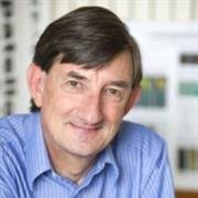 Andrew Amis合作论文数Department of Mechanical Engineering, Faculty of Engineering, Imperial College London9
 Andy Williams合作论文数7
 Simon Ball合作论文数4
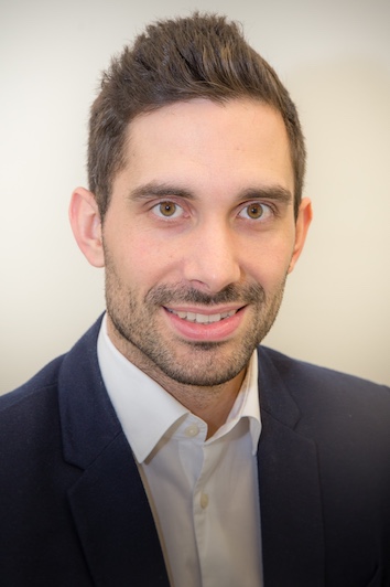 Lukas Willinger合作论文数Department of Trauma Surgery, Technische Universität München3
 Kiron Athwal合作论文数3
 Andreas B. Imhoff合作论文数School of Medicine, Technical University of Munich3Nobuaki Miyaji合作论文数2Jobe Shatrov合作论文数2
 Richard Jan van Arkel合作论文数Department of Mechanical Engineering, Imperial College London2
 Angela Brivio合作论文数1
 Giovanni Milandri合作论文数1Mehdi Saeidi合作论文数1
 Ricardo Bastos合作论文数Clin Dragao Espregueira Mendes Sports Ctr1David Barrett合作论文数1
 Anthony Bull合作论文数Department of Bioengineering, Faculty of Engineering, Imperial College London;Smart Surgical Solutions;GO Assistive Technology;Biomex1
 Michael Berthaume合作论文数Department of Mechanical &Industrial Engineering, University of Massachusetts;Department of Mechanical & Industrial Engineering, University of Massachusetts1
 Jonathan Jeffers合作论文数1
 Joao Espregueira-Mendes合作论文数H.S. Sebastião and Porto University1




