Escherichia coli cells can grow in a DnaA- and oriC-independent manner in rnhA-deficient cells that lack the major RNaseH activity. This mode of replication is named cSDR (constitutive stable DNA replication) and is believed to initiate from the sites of RNA-DNA hybrids, although the detailed mechanisms of cSDR are unclear. In this study, we discovered that dif sequence and XerC/D are essential for cSDR-dependent growth. cSDR was observed in either dif or xerC/D mutant cells as efficiently as in the control cells, as measured by the incorporation of [3H]-thymine. However, we found that these mutants accumulate cells with extra DNA contents in the presence of rifampicin and cefalexin, suggesting accumulation of unresolved dimeric or multimeric chromosomes or that of catenated chromosomes due to potential problems in resolution of the replicated chromosomes or in decatenation process. These results indicate that site-specific recombination mediated by dif-XerC/D is essential for cSDR-dependent growth.
Abstract Guanine-rich nucleic acid sequences can fold into G-quadruplex (G4) structures that regulate DNA replication, transcription, and translation. Fanconi anemia group J helicase (FANCJ) resolves G4 structures at stalled replication forks. Despite its central role in genome maintenance, the molecular basis of G4 recognition and unwinding by FANCJ has remained unclear. Here, we report cryo-EM structures of human FANCJ bound to a G4-containing DNA substrate and ATPγS. The structures reveal direct engagement of the G4 by the Fe–S domain. Structure-guided mutagenesis demonstrates that this interface is essential for G4 binding and unwinding. The structures further capture open and closed conformational states linked to ATP hydrolysis, providing a mechanism for directional translocation along 5′ ssDNA and progressive G4 unwinding. Together, these findings establish the structural basis of G4 recognition by FANCJ and provide mechanistic insights into how disease-associated mutations linked to Fanconi anemia and breast cancer impair helicase function.
Migratory cells tend to have soft nuclei that deform and penetrate narrow spaces1,2. Extensive nuclear deformation during migration can cause nuclear-envelope rupture and DNA damage in cancer cells, which may contribute to malignant transformation during tumour progression3–6. However, the importance of DNA damage in physiological migration is less well understood. Here we demonstrate that the migration of neurons in developing cerebral and cerebellar cortices is accompanied by massive DNA double-stranded breaks (DSBs) due to mechanostress during passage through narrow interstitial spaces. In contrast to many other migratory cells, these DSBs occur without detectable nuclear envelope rupture. Confined migration increases topoisomerase-IIβ covalently bound DSBs, and these lesions are repaired through non-homologous end-joining during brain development without causing cell death. Genome sequencing revealed that DSBs tend to occur at transcriptionally inactive regions. The deletion of ligase IV at the onset of neuronal migration leads to persistent DSB accumulation in cerebellar neurons with moderate transcriptional changes in genes related to synaptic function, neuronal development and stress and immune responses. The mutant mouse develops mild motor deficits in later life, suggesting that the DNA damage generated during normal brain development poses a potential disease risk if left unrepaired. The migration of neurons in developing cerebral and cerebellar cortices is accompanied by massive DNA double-strand breaks due to mechanostress during passage through narrow interstitial spaces.
Abstract BRCA2 plays a central role in maintaining genome integrity through homologous recombination and replication-fork protection, yet the compensatory networks sustaining BRCA2 -deficient cells remain unclear. Here we show BRCA2 enforces a homeostatic mechanism aligning mitochondrial respiration with DNA repair capacity in both cancer and non-cancer contexts. Genome-wide CRISPR screening identified glutathione metabolism and base-excision repair as the key compensatory networks sustaining BRCA2 -deficient cells by detoxifying mitochondria-derived reactive oxygen species. BRCA2 loss provokes an acute mitochondrial ROS surge, causing 8-oxoguanine accumulation and a systemic metabolic crisis marked by NAD + and glutathione depletion. PARP inhibitor targets DNA replication vulnerabilities, increasing the cellular requirement for BRCA2. The resulting oxidative burden primes cells for TP53 -dependent apoptosis in G 1 during olaparib treatment, which extends cytotoxicity beyond canonical S-phase stress. These findings indicate BRCA2 prevents metabolic flux from outpacing repair capacity, providing a rationale for combining PARP inhibition with redox modulation to enhance efficacy and overcome resistance. Highlights Acute BRCA2 loss induces ROS and mitochondrial dysfunction creating a metabolic scar Oxidative lesions drive PARP hyperactivation and precipitate a cellular NAD crisis PARP inhibitors provoke TP53-dependent apoptosis in G 1 beyond replication stress in S phase Glutathione deficiency exacerbates bone marrow failure under BRCA2 depletion BRCA2 tightly couples mitochondrial redox homeostasis to genomic maintenance
Prostate cancers (PCa) harboring CDK12 alterations exhibit aggressive clinical behavior and poor responses to current therapeutic strategies. Although CDK12 is categorized as a homologous recombination repair (HRR)-related gene, clinical evidence indicates limited benefit of poly(ADP-ribose) polymerase (PARP) inhibitor monotherapy in CDK12-defective tumors. To clarify the functional consequences of CDK12 loss and identify a mechanistically rational therapeutic approach, we generated CDK12-knockout (KO) PCa cell lines using CRISPR/Cas9 and assessed cell-cycle regulation, DNA damage repair (DDR), and replication stress by flow cytometry, γH2AX immunofluorescence, and DNA fiber assays. CDK12 knockout abrogated the G0/G1 checkpoint, permitting cell cycle progression into M phase despite the persistence of unrepaired DNA double-strand breaks (DSBs), and reduced ATM transcription, resulting in a DSB response phenotype resembling ATM deficiency. CDK12 loss also increased intrinsic replication stress and sensitized PCa cells to ATR inhibition. In patient-derived xenograft models harboring CDK12 alterations, combined PARP and ATR inhibition led to enhanced DSB accumulation and selectively suppressed tumor growth in CDK12-defective models. These findings highlight the importance of interrogating individual genes within the HRR pathway to define distinct mechanistic vulnerabilities and provide a strong rationale for combined PARP and ATR inhibition as a novel therapeutic strategy for patients with CDK12-altered prostate cancer.
Poly(ADP-ribose) glycohydrolase (PARG) is the primary enzyme responsible for degrading poly(ADP-ribose) (PAR) chains generated by poly(ADP-ribose) polymerases (PARPs) during DNA damage responses. Although pharmacological inhibition of PARG has emerged as a promising anticancer strategy targeting PAR metabolism, its cytotoxic mechanisms and the tumor contexts most susceptible to PARG inhibition remain incompletely understood. Here, we developed the novel PARG inhibitors, MOD000568 and MOD000582, and demonstrated their antitumor efficacy and tolerability in mouse xenograft models. Through an siRNA screen and biological experiments using genetically modified cells, we found that cellular vulnerability to PARG inhibition is strongly associated with elevated PAR metabolism, particularly base excision repair (BER) deficiency. Mechanistically, PARG inhibitors induced progressive accumulation of PARylated proteins, accompanied by nuclear translocation of apoptosis-inducing factor, a hallmark of parthanatos, caspase-dependent apoptosis, and mitochondrial dysfunction, while intracellular NAD⁺ depletion was not the primary determinant of cell death. Importantly, PARG inhibitor–induced cytotoxicity occurred largely independently of DNA replication and extended to G1-arrested and senescence-like cells. Although BER deficiency enhanced sensitivity, genetic defects were not required, indicating that both BER deficiency and PAR-inducing stresses increase susceptibility by elevating PAR metabolism. These findings support a model in which PAR accumulation beyond a critical threshold determines cell fate and identify PARG inhibition as a therapeutic strategy for tumors with elevated PAR metabolism, including non-proliferating and therapy-resistant cell populations.
Ataxia-telangiectasia mutated and RAD3-related (ATR) and its partner ATR-interacting protein (ATRIP) function as a critical proximal sensor and transducer of the DNA damage response (DDR). Several ATR substrates, including p53 and CHK1, are crucial for the coordination of cell cycle phase transitions, transcription, and DNA repair when cells sustain DNA damage. While much is known about ATR activation mechanisms, it is less clear how ATR signaling is negatively regulated in cells. Here, we identify the DNA repair protein REV7 as a novel direct binding partner of ATRIP. We define a REV7-interaction motif in ATRIP, which, when mutated, abrogates the REV7-ATRIP interaction in vitro and in intact cells. Using in vitro kinase assays, we show that REV7 inhibits ATR-mediated phosphorylation of its substrates, including p53. Disruption of the REV7-ATRIP interaction also enhances phosphorylation of CHK1 at Ser317 in intact cells. Taken together, our results establish REV7 as a critical negative regulator of ATR signaling. REV7 has pleiotropic roles in multiple DDR pathways, including Translesion Synthesis, DNA double-strand break resection, and p53 stability and may play a central role in the integration of multiple genome maintenance pathways.
Migratory cells tend to have soft nuclei that deform and penetrate narrow spaces[1][1],[2][2]. Extensive nuclear deformation during migration can cause nuclear envelope rupture and DNA damage in cancer cells, which may contribute to the malignant transformation during tumor progression[3][3],[4][4],[5][5],[6][6]. However, the significance of DNA damage in physiological migration is less well understood. Here, we demonstrate that the migration of neurons in developing cerebral and cerebellar cortices is accompanied by massive DNA double-strand breaks (DSBs) due to mechanostress during passage through narrow interstitial spaces. Confined migration enhances the binding and cleavage of the genome by topoisomerase IIβ, expressed in neuronal nucleus, independently of the nuclear envelope rupture. Genome sequencing revealed that DSBs tend to occur outside of protein-coding regions and transcription regulatory regions. During normal development, DSBs are rapidly repaired by the non-homologous end joining pathway. The deletion of ligase IV at the onset of neuronal migration leads to persistent DSB accumulation in cerebellar neurons with moderate transcriptional changes in genes related to synaptic function, neuronal development, and stress and immune responses. The mutant mouse develops mild motor deficits in later life, suggesting that the DNA damage generated during normal brain development poses a potential disease risk if left unrepaired. ### Competing Interest Statement The authors have declared no competing interest. [1]: #ref-1 [2]: #ref-2 [3]: #ref-3 [4]: #ref-4 [5]: #ref-5 [6]: #ref-6
G-quadruplexes (G4s) regulate multiple biological processes by interacting with G4-binding proteins (G4BPs). We developed a sandwich ELISA system to evaluate G4 ligand effects on G4-G4BP binding. G4 ligands strongly affect G-quartet-protein interactions, demonstrating that high binding affinity of G4 ligands for G4s does not guarantee ternary complex formation with G4BPs.
Integration of DNA replication with DNA repair, cell cycle progression, and other biological processes is crucial for preserving genome stability and fundamentally important for all life. Ataxia-telangiectasia mutated and RAD3-related (ATR) and its partner ATR-interacting protein (ATRIP) function as a critical proximal sensor and transducer of the DNA Damage Response (DDR). Several ATR substrates, including p53 and CHK1, are crucial for coordination of cell cycle phase transitions, transcription, and DNA repair when cells sustain DNA damage. While much is known about ATR activation mechanisms, it is less clear how ATR signaling is negatively regulated in cells. Here, we identify the DNA repair protein REV7 as a novel direct binding partner of ATRIP. We define a REV7-interaction motif in ATRIP, which when mutated abrogates the REV7-ATRIP interaction in vitro and in intact cells. Using in vitro kinase assays, we show that REV7 inhibits ATR-mediated phosphorylation of its substrates, including p53. Disruption of the REV7-ATRIP interaction also enhances phosphorylation of CHK1 at Ser317 (a known ATR target site) in intact cells. Taken together our results establish REV7 as a critical negative regulator of ATR signaling. REV7 has pleiotropic roles in multiple DDR pathways including Trans-Lesion Synthesis, DNA Double-Strand Break resection, and p53 stability and may play a central role in the integration of multiple genome maintenance pathways.
Acetaldehyde, a chemical that can cause DNA damage and contribute to cancer, is prevalently present in our environment, e.g. in alcohol, tobacco, and food. Although aldehyde potentially promotes crosslinking reactions among biological substances including DNA, RNA, and protein, it remains unclear what types of DNA damage are caused by acetaldehyde and how they are repaired. In this study, we explored mechanisms involved in the repair of acetaldehyde-induced DNA damage by examining the cellular sensitivity to acetaldehyde in the collection of human TK6 mutant deficient in each genome maintenance system. Among the mutants, mismatch repair mutants did not show hypersensitivity to acetaldehyde, while mutants deficient in base and nucleotide excision repair pathways or homologous recombination (HR) exhibited higher sensitivity to acetaldehyde than did wild-type cells. We found that acetaldehyde-induced RAD51 foci representing HR intermediates were prolonged in HR-deficient cells. These results indicate a pivotal role of HR in the repair of acetaldehyde-induced DNA damage. These results suggest that acetaldehyde causes complex DNA damages that require various types of repair pathways. Mutants deficient in the removal of protein adducts from DNA ends such as TDP1-/- and TDP2-/- cells exhibited hypersensitivity to acetaldehyde. Strikingly, the double mutant deficient in both TDP1 and RAD54 showed similar sensitivity to each single mutant. This epistatic relationship between TDP1-/- and RAD54-/- suggests that the protein-DNA adducts generated by acetaldehyde need to be removed for efficient repair by HR. Our study would help understand the molecular mechanism of the genotoxic and mutagenic effects of acetaldehyde.
RNA-DNA hybrid is a part of the R-loop which is an important non-standard nucleic acid structure. RNA-DNA hybrid/R-loop causes genomic instability by inducing DNA damages or inhibiting DNA replication. It also plays biologically important roles in regulation of transcription, replication, recombination and repair. Here, we have employed catalytically inactive human RNase H1 mutant (D145N) to visualize RNA-DNA hybrids and map their genomic locations in fission yeast cells. The RNA-DNA hybrids appear as multiple nuclear foci in rnh1∆rnh201∆ cells lacking cellular RNase H activity, but not in the wild-type. The majority of RNA-DNA hybrid loci are detected at the protein coding regions and tRNA. In rnh1∆rnh201∆ cells, cells with multiple Rad52 foci increase during S-phase and about 20% of the RNA-DNA hybrids overlap with Rad52 loci. During S-phase, more robust association of Rad52 with RNA-DNA hybrids was observed in the protein coding region than in M-phase. These results suggest that persistent RNA-DNA hybrids in the protein coding region in rnh1∆rnh201∆ cells generate DNA damages during S-phase, potentially through collision with DNA replication forks.
RecA protein and RecA/Rad51 orthologues are required for homologous recombination and DNA repair in all living creatures. RecA/Rad51 catalyzes formation of the D-loop, an obligatory recombination intermediate, through an ATP-dependent reaction consisting of two phases: homology recognition between double-stranded (ds)DNA and single-stranded (ss)DNA to form a hybrid-duplex core of 6-8 base pairs and subsequent hybrid-duplex/D-loop processing. How dsDNA recognizes homologous ssDNA is controversial. The aromatic residue at the tip of the β-hairpin loop (L2) was shown to stabilize dsDNA-strand separation. We tested a model in which dsDNA strands were separated by the aromatic residue before homology recognition and found that the aromatic residue was not essential to homology recognition, but was required for D-loop processing. Contrary to the model, we found that the double helix was not unwound even a single turn during search for sequence homology, but rather was unwound only after the homologous sequence was recognized. These results suggest that dsDNA recognizes its homologous ssDNA before strand separation. The search for homologous sequence with homologous ssDNA without dsDNA-strand separation does not generate stress within the dsDNA; this would be an advantage for dsDNA to express homology-dependent functions in vivo and also in vitro.
REV7 is an abundant, multifunctional protein that is a known factor in cell cycle regulation and in several key DNA repair pathways including Trans-Lesion Synthesis (TLS), the Fanconi Anemia (FA) pathway, and DNA Double-Strand Break (DSB) repair pathway choice. Thus far, no direct role has been studied for REV7 in the DNA damage response (DDR) signaling pathway. Here we describe a novel function for REV7 in DSB-induced p53 signaling. We show that REV7 binds directly to p53 to block ATM-dependent p53 Ser15 phosphorylation. We also report that REV7 is involved in the destabilization of p53. These findings affirm REV7's participation in fundamental cell cycle and DNA repair pathways. Furthermore, they highlight REV7 as a critical factor for the integration of multiple processes that determine viability and genome stability.
TDP1 expression in human cancer cells. TDP1 expression in the CCLE (A) or TCGA (B) databases. TDP1 messenger RNA (mRNA) expression was determined by Affymetrix's GeneChip array in CCLE (A) or RNA sequencing technology in TCGA (B). TDP1 expression is shown. Each dot represents a single cell line (A) or tumor specimen (B).
Schematic representation of the strategy used to knockout TDP1 by CRISPR/Cas9 in TSCER2 (A) or HCT116 (B) cells. RT-PCR of TDP1 mRNA in TSCER2 is shown (A).
e17042 Background: CDK12 is considered as a gene involved in homologous recombination repair (HRR); however, recent clinical trials have shown poor benefit of poly(ADP-ribose) polymerase(PARP) inhibitors in prostate cancer (PCa) patients with CDK12 alterations. Since CDK12 is the third most commonly altered gene in the HRR pathway in PCa, development of a new treatment strategy is urgently needed. Methods: To understand the biological consequence of CDK12 alteration, CDK12 was knocked out in LNCaP using CRISPR-Cas9 system to generate CDK12KO cells. Cell growth and cell cycle were evaluated as well as HRR efficiency. Downstream genes were evaluated by RT-PCR and western blots. Combination therapy by a PARP inhibitor and ATR inhibitors were tested using two lines of patient derived xenografts (PDX) established from tissues of patients with deleterious CDK12 variants. Three different ATR inhibitors were tested to confirm reproducibility. Results: In LNCaP, CDK12 knockout enhanced cell cycle progression until the M phase, however, cell cycle was stalled at the M phase with accumulation chromosomal aberration, suggesting that rapidly progressive CDK12 altered tumors clinically encountered harbor a mechanism of escaping the M phase checkpoint without DNA repair. γH2AX was accumulated in CDK12KO after treatment with CPT11, consistent with impaired HRR in CDK12 KO cells. Analysis of HRR associated genes showed that in PCa, CDK12 knockout leads to transcriptional downregulation of ATM but not the other HRR associated genes. Expression patterns of downstream genes after double strand break (DSB) induction in CDK12 and ATM knockout cells suggested that in PCa, CDK12 regulates HRR through regulation of ATM. Since synthetic lethality of PARP and ATR inhibition has been reported in ATM deficient PCa, 2 PDX lines with CDK12 alterations were administered the combination therapy. Tumor growth suppression and accumulation of γH2AX in tumor tissue was observed when treated by the combination therapy, but not with either drug alone. Either drug alone or in combination did not affect the growth of a PDX established from a tissue of a patient with wild-type CDK12. Conclusions: In PCa, CDK12 acts through transcriptional regulation of ATM, and combination treatment with PARP inhibitor and ATR inhibitor could be a potential treatment strategy in PCa with CDK12 alteration.
 Shunichi Takeda合作论文数Medicine School, Shenzhen University;Graduate School of Medicine, Kyoto University56
 Masataka Tsuda合作论文数24
 Kouji Hirota合作论文数Genetic Dynamics Research;Cellular & Molecular Biology Laboratory;Department of Biophysics;Graduate School of Science;University of Tokyo;Department of Biochemistry and Molecular Biology;University of Arkansas for Medical Sciences17
 Keizo Tano合作论文数10
 Remi Akagawa合作论文数9
 Masamitsu Honma合作论文数Division of Genetics and Mutagenesis, National Institute of Health Sciences9
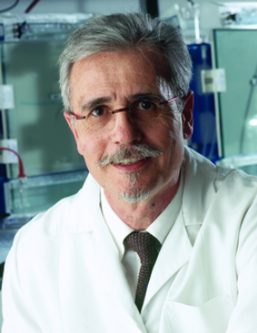 Yves Pommier合作论文数Developmental Therapeutics Branch and Laboratory of Molecular Pharmacology, National Cancer Institute, National Institutes of Health9
 Liton Saha合作论文数Department of Radiation Genetics, Kyoto University, Graduate School of Medicine, Yoshida Konoe, Sakyo-ku, Kyoto, 606-8501, Japan.8Shintaro Yamada合作论文数8
 Kunihiro Ohta合作论文数7
 Shin-Ichiro Masunaga (増永慎一郎)合作论文数Particle Radiation Oncology Research Center, Research Reactor Institute, Kyoto University7
 Masakazu Toi合作论文数Kyoto University Hospital;Tokyo Metropolitan Cancer and Infectious Disease Center, Komagome Hospital7
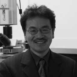 Akira Shinohara合作论文数Inst Prot Res, Osaka Univ7Takehiko Shibata合作论文数6Suguru Morimoto合作论文数6Huang Shar-yin N合作论文数6Junji Itou合作论文数6
 Miki Shinohara合作论文数6
 Minoru Takata (髙田穰)合作论文数Division of Systemic Life Science, Graduate School of Biostudies, Kyoto University;Radiation Biology Center, Kyoto University6
 Tetsushi Sakuma合作论文数5
 Md Maminur Rahman合作论文数5
 Salma Akter合作论文数Dept Radiat Genet, Kyoto Univ5
 Takahito Moriwaki合作论文数Graduate School of Science, Kyoto University5
 Shamima Islam Keka合作论文数5Hiroshi Ide (井出博)合作论文数Graduate School of Integrated Sciences for Life, Hiroshima University5
 Takashi Yamamoto (山本卓)合作论文数Graduate School of Integrated Sciences for Life, Hiroshima University5Kazuto Kugou合作论文数5Tomoyuki Fukuda合作论文数5Hisao Masai合作论文数5
 Nguyen Ngoc Hoa合作论文数4Kei Kadoda合作论文数4Naoto Shimizu合作论文数4Tomoko Ishii合作论文数4Evgeny Kiselev合作论文数4
 Murai Junko合作论文数Department of Radiation Genetics, Kyoto University Graduate School of Medicine4
 Junya Kobayashi合作论文数4Nakada Shinichiro合作论文数4
 Kaoru Sugasawa (菅澤薫)合作论文数Department of Biology, Graduate School of Science, Kobe University;Biosignal Research Center, Kobe University4
 Yoshiaki Matsumoto合作论文数4Takahashi Rei合作论文数4Manabu Yasui合作论文数4
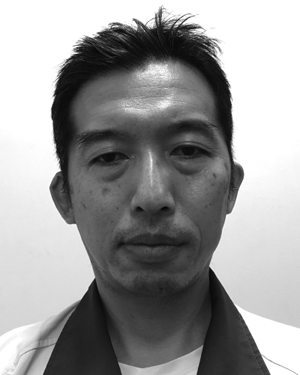 Fumiaki Sato合作论文数Faculty of Veterinary Medicine, Hokkaido University4
 Akira Sassa合作论文数4
 Kiyoe Ura合作论文数4Shusuke Akamatsu合作论文数Kyoto4Haruna Fujiike合作论文数3Takuro Sunada合作论文数3Ayuna Takeishi合作论文数3Muthana Al Abo合作论文数3




