The potassium chloride cotransporters (KCC) family of proteins are widely expressed and are involved in the transepithelial movement of potassium and chloride ions and the regulation of cell volume. KCC activity is high in reticulocytes, and contributes to the dehydration of sickle red blood cells. Because plasma levels of both vascular endothelial growth factor (VEGF) and placental growth factor (PlGF) are elevated in sickle cell individuals, and VEGF has been shown to increase KCC expression in other cells, we hypothesized that VEGF and PlGF influence KCC expression in erythroid cells. Both VEGF and PlGF treatment of human erythroid K562 cells increased both mRNA and protein levels of KCC1, KCC3b, and KCC4. VEGF‐ and PlGF‐mediated cellular signaling involved VEGF‐R1 and downstream effectors, specifically, PI‐3 kinase, p38 MAP kinase, mTOR, NADPH‐oxidase, JNK kinase, and HIF‐1α. VEGF and PlGF‐mediated transcription of KCC3b and KCC4 involved hypoxia response element (HRE) motifs in their promoters, as demonstrated by promoter analysis, EMSA and ChiP. These results were corroborated in vivo by adenoviral‐mediated overexpression of PlGF in normal mice, which led to increased expression of mKCC3 and mKCC4 in erythroid precursors. Our studies show that VEGF and PlGF regulate transcription of KCC3b and KCC4 in erythroid cells via activation of HIF‐1α, independent of hypoxia. These studies provide novel therapeutic targets for regulation of cell volume in RBC precursors, and thus, amelioration of dehydration in RBCs in sickle cell disease. Am. J. Hematol. 89:273–281, 2014. © 2013 Wiley Periodicals, Inc.
Abstract Abstract 3163 The KCl co-transporter (KCC) family of proteins catalyzes the electroneutral, coupled movement of K+ and Cl− ions across the plasma membrane, thereby mediating transepithelial ion transport and regulating cell volume. These proteins play an important role in disease states such as cancer, numerous neurological conditions as well as sickle cell disease (SCD). KCC activity is increased in sickle red blood cells and contributes to their dehydration, which potentiates sickling. The mechanisms of increased KCC activity and its abnormal regulation are not understood. Of the four mammalian KCC isoforms, KCC 1, 3 and 4 are expressed in erythroid cells (Crable et al. Exp Hematol. 2005; 33:624). Hiki et. al. showed that the angiogenic factor vascular endothelial growth factor (VEGF) increased KCC 3a expression in HUVEC cells (J.B.C. 274, 10661–10667, 1999). As levels of VEGF and related family member, placenta growth factor (PlGF) are elevated in sickle cell patients, we hypothesized that VEGF and PlGF may influence KCC expression in erythroid cells. RT-PCR revealed that erythroid K562 cells expressed the VEGF receptor-1 (VEGF-R1, or Flt-1) but not VEGF receptor-2, (VEGF-R2 or Flk-1). Additionally, flow cytometric analysis of WT C57Bl6 mouse bone marrow showed the presence of the Flt-1 receptor, but not Flk-1 or Flk-3 in erythroid progenitors and expression decreased with maturation. VEGF treatment (50 ng/ml) of K562 cells increased KCC 1, 3a, 3b and 4 mRNA levels; PlGF treatment increased KCC 1, 3a and 4 mRNA levels but not KCC 3b. The VEGF receptor inhibitor, SU5416, ablated the effect of VEGF. VEGF-stimulated KCC 4 expression was blocked by pharmacological inhibitors that implicated PI3 kinase, p38 MAP kinase, mTOR, JNK kinase and the transcription factor hypoxia inducible factor-1α (HIF-1α), as with other VEGF effects. Analysis of the KCC 4 promoter showed that the −875 and −90 bp promoter luciferase constructs exhibited similar levels of activity as the −1200 bp promoter construct, when compared to the promoterless reporter plasmid. Deleted constructs corresponding to −65 bp from transcription start site showed ∼90% reduced promoter activity. In silico analysis of the −90 bp region of the KCC 4 promoter showed potential binding sites for transcription factor SP-1 and HIF-1α. Binding sites for transcription factor SP-1 at positions −35 to −44 bp and −56 to −64 bp were shown to be active by site directed mutagenesis. Mutation of the HIF-1α binding site at −73 to −76 bp significantly inhibited promoter activity, whereas mutation of the HIF-1α binding site at position −21 bp to −18 bp did not have any effect on activity. Similar analysis of the KCC 3a promoter indicate potential binding sites for SP-1 at positions −8 to −4 bp and a HIF-1α binding site at position −23 to −20 bp, and the KCC 3b promoter has binding sites for HIF-1α at –9 to −6 bp and −49 to −46 bp and an AP-1 binding site at position −13 to −10 bp. Luciferase assays with KCC 3b promoter constructs indicated that the −190 bp promoter region containing HIF-1α sites at –9 to −6 bp and −49 to −46 bp and an AP-1 binding site at −13 to −10 bp contained minimal promoter required for transcription activity. Mutations within both HIF-1α binding sites attenuated promoter activity indicating a role for HIF-1α in regulating KCC 3b activity, as well. EMSA and ChIP assays with the KCC 4 promoter demonstrated that VEGF treatment of K562 cells increased HIF-1α binding to the HIF-1α sites, which was abrogated by mutating these sites. Similar results were obtained for the KCC 3a and 3b promoters.These results suggest that activation of VEGF-R1 by VEGF, and presumably its other ligand, PlGF, leads to non-hypoxic activation of HIF-1α and SP-1-mediated up-regulation of KCC3a, 3b and 4 expressions in erythroid K562 cells via its canonical signaling pathways. Variation in KCC gene expression and its modulation by cytokines and growth factors may be a source of inter-individual variation in SS RBC volume regulation and thus of phenotypic variability of SCD. Identifying the factors that modulate transcriptional control of KCC expression is important to understanding volume regulation in reticulocytes and its dysregulation in SS RBC. Disclosures: No relevant conflicts of interest to declare.
The K-Cl cotransporter (KCC) regulates red blood cell (RBC) volume, especially in reticulocytes. Western blot analysis of RBC membranes revealed KCC1, KCC3, and KCC4 proteins in mouse and human cells, with higher levels in reticulocytes. KCC content was higher in sickle versus normal RBC, but the correlation with reticulocyte count was poor, with inter-individual variability in KCC isoform ratios. Messenger RNA for each isoform was measured by real time RT-quantitative PCR. In human reticulocytes, KCC3a mRNA levels were consistently the highest, 1-7-fold higher than KCC4, the second most abundant species. Message levels for KCC1 and KCC3b were low. The ratios of KCC RNA levels varied among individuals but were similar in sickle and normal RBC. During in vivo maturation of human erythroblasts, KCC3a RNA was expressed consistently, whereas KCC1 and KCC3b levels declined, and KCC4 message first increased and then decreased. In mouse erythroblasts, a similar pattern for KCC3 and KCC1 expression during in vivo differentiation was observed, with low KCC4 RNA throughout despite the presence of KCC4 protein in mature RBC. During differentiation of mouse erythroleukemia cells, protein levels of KCCs paralleled increasing mRNA levels. Functional properties of KCCs expressed in HEK293 cells were similar to each other and to those in human RBC. However, the anion dependence of KCC in RBC resembled most closely that of KCC3. The results suggest that KCC3 is the dominant isoform in erythrocytes, with variable expression of KCC1 and KCC4 among individuals that could result in modulation of KCC activity.
Abstract 3163 The KCl co-transporter (KCC) family of proteins catalyzes the electroneutral, coupled movement of K + and Cl − ions across the plasma membrane, thereby mediating transepithelial ion transport and regulating cell volume. These proteins play an important role in disease states such as cancer, numerous neurological conditions as well as sickle cell disease (SCD). KCC activity is increased in sickle red blood cells and contributes to their dehydration, which potentiates sickling. The mechanisms of increased KCC activity and its abnormal regulation are not understood. Of the four mammalian KCC isoforms, KCC 1, 3 and 4 are expressed in erythroid cells (Crable et al. Exp Hematol. 2005; 33:624). Hiki et. al. showed that the angiogenic factor vascular endothelial growth factor (VEGF) increased KCC 3a expression in HUVEC cells (J.B.C. 274, 10661–10667, 1999). As levels of VEGF and related family member, placenta growth factor (PlGF) are elevated in sickle cell patients, we hypothesized that VEGF and PlGF may influence KCC expression in erythroid cells. RT-PCR revealed that erythroid K562 cells expressed the VEGF receptor-1 (VEGF-R1, or Flt-1) but not VEGF receptor-2, (VEGF-R2 or Flk-1). Additionally, flow cytometric analysis of WT C57Bl6 mouse bone marrow showed the presence of the Flt-1 receptor, but not Flk-1 or Flk-3 in erythroid progenitors and expression decreased with maturation. VEGF treatment (50 ng/ml) of K562 cells increased KCC 1, 3a, 3b and 4 mRNA levels; PlGF treatment increased KCC 1, 3a and 4 mRNA levels but not KCC 3b. The VEGF receptor inhibitor, SU5416, ablated the effect of VEGF. VEGF-stimulated KCC 4 expression was blocked by pharmacological inhibitors that implicated PI3 kinase, p38 MAP kinase, mTOR, JNK kinase and the transcription factor hypoxia inducible factor-1α (HIF-1α), as with other VEGF effects. Analysis of the KCC 4 promoter showed that the −875 and −90 bp promoter luciferase constructs exhibited similar levels of activity as the −1200 bp promoter construct, when compared to the promoterless reporter plasmid. Deleted constructs corresponding to −65 bp from transcription start site showed ∼90% reduced promoter activity. In silico analysis of the −90 bp region of the KCC 4 promoter showed potential binding sites for transcription factor SP-1 and HIF-1α. Binding sites for transcription factor SP-1 at positions −35 to −44 bp and −56 to −64 bp were shown to be active by site directed mutagenesis. Mutation of the HIF-1α binding site at −73 to −76 bp significantly inhibited promoter activity, whereas mutation of the HIF-1α binding site at position −21 bp to −18 bp did not have any effect on activity. Similar analysis of the KCC 3a promoter indicate potential binding sites for SP-1 at positions −8 to −4 bp and a HIF-1α binding site at position −23 to −20 bp, and the KCC 3b promoter has binding sites for HIF-1α at –9 to −6 bp and −49 to −46 bp and an AP-1 binding site at position −13 to −10 bp. Luciferase assays with KCC 3b promoter constructs indicated that the −190 bp promoter region containing HIF-1α sites at –9 to −6 bp and −49 to −46 bp and an AP-1 binding site at −13 to −10 bp contained minimal promoter required for transcription activity. Mutations within both HIF-1α binding sites attenuated promoter activity indicating a role for HIF-1α in regulating KCC 3b activity, as well. EMSA and ChIP assays with the KCC 4 promoter demonstrated that VEGF treatment of K562 cells increased HIF-1α binding to the HIF-1α sites, which was abrogated by mutating these sites. Similar results were obtained for the KCC 3a and 3b promoters.These results suggest that activation of VEGF-R1 by VEGF, and presumably its other ligand, PlGF, leads to non-hypoxic activation of HIF-1α and SP-1-mediated up-regulation of KCC3a, 3b and 4 expressions in erythroid K562 cells via its canonical signaling pathways. Variation in KCC gene expression and its modulation by cytokines and growth factors may be a source of inter-individual variation in SS RBC volume regulation and thus of phenotypic variability of SCD. Identifying the factors that modulate transcriptional control of KCC expression is important to understanding volume regulation in reticulocytes and its dysregulation in SS RBC. Disclosures: No relevant conflicts of interest to declare.
The KCl cotransporter (KCC) mediates volume reduction in normal reticulocytes and exaggerated KCC activity in sickle red blood cells (SS RBC) (Joiner et al, Blood 109:1728, 2007) contributes to pathological dehydration that potentiates sickling. Three separate genes (KCC1, KCC3, KCC4) are expressed in RBC (Crable et al, Exp. Hem. 33:624, 2005). KCC1 and KCC3 proteins have been shown to interact in ex vivo expression systems (Simard et al, JBC 282(25):18083, 2007), and co-expression of an N-terminal truncation of KCC1 reduces KCC activity mediated by full-length KCC1 or KCC3 (Casula et al. JBC 276:41870, 2001), suggesting functional interaction. We show here via western blot analysis that SS RBC membranes contain more KCC1 protein (relative to KCC3) than AA RBC, independent of the reticulocytosis of sickle blood. Immunoprecipitation of solubilized SS RBC membranes with KCC3-specific antibody yielded a band at 125 kD on SDS PAGE which contained KCC1, as identified by western blotting with KCC1-specific antibody and by TOF mass spectroscopy. The effect of co-expression of KCC1 and KCC3 on KCC activity was assessed by measuring NEM-stimulated, Cl-dependent, (ouabain + bumetanide)-insensitive Rb uptake in HEK 293 cells. The Flip-In T-rex HEK 293 cell line (Invitrogen) containing a tetracycline-response promoter was transfected with a pcDNA5a plasmid containing KCC3a cDNA. Recombination of the plasmid with the integrated tet-promoter construct inserts the KCC3a gene under control of a tetracycline-responsive promoter. These cells were subsequently transduced with a retroviral vector (SF-91. Hildinger et at, Gene Ther . 5:1575, 1998) containing KCC1 cDNA linked to a GFP cassette. Control cells contained SF-91 vector lacking KCC1. Cells were selected for GFP expression and grown in the absence (un-induced, no KCC3a expression) or presence of tetracycline (induced, KCC3a expression). From this binary matrix, four types of cells were obtained: Cells with no additional KCC expression, representing endogenous KCC activity; cells with only KCC1 or KCC3a expression; cells with both KCC1 and KCC3a expression. Western blots indicated similar KCC1 expression in cells with KCC1 only and [KCC1 + KCC3] and similar KCC3 expression in cells with KCC3 only and [KCC1 + KCC3]. Thus, the expression of neither isoform was affected by the presence of the other. KCC activity in cells overexpressing KCC1 only was similar to endogenous activity in HEK 293 cells; i.e., transport activity of KCC1 alone was minimal. Cells overexpressing KCC3 only had a 5-fold increase in KCC activity over endogenous levels. When KCC1 was co-expressed with KCC3 in [KCC1 + KCC3] cells, an additional 50% increase in KCC activity was observed (p < 0.05 by paired t-test, N=4), despite similar levels of KCC3 expression by western blot analysis. This synergistic effect was dependent on the cytoplasmic N-terminus of KCC1, as it was not seen when the first 39 amino acids of KCC1 were removed. Interestingly, removal of the entire cytoplasmic N-terminal domain (117 aa) produced an inhibitory effect when co-expressed with KCC3a in HEK cells, as previously reported in Xenopus oocytes (Casula et al.). These data indicate that KCC1 and KCC3 interact structurally and functionally in RBC membranes, and provide another potential mechanism for regulation of KCC activity via multimeric associations between KCC isoforms. Thus, KCC activity could be modulated not only by transcriptional mechanisms and post-translational modification (phosphorylation), but also by altering the ratios of KCC isoforms or the kinetics of their association. We speculate that higher levels of KCC1 protein relative to KCC3 in SS RBC membranes could account for higher KCC activity in these cells relative to AA RBC.
The KCl Cotransporter (KCC) is a key component of the volume regulation system of human reticulocytes, and its excessive activity in sickle cells contributes to cellular dehydration and therefore to sickling pathology. Three of the four KCC genes, are expressed in erythroid cells (Crable et al. Exp. Hematol. 2005; 33:624). Although the relative contribution of the three KCC isoforms to KCC fluxes and volume regulation in red cells remains unknown, KCC3 appears to be the dominant transcript in late erythroid cells. Heterogenity of the 5′ ends of the KCC3 mRNA transcripts has been described by Mount and colleagues (Mercado et al. Am J Physiol 289:F1246, 2005), including two untranslated exons (ex1D and 1C) 1.2 kb upstream from ex1A of KCC3a, which contains the originally described translation initiation site. We used RT-PCR, 5′ RACE (rapid amplification of cDNA ends), and primer extension analyses to study the 5′ ends of KCC3 cDNA transcripts in erythroid cells. The large exon 1 of KCC3 was identified in fetal liver and bone marrow RNA as a 1718bp region (containing exons 1A, 1C, and 1D described by Mercado et al) that undergoes complex patterns of alternate splicing to generate 10 different transcripts. Six major splicing isoforms are expressed in hematopoietic cell RNA. One isoform incorporates the first 167bp of the exon (exon 1D) as 5′ untranslated sequence which splices to the last 208bp of exon 1, including additional 5′ untranslated sequence and an alternative in-frame initiator methionine. The translated protein exhibits a 59 amino acid N-terminal truncation of KCC3a lacking several potential phosphorylation sites (called KCC3a-Short by Mercado et al). This transcript was the most abundant isoform in hematopoietic cell RNA. A second major isoform contains 629bp of the exon immediately 3′ of exon 1D as 5′ untranslated sequence (exon 1C), then splices out the next 735bp to join the last 243bp of the exon, including additional 5′ untranslated sequence and the alternate initiator methionine of KCC3a-Short. A third novel transcript includes the entire exon, utilizing the first initiator methionine of full-length KCC3a. Two other novel transcripts were found, both of which code for KCC3a-Short. These transcripts were also identified in EST databases. We examined the genomic region around exon 1 for promoter activity using luciferase promoter constructs expressed in erythroid K562 cells. Promoter activity was minimal with constructs which spanned from exon 1D to the beginning of exon 1A, but increased substantially in constructs that included this region plus 900 bp 5′ sequence. Further deletion analysis shows that a minimal promoter containing less than 125 base pairs yields full promoter activity. A mutation of a GC box within this region reduces activity over 15 fold, implicating SP-1 as a trans-regulatory factor. Thus, KCC3 exhibits a complex pattern of alternative splicing in erythroid cells, producing several novel transcripts, some of which encode an N-terminal truncation of KCC3a. Identifying the factors modulating transcriptional and translational control of KCC3 expression and the functional behavior of this truncated protein in erythroid cells is important to understanding volume regulation in reticulocytes and its abnormalities in sickle cells.
Maintenance of cell volume by regulated cation transport is a fundamental cellular process. The KCl cotransporter (KCC) contributes to red blood cell (RBC) volume regulation, especially in reticulocytes. Erythroid K-Cl cotransport activity is increased in sickle cells (SS RBC) and contribute to SS RBC dehydration, which potentiates sickling. Three cation cotransporter genes, SLC12A4 (KCC1), SLC12A6 (KCC3) and SLC12A7 (KCC4), and several splicing variants, mediate KCC activity in non-neuronal tissues. To determine which KCC isoform(s) predominates in human RBC we examined the quantitative expression patterns of KCC isoforms during erythroid differentiation. We developed a set of real-time RT-QPCR assays specific for KCC1, KCC1b, KCC3a, KCC3b or KCC4, over a 7-log quantitation range and sensitivity of 10 copies per reaction using multiplex amplification of GAPDH as internal controls. In human reticulocytes isolated by magnetic separation using anti-transferrin receptor coated beads, KCC3a mRNA levels were consistently the highest (4–24 fold of KCC1), while KCC4 levels varied from 1 to 7-fold of KCC1 levels (n=8). Message levels for KCC3b were relatively low (20–80% of KCC1), and for KCC1b were negligible (1–2% of KCC1). Substantial variability in the relative levels of KCC1, KCC3a, and KCC4 mRNA was observed among individual samples, but no consistent difference was apparent comparing sickle and normal reticulocytes. Western blot analysis of sickle and normal RBC ghost membranes confirmed the presence of KCC1, KCC3 and KCC4 at the protein level. To evaluate cells at various erythroid differentiation stages, human CD34+ cells were cultured under conditions favoring erythroid differentiation for 26 days. During early in vitro differentiation, KCC1 was the main mRNA species, followed by KCC4, with similar levels of KCC3a and KCC3b. RNA levels for KCC3a and KCC4 increased during maturation and became the most abundant at later stages. KCC1b mRNA remained low, and KCC3b levels decreased during erythroid development. To further define this temporal sequence of KCC expression, cells cultured for 10–17 days were sorted by FACS into four subpopulations (I to IV), characterized by immunostaining for relative expression of CD71 and glycophorin A, with enrichment of pronormoblasts
The KCl cotransporter mediates volume reduction in normal (AA) reticulocytes, and its abnormal regulation in sickle (SS) reticulocytes contributes to cellular dehydration that facilitates Hb S sickling. mRNA for three KCC genes - KCC1, KCC3, and KCC4 - as well as a splicing isoform, KCC1ex1b, is present in reticulocytes (Exp. Hem. 2005; 33:624). Western blotting has demonstrated KCC1 in human RBC membranes (Su et al, AJPhysiol. 1999; 277:C899) and KCC3 in sheep (Lauf et al, CompBiochemPhysiol 2001; 130:499); KCC4 protein was found in hRBC membranes by proteomic analysis (Pasini et al, Blood 2006; 108:791). We confirm here the presence of KCC3 protein in hRBC via western blotting using an antibody to an exon 3 epitope distal to known N-terminal splicing sites. We sought to characterize and compare human KCC isoforms expressed in human cells (HEK 293) and assess their similarity to KCC activity in RBC. cDNAs for human KCC1, KCC1ex1b, KCC3a, and KCC4, with N-terminal c-myc epitope tags were expressed in HEK 293 cells. Stable cell lines were selected by growth in neomycin, and expression monitored by quantitative PCR analysis of the expressed construct and other endogenous isoforms and by western blotting (anti-myc) of plasma membranes. KCC activity was measured as N-ethylmaleimide(NEM)-stimulated, Cl-dependent Rb uptake in cells grown to 75–90 % confluency. Cells were incubated at 37°C in isotonic saline media with various concentrations of Rb (Na replacement), plus 0.1 mM ouabain and 0.01 mM bumetanide. At 2 and 4 min cells were washed with iced Rb-free media, then lysed and assayed for Rb by flame emission, normalized to sample protein. Flux rates were calculated from Rb uptake at 2 and 4 min. The flux rate in Cl-free sulfamate media was subtracted from that in Cl-media to yield the Cl-dependent flux rate. Wild-type HEK cells showed no increase in Cl-dependent Rb influx when exposed to 1 mM NEM, but Cl-dependent, NEM-stimulated Rb uptake was apparent in cells expressing KCC isoforms. All isoforms were stimulated by hypotonic conditions (75 mOsm), but relative to NEM-stimulated activity, KCC3 was most responsive. Kinetic characteristics of Cl-dependent, NEM-stimulated Rb influx in HEK cell expressing human KCC1, KCC3a, and KCC4 isoforms isoforms are given below, compared to RBC KCC fluxes. A splice variant, KCC1ex1b, coding for a protein with a truncated N-terminus, exhibited 48 ± 7% of the activity of the full-length KCC1 isoform, but did not alter transport activity of coexpressed KCC3a. Thus, KCC isoforms expressed in HEK cells share some, but not all, characteristics with RBC KCC fluxes. The isoform kinetics also differ from those previously published in xenopus oocyte expression systems, suggesting that the membrane milieu in which KCC proteins are expressed may influence their functional characteristics. Likewise, KCC activity in RBC may represent an average of several transporters operating in parallel. The truncated KCC1ex1b isoform, expressed at higher levels in normal than in sickle reticulocytes, has lower activity than KCC1. KCC3 and KCC4 exhibit more robust transport activity than KCC1 and may mediate a substantial part of KCC activity in RBC.
T cell acute lymphoblastic leukemia (T-ALL) is frequently associated with overexpression of the oncogenes LMO2 and SCL(TAL1) which are normally down regulated following the double negative stage of T cell development. Our goal is to decipher the molecular and cellular mechanisms leading to the onset of LMO2 associated T-ALL. We were able to isolate a complex containing the transcription factors LMO2, SCL(TAL1) and E47 from primary human T-ALL cells with proven aberrant expression of LMO2 and SCL(TAL1) by applying immunoprecipitation and Western blotting techniques. This protein complex regulates the transcription of a truncated form of RALDH2 (retinaldehyde dehydrogenase) in T-ALL cells as shown by gene transcription profiling in conjunction with RT-PCR and siRNA approaches. To monitor the effect of LMO2 expression on T cell development and leukemogenesis, lethally irradiated mice (C57BL/6) were transplanted with bone marrow cells that had been transduced with a retrovirus carrying LMO2 as the transgene. One year later, 88% of the cells in the thymus expressed LMO2 and a shift towards CD3−/CD44+/CD25+ cells was observed (an 88% increase compared to normal thymocytes), suggesting a differentiation block caused by LMO2 leading to an accumulation of immature T cells. To test and identify cooperating genes in T-ALL development, bone marrow cells of LMO2 double transgenic mice in which tet-inducible LMO2 is controlled by a thymic specific promoter, were retrovirally transduced with SCL(TAL1). So far, none of the control animals, transplanted with bone marrow cells transduced with a vector only containing EGFP, developed T-ALL. However, six out of the seven test animals developed T-ALL exhibiting enlargement of the spleen, liver and thymus between seven and nine months after transplantation. Organs and blood of the diseased animals were infiltrated with T-ALL cells of the immature phenotype CD8+/CD4+ in five cases and of the CD3−/CD44+/CD25+ phenotype in one case. This indicates that the differentiation block caused by a lack of down-regulation of LMO2 and SCL(TAL1) in maturing T cells leads to a block in T cell differentiation and precedes T-ALL. These models will be used to examine the involvement of other cooperating genes in T-ALL development as well as downstream target genes of LMO2/SCL(TAL1), such as RALDH2, in the onset of T-ALL. We conclude that aberrant expression of LMO2 in T cells leads to a block in T cell maturation and, in conjunction with up-regulation of secondary genes like SCL(TAL1), triggers deregulation of genes in immature T cells leading to impaired T cell development and the onset of T-ALL. The described model will help to identify cooperating genes in LMO2 associated T-ALL as well as the chain of events leading to malignancy.
The KCl Cotransporter (KCC) is a key component of the volume regulation system of human reticulocytes, and its excessive activity in sickle cells contributes to cellular dehydration and therefore to sickling pathology. Three of the four KCC genes, including KCC3, are expressed in erythroid cells (Exp.Hematol. 2005; 33:624), but their relative contribution to KCC fluxes and volume regulation in red cells remains unknown. Heterogenity of the 5′ ends of the KCC3 mRNA transcripts has been described by Mount and colleagues (Mercado et al. Am J Physiol 289:F1246, 2005), including two untranslated exons (ex1D and 1C) 1.2 kb upstream from ex1A of KCC3a, which contains the originally described translation initiation site. We used RT-PCR, 5′ RACE (rapid amplification of cDNA ends), and primer extension analyses to study the 5′ ends of KCC3 cDNA transcripts in erythroid cells. The large exon 1 of KCC3 was identified in fetal liver and bone marrow RNA as a 1646 bp region (containing exons 1A, 1C, and 1D described by Mercado et al) that undergoes complex patterns of alternate splicing to generate 5 different transcripts. Three major splicing isoforms are expressed in hematopoietic cell RNA. One isoform incorporates the first 103bp of the exon (exon 1D) as 5′ untranslated sequence which splices to the last 208bp of the exon, including additional 5′ untranslated sequence and an alternative in-frame initiator methionine. The translated protein exhibits a 59 amino acid N-terminal truncation of KCC3a lacking several potential phosphorylation sites (KCC3a-Short of Mercado et al). This transcript was the most abundant isoform in hematopoietic cell RNA. A second major isoform contains the first 668bp of the exon (including both exons 1D and 1C) as 5′ untranslated sequence, then splices out the next 735bp to join the last 243bp of the exon, including additional 5′ untranslated sequence and the alternate initiator methionine of KCC3a-Short. A third novel transcript includes the entire exon, utilizing the first initiator methionine of ‘full-length' KCC3a. Two other novel transcripts were found, both of which code for KCC3a-Short. These transcripts were also identified in EST databases. We examined the genomic region around exon 1 for promoter activity using luciferase promoter constructs expressed in erythroid K562 cells. Promoter activity was minimal with constructs which spanned from exon 1D to the beginning of exon 1A, but increased substantially in constructs that included this region plus 900 bp 5′ sequence. Reporter activity increased 3 fold upon removal of the 590 bp 3′ segment of such constructs, suggesting the presence of negative regulatory elements within the exon immediately upstream of exon 1A. Thus, KCC3 exhibits a complex pattern of alternative splicing in erythroid cells, producing several novel transcripts, some of which encode an N-terminal truncation of KCC3a. Identifying the factors modulating transcriptional control of KCC3 expression and the functional behavior of this truncated protein in erythroid cells is important to understanding volume regulation in reticulocytes and its abnormalities in sickle cells.
Our interest in the cis-acting elements that promote the up-regulation of the beta globin gene has led to a systematic deletion analysis of portions of the beta globin gene in the context of the HS2 and gamma globin gene using transgenic mice. In constructs that delete the 5' region to only 265 bp, high-level, erythroid-specific expression was observed. Further deletion to 122 bp, however, results in significantly reduced expression levels. A substitution of a minilocus control region for the single HS2 site was also produced, resulting in increased beta globin expression over that seen with the HS2 alone. These results are consistent with the presence of an enhancer-like element between -122 and -265. In addition, a construct in which the entire beta globin gene promoter was replaced by a thymidine kinase promoter was tested. Interestingly, no expression was detected in these transgenic mice. This may indicate the requirement for an erythroid-specific promoter to drive this gene. Finally, the 3' region of the beta globin gene was deleted in order to examine the effect of a previously defined 3' enhancer region. With deletion of this region, the expression of the human beta globin gene in transgenic mice is unchanged relative to the parental constructs.
 Clinton H Joiner合作论文数Cincinnati Comprehensive Sickle Cell Center, Children's Hospital Research Foundation and Children's Hospital Medical Center7
 Patrick Gallagher合作论文数School of Medicine, Yale University4
 Dao Pan合作论文数4
 Sharad Chandra合作论文数4
 Gonsalves Caryn S合作论文数3Mary Risinger合作论文数3Anna Ottlinger合作论文数2Daren Wang合作论文数2Franco R S合作论文数2
 David Bruce Mount合作论文数Division of Renal Medicine, Brigham and Women's Hospital, Harvard Medical School2Christian A. Hübner合作论文数Inst Human Genet, Jena Univ Hosp2
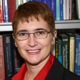 Theodosia A. Kalfa合作论文数Children’s Hospital Medical Center Department of Pediatrics, University of Cincinnati2Kathleen P. Anderson合作论文数2Jesse Rinehart合作论文数Molecular Medicine, Pharmacology, and Physiology, Biological & Biomedical Sciences, Yale University1
 Kalra Vijay K合作论文数Dept Biochem & Mol Biol, Univ So Calif1Neumann Jon C合作论文数1Li Wei合作论文数1
 Christof von Kalle合作论文数Berlin Institute of Health, Charité – Universitätsmedizin Berlin;GeneWerk;Clinical Study Center, Charité – Universitätsmedizin Berlin1Peter Ciraolo合作论文数1Clara Wong合作论文数1Gerlinde Layh-Schmitt合作论文数1Charles A. Klanke合作论文数1Jeff Bailey合作论文数1
 Christine B Kern合作论文数1
 Jerry B. Lingrel合作论文数Department of Molecular Genetics, Biochemistry and Microbiology, College of Medicine, University of Cincinnati1Robert Su合作论文数1Guoping Zhou合作论文数1




