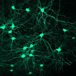
This is a maximal intensity projection of CA1 pyramidal cell transfected with plasmid with the reporter GFP using single cell electroporation technique. In this particular case the organotypic slices were prepared from p5-7 pups in a tissue chopper (McIlwain). And maintained in MEM bases media with added glutamax with a change in 2 alternative dyas at 37°C and 5% CO 2 for 4 days in-vitro (DIV) before electroporating with a glass pipette of 7-10mΩ resistance by applying 4 square pulses of -ve voltage of −2.5 V, for 25 ms duration at 1 Hz frequency. Then maintained in the same medium for one four more days before fixing them in PFA(4‰ v/v) and amplified with anti-GFP antibody (ab290, 1:1000 dil) and imaged in SP2 confocal microscope (Leica) for z-stack.
PhospholipaseC γ2 (PLCG2) is known to have direct link with genetic risk factors for Alzheimer’s like dementia (AD). PLCG2 has been previously demonstrated to have association with Aß uptake through microglia. And mostly expressed in dentate gyrus (DG) network of hippocampus. WT mouse were transfected with sh-RNA of LV-PLCG2 (U6 promotor, GFP as reporter; shRNA) or scrambled (used as control; shNT). Whole-cell patch-clamp recordings were performed on acute slices with glass pipette (<3-4MΩ) at -70mv, which was accompanied with morphometry of perforant pathway (PP)-DG circuitry. Two-tailed Mann-Whitney test was used for statistics unless otherwise mentioned, p<0.05 was considered to be significant. We found significant increase in AP half width (p = 0.0274), rheobase (p = 0.0046) in shRNA on the other hand, input resistance (p = 0.0159), number of spike vs current injection (p = 0.0268) was significantly decreased. However, there were no significant change was observed in AP onset (p = 0.6105), AP amplitude (p = 0.7345), RMP (p = 0.7591) and firing threshold (p = 0.5247) in shRNA group when compared to shNT. mEPSC amplitude was found to be reduced (p = 0.0288) in shRNA condition, even the cumulative probability of mEPSC amplitude was also found to be significantly altered after silencing of PLCG2 gene in DG cells (p = 0.0014; Kolmogorof-Smirnov test). mEPSC frequency of the miniature activity of transfected DG cells were did not show significant change (p = 0.7707). Sholl analysis of the DG cells expressing shRNA showed a decrease in the number of intersections vs distance from soma (P<0.05), accompanied by significant decrease in dendritic length (p = 0.0501), dendritic volume (p<0.0001) and dendritic surface area (p = 0.0273). When spine density was measured we have found a significant decrease in cumulative distribution of spine density (p<0.0001; Kolmogorof-Smirnov test), which is tightly associated with decrease in spine length (p = 0.0209) and spine head diameter (P<0.0001). Furthermore, these dendritic and spine morphometric alteration is also accompanied with the decrease in the volume of mossy fiber gigantic bouton (mfB) with alteration in the number of filopodia per mfBs (p<0.05). Results from the study, reflect critical insight of PLCG2 regulating neuronal and/or synaptic activity, which may underlie the cognitive dysfunction (viz. recognition memory) and synaptic alteration to the PP-DG circuitry seen in AD.
Forming new memories after a one-time experience requires initial encoding then consolidation over time. During learning, multimodal information converges onto the hippocampus, activating sparse neuronal assemblies which are thought to form a memory representation through concerted activity and synaptic interconnectivity. In this work, we use a novel tool for fast fluorescent labeling of engram neurons (FLEN). FLEN is based on c-Fos activity-dependent transient expression of a destabilized fluorescent marker ZsGreen1 rapidly after one-trial learning. With FLEN, we explore the electrophysiological properties of c-Fos activated CA3 pyramidal neurons (PNs) a few hours following one-trial learning of an episodic-like memory. In parallel, we employ the Robust Activity Marker (RAM) system, which provides activity-dependent labeling 24 hr following a novel experience. Comparing FLEN+ and RAM+ neurons allows us to characterize how the properties of neuronal assemblies evolve during an initial phase of consolidation. Whereas no difference was observed in the excitability of FLEN+ vs. FLEN− neurons, RAM+ neurons were more excitable than RAM− neurons. This suggests that CA3 PNs recruited in an engram progressively acquire increased excitability as compared to neurons which were not activated by the one-trial contextual memory task. On the other hand, like RAM+ neurons, FLEN+ CA3 neurons show an increased number of excitatory inputs. Overall, with the FLEN strategy, we can show that both the intrinsic excitability and the synaptic properties of CA3 PNs undergo progressive plastic changes over the first day following a one-trial memory task.
We developed a high content screening to investigate how Alzheimer disease (AD) genetic risk factors may impair synaptic mechanisms in rat primary neuronal cultures. Out of the gene targets identified, we found that shRNA-mediated downregulation of Plcg2 in mouse dentate gyrus neurons consistently impaired dendritic morphology and synaptic function. In human neuronal cultures (hNCs), PLCG2 downregulation also impaired synaptic function and was associated with increased levels of Aβ and Tau phosphorylation, potentially via the AKT/GSK3β axis. Very rare PLCG2 loss-of-functon(LoF) variants were associated with a 10-fold increased AD risk. PLCG2 LoF carriers exhibit low mRNA/protein PLCG2/PLCγ2 levels, consistent with nonsense-mediated mRNA decay mechanisms. Restoring PLCγ2 levels in shPLCG2-hNCs fully reversed the disease-related phenotypes. Our findings indicate that the downregulation of PLCγ2 increases the risk of AD by impairing synaptic function and increasing the levels of Aβ and Tau phosphorylation in neurons.
Mesial temporal lobe epilepsy (mTLE) is the most prevalent type of epilepsy in adults. First and subsequent generations of anti-epileptic therapy regimens fail to decrease seizures in a large number of patients suffering from mTLE, leaving surgical ablation of part of the hippocampus as the only therapeutic option to potentially reach seizure freedom. GluK2 has recently been identified as a promising target for the treatment of mTLE using gene therapy. Here, we engineered an adeno-associated virus serotype 9 vector expressing a cluster of two synthetic microRNAs (miRNAs), expressed from the human synapsin promoter, that target GRIK2 mRNA. Intra-hippocampal delivery of this vector in a mouse model of mTLE significantly reduced GRIK2 expression and daily seizure frequency. This treatment also improved the animals' health, reduced their anxiety, and restored working memory. Focal administration of the vector to the hippocampus of cynomolgus monkeys in GLP toxicology studies led to the selective transduction of hippocampal neurons with little exposure elsewhere in the brain and no transduction outside the central nervous system. Expression of miRNAs in hippocampal neurons resulted in substantially decreased GRIK2 mRNA expression. These data suggest that the intra-hippocampal delivery of a GMP-grade AAV9 encoding a synthetic miRNAs targeting GRIK2 is a promising treatment strategy for mTLE.
Objective Temporal lobe epilepsy (TLE) is characterized by recurrent seizures generated in the limbic system, particularly in the hippocampus. In TLE, recurrent mossy fiber sprouting from dentate gyrus granule cells (DGCs) creates an aberrant epileptogenic network between DGCs which operates via ectopically expressed GluK2/GluK5-containing kainate receptors (KARs). TLE patients are often resistant to anti-seizure medications and suffer significant comorbidities; hence there is an urgent need for novel therapies. Previously we have shown that GluK2 knockout mice are protected from seizures. This study aims at providing evidence that downregulating KARs in the hippocampus using gene therapy reduces chronic epileptic discharges in TLE. Methods We combined molecular biology and electrophysiology in rodent models of TLE and in hippocampal slices surgically resected from patients with drug-resistant TLE. Results Here we confirmed the translational potential of KAR suppression using a non-selective KAR antagonist that markedly attenuated Interictal-like Epileptiform Discharges (IEDs) in TLE patient-derived hippocampal slices. An adeno-associated virus (AAV) serotype-9 vector expressing anti- grik2 miRNA was designed to specifically downregulate GluK2 expression. Direct delivery of AAV9-anti grik2 miRNA into the hippocampus of TLE mice led to a marked reduction in seizure activity. Transduction of TLE patient hippocampal slices reduced levels of GluK2 protein and, most importantly, significantly reduced IEDs. Interpretation Our gene silencing strategy to knock down aberrant GluK2 expression demonstrates inhibition of chronic seizure in a mouse TLE model and IEDs in cultured slices derived from TLE patients. These results provide proof-of-concept for a gene therapy approach targeting GluK2 KARs for drug-resistant TLE patients.
Intracellular calcium signaling underlies the astroglial control of synaptic transmission and plasticity. Mitochondria-endoplasmic reticulum contacts (MERCs) are key determinants of calcium dynamics, but their functional impact on astroglial regulation of brain information processing is unexplored. We found that the activation of astrocyte mitochondrial-associated type-1 cannabinoid (mtCB(1)) receptors determines MERC-dependent intracellular calcium signaling and synaptic integration. The stimulation of mtCB(1) receptors promotes calcium transfer from the endoplasmic reticulum to mitochondria through a specific molecular cascade, involving the mitochondrial calcium uniporter (MCU). Physiologically, mtCB(1)-dependent mitochondrial calcium uptake determines the dynamics of cytosolic calcium events in astrocytes upon endocannabinoid mobilization. Accordingly, electrophysiological recordings in hippocampal slices showed that conditional genetic exclusion of mtCB(1) receptors or dominant-negative MCU expression in astrocytes blocks lateral synaptic potentiation, through which astrocytes integrate the activity of distant synapses. Altogether, these data reveal an endocannabinoid link between astroglial MERCs and the regulation of brain network functions.
Alzheimer's disease (AD) is a progressive neurodegenerative disease characterized by cognitive decline related to deficits in synaptic transmission and plasticity. We report in APP/PS1 mice, a double transgenic mouse model of AD, that females displayed an early burden of Aβ plaques load in the stratum moleculare of the dentate gyrus (DG) together with prominent neuroinflammatory activation of astrocytes and microglia. Robust deficits in hippocampus-dependent memory tasks were observed in APP/PS1 female mice as early as 3 months of age. We then studied the functional properties of the lateral perforant path (LPP) to DG granule cells. Remarkably DG granule cells displayed higher intrinsic excitability in APP/PS1 female mice. We showed that the long term potentiation of population spike amplitude induced by high frequency stimulation (HFS) at LPP-DG granule cells synapse is impaired in APP/PS1 female mice. HFS induced plasticity of intrinsic excitability in DG granule cells without inducing noticeable modification of synaptic strength. Furthermore, the enhanced intrinsic excitability was potentiated to a greater extent in APP/PS1 as compared to control mice following HFS. Our study shows that changes in the intrinsic excitability of DG granule cells in AD contribute to the dysfunctional transfer of information from the entorhinal cortex to the hippocampus.
Intracellular calcium signaling underlies the astroglial control of brain functions. However, the cellular mechanisms regulating calcium handling by astrocytes are far from being understood. Mitochondria-endoplasmic reticulum contacts (MERCs) are key determinants of calcium dynamics, but their functional impact on astroglial regulation of brain information processing is currently unexplored. Here we show that the activation of astrocyte mitochondrial-associated CB1 receptors (mtCB1) regulates MERCs-dependent intracellular calcium signaling, thereby determining the synaptic functions of these cells. In vitro and in vivo stimulation of mtCB1 receptors promotes calcium transfer from the endoplasmic reticulum to mitochondria through a specific molecular cascade, involving AKT signaling, IPR3 receptors and different components of the mitochondrial calcium uniporter complex (MCU). Physiologically, mtCB1-dependent mitochondrial calcium uptake determines the dynamics of cytosolic calcium events in astrocytes upon endocannabinoid mobilization. Accordingly, electrophysiological recordings in hippocampal slices showed that astrocyte-specific mtCB1 receptors exclusion or dominant negative MCU expression blocks lateral synaptic potentiation, through which astrocytes integrate the activity of distant synapses. Altogether, these data reveal a cellular endocannabinoid link between astroglial MERCs and the regulation of brain network functions.
Summary Intracellular calcium signaling underlies the astroglial control of synaptic transmission and plasticity. Mitochondria-endoplasmic reticulum contacts (MERCs) are key determinants of calcium dynamics, but their functional impact on astroglial regulation of brain information processing is currently unexplored. We found that the activation of astrocyte mitochondrial-associated CB1 receptors (mtCB1) determines MERCs-dependent intracellular calcium signaling and synaptic integration. The stimulation of mtCB1 receptors promotes calcium transfer from the endoplasmic reticulum to mitochondria through specific mechanisms regulating the activity of the mitochondrial calcium uniporter (MCU) channel. Physiologically, mtCB1-dependent mitochondrial calcium uptake determines the precise dynamics of cytosolic calcium events in astrocytes upon endocannabinoid mobilization. Accordingly, electrophysiological recordings in hippocampal slices showed that genetic exclusion of mtCB1 receptors or specific astroglial MCU inhibition blocks lateral synaptic potentiation, a key example of astrocyte-dependent integration of distant synapses activity. Altogether, these data reveal an unforeseen link between astroglial MERCs and the regulation of brain network functions.
Amyotrophic Lateral Sclerosis is an adult-onset neurodegenerative disease characterized by the specific loss of motor neurons, leading to muscle paralysis and death. Although the cellular mechanisms underlying amyotrophic lateral sclerosis (ALS)-induced toxicity for motor neurons remain poorly understood, growing evidence suggest a defective energetic metabolism in skeletal muscles participating in ALS-induced motor neuron death ultimately destabilizing neuromuscular junctions. In the present study, we report that a specific exercise paradigm, based on a high intensity and amplitude swimming exercise, significantly improves glucose metabolism in ALS mice. Using physiological tests and a biophysics approach based on nuclear magnetic resonance (NMR), we unexpectedly found that SOD1(G93A) ALS mice suffered from severe glucose intolerance, which was counteracted by high intensity swimming but not moderate intensity running exercise. Furthermore, swimming exercise restored the highly ALS-sensitive tibialis muscle through an autophagy-linked mechanism involving the expression of key glucose transporters and metabolic enzymes, including GLUT4 and glyceraldehyde-3-phosphate dehydrogenase (GAPDH). Importantly, GLUT4 and GAPDH expression defects were also found in muscles from ALS patients. Moreover, we report that swimming exercise induced a triglyceride accumulation in ALS tibialis, likely resulting from an increase in the expression levels of lipid transporters and biosynthesis enzymes, notably DGAT1 and related proteins. All these data provide the first molecular basis for the differential effects of specific exercise type and intensity in ALS, calling for the use of physical exercise as an appropriate intervention to alleviate symptoms in this debilitating disease.
Alzheimer's disease (AD) is a progressive neurodegenerative disease characterized by early cognitive deficits linked to synaptic dysfunction and loss. Considerable evidence suggests that neuroinflammation contributes to AD. Prostaglandin E2 (PGE2), a key neuroinflammatory molecule, modulates hippocampal synaptic transmission and plasticity. We investigated the effect of PGE2 on synaptic transmission and presynaptic plasticity at synapses between mossy fibers from the dentate gyrus and CA3 pyramidal cells (Mf-CA3 synapse). These synapses are involved in mnemonic processes and consequently may be of relevance for AD. We provide evidence that although PGE2 had no effect both on either basal transmission or short-term plasticity, it strongly impaired presynaptic Mf-CA3 long-term potentiation (LTP) by acting on PGE2 receptor 3 (EP3) receptors. During aging, hippocampal levels of PGE2 markedly increased in the APP/PS1 mouse model of AD and impaired specifically presynaptic LTP via a PGE2-EP3 signaling pathway. In summary, the building up of PGE2 during the progression of AD leads to specific impairment of hippocampal presynaptic plasticity and highlights EP3 receptors as a potential target to alleviate cognitive deficits in AD.
Spinal muscular atrophy (SMA), a lethal neurodegenerative disease that occurs in childhood, is caused by the misexpression of the survival of motor neuron (SMN) protein in motor neurons. It is still unclear whether activating motor units in SMA corrects the delay in the postnatal maturation of the motor unit resulting in an enhanced neuroprotection. In the present work, we demonstrate that an adequate NMDA receptor activation in a type 2 SMA mouse model significantly accelerated motor unit postnatal maturation, counteracted apoptosis in the spinal cord, and induced a marked increase of SMN expression resulting from a modification of SMN2 gene transcription pattern. These beneficial effects were dependent on the level of NMDA receptor activation since a treatment with high doses of NMDA led to an acceleration of the motor unit maturation but favored the apoptotic process and decreased SMN expression. In addition, these results suggest that the NMDA-induced acceleration of motor unit postnatal maturation occurred independently of SMN. The NMDA receptor activating treatment strongly extended the life span in two different mouse models of severe SMA. The analysis of the intracellular signaling cascade that lay downstream the activated NMDA receptor revealed an unexpected reactivation of the CaMKII/AKT/CREB (cAMP response element-binding protein) pathway that induced an enhanced SMN expression. Therefore, pharmacological activation of spinal NMDA receptors could constitute a useful strategy for both increasing SMN expression and limiting motor neuron death in SMA spinal cord.
Mutant mice are a good model to study to what extent the postnatal activity of sensory afferents is necessary for the maturation of central neurons. In particular, the question arises whether the signals carried by the first‐order vestibular neurons, which encode information on the head movement of pups, are necessary for the maturation of second‐order vestibular neurons. To address that question, juvenile and adult transgenic, vestibular‐deficient mutants where a null mutation of the KCNE1 potassium‐channel gene leads to degeneration of all hair cells of the inner ear just after birth were studied. These KCNE1−/− mutants are deaf and show quasi‐constant head bobbing and a permanent shaker/waltzer phenotype. This behavior is not due to persistent abnormalities of the membrane properties of central vestibular neurons, because their maturation is delayed but not impaired by the absence of sensory vestibular information. On the other hand, the data shed light on how the membrane properties of vestibular neurons might be modified according to functional requirements or following lesions. The expression levels of the protein calretinin that regulates the intracellular free‐calcium concentration in central vestibular neurons could play a major role both in intact animals and following labyrinthectomy. By comparing the KCNE1−/− mutant mice to other vestibular‐deficient animals, it was concluded that the suppression of vestibular inputs during a “critical period” of postnatal development can induce a permanent circling behavior, but that this phenotype is not always due to congenital vestibular deficiency.
Several studies using transgenic mouse models of familial amyotrophic lateral sclerosis (ALS) have reported a life span increase in exercised animals, as long as animals are submitted to a moderate-intensity training protocol. However, the neuroprotective potential of exercise is still questionable. To gain further insight into the cellular basis of the exercise-induced effects in neuroprotection, we compared the efficiency of a swimming-based training, a high-frequency and -amplitude exercise that preferentially recruits the fast motor units, and of a moderate running-based training, that preferentially triggers the slow motor units, in an ALS mouse model. Surprisingly, we found that the swimming-induced benefits sustained the motor function and increased the ALS mouse life span by about 25 days. The magnitude of this beneficial effect is one of the highest among those induced by any therapeutic strategy in this disease. We have shown that, unlike running, swimming significantly delays spinal motoneuron death and, more specifically, the motoneurons of large soma area. Analysis of the muscular phenotype revealed a swimming-induced relative maintenance of the fast phenotype in fast-twitch muscles. Furthermore, the swimming programme preserved astrocyte and oligodendrocyte populations in ALS spinal cord. As a whole, these data are highly suggestive of a causal relationship not only linking motoneuron activation and protection, but also motoneuron protection and the maintenance of the motoneuron surrounding environment. Basically, exercise-induced neuroprotective mechanisms provide an example of the molecular adaptation of activated motoneurons.
This study establishes a causal link between the limitation of myofibre transitions and modulation of calcineurin activity, during different exercise paradigms. We have designed a new swimming‐based training protocol in order to draw a comparison between a high frequency and amplitude exercise (swimming) and low frequency and amplitude exercise (running). We initially analysed the time course of muscle adaptations to a 6‐ or 12‐week swimming‐ or running‐based training exercise program, on two muscles of the mouse calf, the slow‐twitch soleus and the fast‐twitch plantaris . The magnitude of exercise‐induced muscle plasticity proved to be dependent on both the muscle type and the exercise paradigm. In contrast to the running‐based training which generated a continuous increase of the slow phenotype throughout a 12‐week training program, swimming induced transitions to a slower phenotype which ended after 6 weeks of training. We then compared the time course of the exercise‐induced changes in calcineurin activity during muscle adaptation to training. Both exercises induced an initial activation followed by the inhibition of calcineurin. In the muscles of animals submitted to a 12‐week swimming‐based training, this inhibition was concomitant with the end of myofibre transition. Calcineurin inhibition was a consequence of the inhibition of its catalytic subunit gene expression on one hand, and of the expression increase of the modulatory calcineurin interacting proteins 1 gene (MCIP1), on the other. The present study provides the first experimental cues for an interpretation of muscle phenotypic variation control. J. Cell. Physiol. 214:126–135, 2008. © 2007 Wiley‐Liss, Inc.
Spinal muscular atrophy (SMA) is an inborn neuromuscular disorder caused by low levels of survival motor neuron protein, and for which no efficient therapy exists. Here, we show that the slower rate of postnatal motor-unit maturation observed in type 2 SMA-like mice is correlated with the motor neuron death. Physical exercise delays motor neuron death and leads to an increase in the postnatal maturation rate of the motor-units. Furthermore, exercise is capable of specifically enhancing the expression of the gene encoding the major activating subunit of the NMDA receptor in motor neurons, namely the NR2A subunit, which is dramatically downregulated in the spinal cord of type 2 SMA-like mice. Accordingly, inhibiting NMDA-receptor activity abolishes the exercise-induced effects on muscle development, motor neuron protection and life span gain. Thus, restoring NMDA-receptor function could be a promising therapeutic approach to SMA treatment.
 Christophe Mulle合作论文数Bordeaux School of Neuroscience, Université de Bordeaux11
 O. Biondi合作论文数5
 Philippe Lopes合作论文数Université d’Evry Laboratoire d’Etudes en Physiologie de l’Exercice (LEPHE) E.A. 3872, Genopole. Boulevard F. Mitterrand Evry cedex 91025 France5Frédéric Charbonnier合作论文数Centre Universitaire des Saints-Pères, Université Paris Descartes5
 Christophe Chanoine合作论文数5
 Sylvie Lecolle合作论文数4Claude Pariset合作论文数4Carol Stella合作论文数3Guillaume Penchet合作论文数3Corinne Blancard合作论文数3Francisca Julio-Kalajzic合作论文数3Marjorie Varilh合作论文数3
 Laurie M Robin合作论文数3Richard Porter合作论文数3
 Roman Serrat合作论文数3Nicolas Partouche合作论文数3
 Vladimir Kouskoff合作论文数CNRS, Univ Bordeaux3
 Avishek Roy合作论文数All India Institute of Medical Sciences3Federico Massa合作论文数3
 Diego De Stefani合作论文数Dept Biomed Sci, Univ Padua3Boileau Céline合作论文数3
 Ana Covelo合作论文数Department of Neuroscience, University of Minnesota3
 Sebastien Delcasso合作论文数Center for Integrative and Cognitive Neuroscience, CNRS UMR 52283
 Anna Beyeler合作论文数(Unité Mixte de Recherche 5227), Centre National de la Recherche Scientifique3Astrid Cannich合作论文数3Sandrine Pouvreau合作论文数Universite Claude-Bernard Lyon 13C. Grondard合作论文数3
 Didier Scavarda合作论文数SFNCP French Society of Pediatric Neurosurgery3Bénédicte Salin合作论文数3
 Valerie Crepel合作论文数3
 Frédéric Gambino合作论文数Institut des Neurosciences Cellulaires et Intégratives (UPR-3212), Université de Strasbourg3
 Nicolas Chenouard合作论文数Institut Pasteur, CNRS3
 Arnau Busquets-Garcia合作论文数Facultat de Ciències de la Salut i de la Vida, Universitat Pompeu Fabra3
 Giovanni Marsicano合作论文数Université de Bordeaux3
 Fabrice Bartolomei合作论文数Aix-Marseille University3Cécile Marchal合作论文数2
 Julien Branchu合作论文数2Noelle Grosjean合作论文数2Andreas Borta合作论文数2Nan Jiang合作论文数2Dario Cupolillo合作论文数2Andrew C. Mercer合作论文数Res & Early Dev, REGENXBIO Inc2
 Jared Brent Smith合作论文数Res & Early Dev, REGENXBIO Inc2
 Stephane Baudouin合作论文数2
 Pierre-Paul JL Vidal合作论文数2
 Andrea Ruiz合作论文数2Giles April R合作论文数2Amédée Thierry合作论文数2Olivier Danos合作论文数2




