Background Chronic diabetic wounds are characterized by persistent inflammation and impaired macrophage transition from the pro-inflammatory M1 phenotype to the pro-regenerative M2 phenotype. To overcome these limitations, we engineered a sequential exosome delivery hydrogel designed to provide phase-specific release of Wharton's Jelly mesenchymal stem cell-derived exosomes (WJ-MSC-Exos) for controlled immune regulation and accelerate wound regeneration. Methods A hybrid polyvinyl alcohol–borax–glycerol–carboxymethyl cellulose hydrogel was synthesized to provide controlled porosity, elasticity, and degradability for phase-specific exosome release. Physicochemical characterization was performed using FTIR, SEM, rheological, and swelling analyses to confirm crosslinking integrity and viscoelastic behavior. In vitro studies were conducted to evaluate cytocompatibility and migration assay, followed by in vivo therapeutic evaluation in a streptozotocin-induced diabetic rat wound model. Results The hydrogel maintained a stable network within its linear viscoelastic range and enabled sustained exosome release over 14 days. In vitro results confirmed high cell viability and enhanced migration. In vivo, wounds treated with the exosome-loaded hydrogel exhibited significantly accelerated closure, and well-organized collagen alignment. Gene expression revealed downregulation and upregulation of genes related to M1-to-M2 macrophage polarization and improved angiogenesis. Histological evaluation showed complete re-epithelialization and a balanced Col-1/Col-3 ratio, indicating scar-minimized dermal regeneration. Conclusion This engineered sequential delivery system provides temporally controlled exosome release synchronized with wound-healing phases. By integrating material design with immunomodulatory drug delivery principles, the platform represents a promising next-generation therapeutic approach for chronic diabetic wound management.
INTRODUCTION:This study aimed to enhance wound healing using a novel natural cocktail gel dressing composed of purslane, human amniotic membrane (hAM), and platelet-rich plasma (PRP). In addition, a new ratio-based analytical approach was applied to evaluate the healing dynamics in each treatment group, revealing correlations between the healing rate and the respective treatment compound. METHODS:Under aseptic conditions, specific amounts of freeze-dried decellularized hAM, PRP, and hydroalcoholic extract of purslane (HAEP) powder were prepared. The study groups included hAM gel, PRP gel, HAEP gel, a cocktail gel (HAEP + PRP + hAM), and phenytoin gel (positive control). Cytotoxicity was evaluated using the MTT assay. In vivo, seven groups were assessed on days 7, 14, and 21. Wound closure rate was analyzed via photographic imaging, and tissue samples were collected for H&E staining. Wound healing dynamics were further evaluated using 14/7-day and 21/14-day ratios. RESULTS:The cocktail gel significantly enhanced wound healing compared with other groups (p < 0.05), improving cell migration, M2 macrophage polarization, and angiogenesis. The ratio-based analysis indicated that the cocktail group exhibited the fastest healing between days 7 and 14, while purslane and hAM groups showed superior healing between days 14 and 21. CONCLUSIONS:This study introduces the use of 14/7 and 21/14-day ratios for the first time as a quantitative measure of healing progression, bridging macroscopic closure rates with underlying cellular and molecular changes. The combination of purslane, PRP, and hAM significantly accelerated healing and reduced closure time, suggesting a synergistic effect. The proposed ratio-based approach provides a more accurate evaluation of wound healing phases.
Background and purpose: Regenerative medicine focuses on replacing dysfunctional cells with mesenchymal stem cells (MSCs) to repair damaged tissues. However, genomic instability and the potential risk of tumor formation remain significant challenges in the use of these cells. Cell-free therapies, such as those based on conditioned media (CM), have emerged as promising alternatives due to their role in promoting angiogenesis. Since malignant solid tumors exhibit the most robust angiogenic activity, this study aimed to compare the angiogenic potential of CM derived from menstrual blood-derived MSCs (MenSCs), umbilical cord-derived MSCs (UC-MSCs), ovarian cancer cells (SKOV-3), and cancer-associated fibroblasts (CAFs). Materials and methods: UC-MSCs were extracted and passaged, and their mesenchymal nature was confirmed by flow cytometry using CD markers. Isolation and culture of MenSCs, UC-MSCs, CAFs, and tumor cell lines were performed, and CM was subsequently prepared from each. The vascular endothelial growth factor (VEGF) concentration in the CMs was measured by ELISA. Human umbilical vein endothelial cells (HUVECs) were treated with the CMs, and the effects on their proliferation were evaluated using the XTT assay. The angiogenic potential of the CMs was compared using the chicken embryo chorioallantoic membrane (CAM) model. Results: The results showed that the VEGF concentration in the CMs derived from tumor cells and menstrual blood-derived stem cells was higher than in other CMs. Similarly, the proliferation and migration of HUVECs treated with these CMs were greater than those treated with CMs from other cell types. Overall, the findings indicate that different CMs can promote vascular endothelial cell proliferation and enhance angiogenesis. Conclusion: The angiogenic potential of tumor cell-derived conditioned medium (CM) is considerably higher than that of menstrual blood-derived stem cell CM. These findings suggest that tumor cell-derived CM could serve as a potent and cost-effective alternative in regenerative medicine, particularly for paracrine-based therapeutic strategies.
Fibromodulin, a small leucine rich proteoglycan has been suggested to have prominent role in wound healing. On the other hand, fibroblast cells, due to their ability to secrete growth factors and control inflammation in the wound area, have been proposed as effective approaches in cell therapy for wounds. In the current study we attempted to improve treatment results using a combination of fibroblast and fibromodulin features. Fibroblast cells were isolated from the skin and transfected with a vector carrying the fibromodulin gene. Following the assessment of fibromodulin protein production, the effect of transfected fibroblast cells was studied in an animal wound model. Flow cytometry analysis showed high expression of the CD90 marker (97.2
Background: Menstrual blood has been identified as an important source for the isolation of mesenchymal stem cells (MSCs). These stem cells can easily and non-invasively be harvested from menstrual blood during menstrual shedding. There has been extensive research on the differentiation potential of menstrual blood-derived stem cells (MenSCs) and their application in regenerative medicine and the treatment of diseases. Materials and Methods: The aim of this paper was to review the current and future application of MenSCs in the field of regenerative medicine and cell therapy based on an electronic search in various databases, like Scopus, PubMed, and Google Scholar for English-language studies. The application of MenSCs in regenerative medicine can be the window of hope for the treatment of various diseases and disabilities, such as female infertility, type 1 diabetes, myocardial infarction, wound healing, neurodegenerative diseases, and many other conditions that were thought to be incurable in the past. Conclusion: Nowadays, the use of MenSC as a novel source of MSCs has garnered special interest among scientists due to two important factors: Non-invasive accessibility and the immunomodulatory potential of these cells, which have historically posed significant obstacles in the field of stem cell therapy. To date, there has been substantial research on these cells, and many studies are ongoing, as scientists seek to leverage their differentiation potential and optimize differentiation conditions and protocols for their application in regenerative medicine.
Cold atmospheric plasma (CAP) is increasingly recognized for its potential in melanoma cancer treatment due to its ability to inducecell death selectively in cancer cells while sparing normal L929 fibroblast cells. In this study, culturing B16-F10 melanoma andL929 fibroblast cell lines on polycaprolactone (PCL) nanofiber scaffolds, the penetration depth of CAP effects, especially cytotoxicityand selectivity, in a three-dimensional (3D) scaffold has been examined. Results from MTT assays, ROS measurements, and flowcytometry confirmed that CAP effectively reduces the viability of melanoma cells across scaffold layers, with a higher impact onsurface layers and a gradual decline in deeper layers. ROS production followed a similar trend, demonstrating CAP penetrationthrough multiple layers while retaining its selective cytotoxic effects. Flow cytometry analysis further supported CAP-induced apo-ptosis in melanoma cells, with reduced effectiveness in deeper layers. Finally, a strong linear regression model (R2>0.97) enabledextrapolation of cell viability across scaffold layers, yielding an estimated effective plasma penetration depth of 116-166 mu m based onthe 50% viability threshold (IC50). These findings highlight the suitability of PCL-based 3D scaffolds as in vitro models for studyingCAP penetration and reinforce CAP's potential as a targeted therapy for melanoma
Cancer is one of the leading causes of death worldwide. Recent advancements in chemotherapy, particularly using natural drug-based strategies, have shown promise. This study evaluated the antitumor and anti-inflammatory effects of a combination of the brown alga Sargassum ilicifolium with cisplatin in vitro. After collecting and identifying the algae, fucoidan and alginate were extracted. The antioxidant activity was assessed using DPPH and Ferric Reducing Ability of Plasma (FRAP) assays. The cytotoxic effects on the MDA-MB-231 breast cancer cell line were evaluated in both 2D and 3D cultures using the XTT assay. Fucoidan and alginate yielded 7203 and 441 mg per 100 grams of dried algae, respectively. Total polysaccharide content in the fraction was estimated to be 89.39 mg/g dried algae weight. Fucoidan-rich extract exhibited higher antioxidant properties compared to alginate-rich extract. Also, the combination treatment significantly enhanced cytotoxicity, with the IC50 of fucoidan-rich extract reduced from 177.9 ± 8.7 to 79.3 ± 4.6 μg/ml in 2D culture when combined with cisplatin. In 3D culture, the IC50 decreased from over 800 ± 78.5 to 364.5 ± 41.7 μg/ml. Additionally, the fucoidan-rich extract significantly decreased IL-1β and IL-6 cytokine secretion in macrophages. These findings suggest combining cisplatin with fucoidan-rich extract enhances antitumor efficacy and may reduce chemotherapy side effects.
Infertility affects 10-15 % of reproductive-age couples worldwide, with male infertility linked to sperm dysfunction and female infertility caused by ovulation disorders and reproductive abnormalities. Stem cell research presents a promising avenue for infertility treatment through germ cell differentiation. However, standardizing differentiation protocols and ensuring the functionality of in vitro-derived gametes remain significant challenges before clinical application becomes feasible.
3D Scaffold is an evolutionary technology that brings about the development and advancement of tissue engineering processes, restorative medicine, and thus the future of medicine. Bioprinting technologies can make a suitable 3D substrate for adhesion, migration, extracellular matrix components, different phenotypes of human cells, signaling proteins Creating polarity, cell division, and other scaffold-like biomaterials for the manufacture of human skin in regenerative medicine. In this article, some natural polymers used in the field of scaffolding with the help of bio-printing machines have been introduced and studied. In addition, methods of improving and modifying their properties to achieve more desirable properties minimize their weaknesses and produce scaffolding with the highest efficiency through combination with other synthetic polymers and composite production. Natural polymers and polymer composites used in the manufacture of scaffolds and the field of tissue engineering, such as organic scaffolds including collagen, fibronectin, chitosan, gelatin, fibrin, alginates, etc. Chemically conform to the extracellular matrix. These materials should have properties such as biocompatibility, biodegradability, and desirable mechanical properties to play a supporting role for damaged tissues to provide the necessary conditions for proliferation, growth, accumulation, and cell migration to improve and regenerate tissue damaged items. [GRAPHICS]
Diabetes is a global problem that threatens human health. Cell therapy methods using stem cells, and tissue engineering of pancreatic islets as new therapeutic approaches have increased the chances of successful diabetes treatment. In this study, to differentiate Wharton's Jelly-derived mesenchymal stem cells (WJ-MSCs) into insulin-producing cells (IPCs) with improved maturity, and function, platelet-rich plasma (PRP)-Polyvinylpyrrolidone (PVP)-Polycaprolactone (PCL)/PCL scaffold was designed. The two-dimensional (2D) control group included cell culture without differentiation medium, and the experimental groups included 2D, and three-dimensional (3D) groups with pancreatic beta cell differentiation medium. WJ-MSCs-derived IPCs on PRP-PVP-PCL/PCL scaffold took round cluster morphology, the typical pancreatic islets morphology. Real-time PCR, immunocytochemistry, and flowcytometry data showed a significant increase in pancreatic marker genes in WJ-MSCs-derived IPCs on the PRP-PVP-PCL/PCL scaffold compared to the 2D-experimental group. Also, using the ELISA assay, a significant increase in the secretion of insulin, and C-peptide was measured in the WJ-MSCs-derived IPCs of the 3D-experimental group compared to the 2D experimental group, the highest amount of insulin (38 µlU/ml), and C-peptide (43 pmol/l) secretion was in the 3D experimental group, and in response to 25 mM glucose solution, which indicated a significant improvement in the functional level of the WJ-MSCs-derived IPCs in the 3D group. The results showed that the PRP-PVP-PCL/PCL scaffold can provide an appropriate microenvironment for the engineering of pancreatic islets, and the generation of IPCs.
Diabetes Mellitus (DM) disrupts the body's capability to control blood glucose statuses. Type 1 diabetes mellitus (T1DM) arises from inadequate insulin production and is treated with insulin replacement therapy. Stem cell therapy is a hopeful treatment for T1DM that involves using adult stem cells to generate insulin-producing cells (IPCs). Mesenchymal stem cells (MSCs) are particularly advantageous for generating IPCs. The islet cells require interactions with the extracellular matrix for survival, which is lacking in conventional 2D culture systems. Natural or synthetic polymers create a supportive 3D microenvironment in tissue engineering. We aim to construct superior differentiation conditions employing polyethersulfone (PES)/Fish gelatin scaffolds to differentiate Wharton's jelly-derived mesenchymal stem cells (WJ-MSCs) to IPCs. In this study, the PES/fish gelatin scaffold (3D) was manufactured by electrospinning, and then its biocompatibility and non-toxicity were investigated by MTT assay. After that, scaffold-supportive effects on WJ-MSCs differentiation to IPCs were studied at the gene and protein levels. After exposure to the differentiation media, 2D and 3D (PES/Fish gelatin) cultured cells were slowly aggregated and developed spherical-shaped clusters. The viability of cells was found to be comparable in both 2D and 3D cultures. The gene expression analysis showed that efficiency of differentiation was more elevated in 3D culture. Additionally, ELISA results indicated that C-peptide and insulin release were more significant in 3D than in 2D culture. In conclusion, the PES/fish gelatin scaffold is highly promising for pancreatic tissue engineering because it supports the viability, growth, and differentiation of WJ-MSCs into IPCs.
Background Breast cancer remains a primary global health concern due to its limited treatment options, frequent disease recurrence, and high rates of morbidity and mortality. Thereby, there is a need for more effective treatment approaches. The proposal suggests that the combination of targeted therapy with other antitumoral agents could potentially address drug resistance. In this study, we examined the antitumoral effect of combining metformin, an antidiabetic drug, with targeted therapies, including tamoxifen for estrogen receptor-positive (MCF-7), trastuzumab for HER2-positive (SKBR-3), and antibody against ROR1 receptor for triple-negative breast cancer (MDA-MB-231). Methods Once the expression of relevant receptors on each cell line was confirmed and appropriate drug concentrations were selected through cytotoxicity assays, the antitumor effects of both monotherapy and combination therapy on colony formation, migration, invasion were assessed in in vitro as well as tumor area and metastatic potential in ex ovo Chick chorioallantoic membrane (CAM) models. Results The results exhibited the enhanced effects of tamoxifen when combined with targeted therapy. This combination effectively inhibited cell growth, colony formation, migration, and invasion in vitro. Additionally, it significantly reduced tumor size and metastatic potential in an ex ovo CAM model. Conclusions The findings indicate that a favorable strategy to enhance the efficacy of breast cancer treatment would be to combine metformin with targeted therapies. Graphical Abstract
In situ-forming hydrogels that possess the ability to be injected in a less invasive manner and mimic the biochemical composition and microarchitecture of the native cartilage extracellular matrix are desired for cartilage tissue engineering. Besides, gelation time and stiffness of the hydrogel are two interdependent factors that affect cells' distribution and fate and hence need to be optimized. This study presented a bioinspired in situ-forming hydrogel composite of hyaluronic acid (HA), chondroitin sulfate (CS), and collagen short nanofiber (CSNF). HA and CS were functionalized with aldehyde and amine groups to form a gel through a Schiff-base reaction. CSNF was fabricated via electrospinning, followed by fragmentation by ultrasonics. Gelation time (11-360 s) and compressive modulus (1.4-16.2 kPa) were obtained by varying the concentrations of CS, HA, CSNFs, and CSNFs length. The biodegradability and biocompatibility of the hydrogels with varying gelation and stiffness were also assessed in vitro and in vivo. At three weeks, the assessment of hydrogels' chondrogenic differentiation also yields varying levels of chondrogenic differentiation. The subcutaneous implantation of the hydrogels in a mouse model indicated no severe inflammation. Results demonstrated that the injectable CS/HA@CSNF hydrogel was a promising hydrogel for tissue engineering and cartilage regeneration.
Introduction: Cancer is the second cause of death in the world, and among all types of cancer, triple-negative breast cancer (TNBC) has one of the worst prognoses. Repeated use of chemotherapy drugs is associated with drug resistance. This study aims to investigate the effect of jujube alkaloid-rich extract on drug sensitivity and its anti-tumor effects on paclitaxel-resistant MDA-MB-231 cells in vitro. Materials and methods: The MDA-MB-231 breast cancer cell line and its paclitaxel-resistant variant were used. The XTT method was employed to measure cell viability, while Annexin-PI assays were used to detect apoptosis. Various concentrations of the jujube extract alone and in combination with paclitaxel assessed synergistic effects in 2D and 3D cell culture models. Results: The presence of alkaloids in the extract obtained from the jujube seed was confirmed by the dragendorff test and its amount was 0.395 g in 5.0 g of jujube seed powder. Paclitaxel and jujube extract exhibited dosedependent cytotoxic effects on paclitaxel-resistant MDA-MB-231 cells. The combination of jujube extract and paclitaxel significantly decreased IC50 from 541.3 mu g/mL to 124.3 mu g/mL, and the IC20 decreased from 92.1 mu g/ mL to 23.9 mu g/mL when combined with paclitaxel in 2D culture model. The combination's toxicity was reduced in the 3D culture model. Apoptosis rates were 0.75 % in the control group, 13.89 % in the alkaloid-rich extract group, 2.59 % in the paclitaxel group, and 42.90 % in the combination group. Conclusion: Combining paclitaxel with alkaloid-rich jujube seed extract enhances cytotoxicity and apoptosis in paclitaxel-resistant MDA-MB-231 breast cancer cells in both 2D and 3D culture models. This suggests that such combinations might be a viable strategy to overcome drug resistance in TNBC treatments.
Cancer remains a major global health concern, often challenging traditional treatments. Natural compounds like fungal polysaccharides have gained attention for their immune-modulatory properties. This study evaluates the phytochemical properties of the n-hexane fraction of Trichaptum biforme and explores its immune-enhancing effects. The study involved isolating three sterol derivatives using column chromatography and purifying polysaccharides from T. biforme (TBP) through hot aqueous extraction. TBP content was quantified via the phenol‑sulfuric acid method, and antioxidant activity was assessed using DPPH and FRAP assays. Cytotoxicity of TBP on THP-1 cells and the impact on IL-1β and TNF-α secretion were evaluated through the XTT assay. Flow cytometry and ELISA assessed cytotoxic activity and IFN-γ secretion in NK cells. The compound 9, 11-Dehydroergosterol peroxide was identified for the first time in T. biforme. The total polysaccharide content was 78.18 ± 0.81 %. The TBP significantly increased IL-1β and TNF-α secretion from THP-1 cells at concentrations of 10 and 320 μg/mL (p < 0.01). Treatment of NK cells with the extract (320 μg/mL) and IL-2 (100 units/mL) significantly enhanced cytotoxic activity and IFN-γ secretion compared to the control group (p < 0.01). These findings suggest that TBP holds promise as a candidate for bolstering anticancer immune responses.
Objective: Infertility is indeed a significant global health concern. The quality of gametes plays a pivotal role in determining the success rates of assisted reproductive technology (ART) cycles. In contemporary fertility and reproductive medicine, the utilization of machine learning (ML) has emerged as a powerful tool for processing large datasets, offering the potential to enhance existing ART practices. The objective of this review study was to assess sperm and oocyte characteristics in humans using ML techniques. This approach can contribute to a more precise evaluation of the gamete, leading to improved decision-making and potentially higher success rates in ART procedures. Using of ML abilities, researchers can obtain valuable insights into the quality of gametes, thereby optimizing fertility treatments for individuals and couples experiencing infertility issues. Materials and Methods: We conducted a comprehensive search on PubMed, Google Scholar, and Scopus using the keywords “Machine Learning AND Quantification AND IVF.” Eligible articles were initially screened based on their titles. After the title screening, a second screening was performed based on the abstracts of the selected articles. Finally, the full articles of the remaining studies were reviewed to ensure they met our inclusion criteria. From each eligible study, we extracted the following information: author(s) of the study, publication year, and the method employed to evaluate human oocyte quality. Conclusion: The development of a properly trained ML system will require careful attention to data quality, measurement, sample size, and ethics issues agreement.
Skin injuries are a global healthcare problem. Chronic ulcers do not heal in a timely fashion, so it is essential to help the body with skin repair. There are some treatments that have been applied to chronic ulcers. One of these treatments is growth factor (GF) therapy. Platelet-rich plasma (PRP) and Platelet-poor plasma (PPP) are two types of plasma derivatives containing many GFs important for wound healing. Several works have reported their application in wound healing and tissue regeneration. The use of autologous PRP is now an adequate alternative in regenerative medicine. It was also demonstrated that PPP is a hemostatic agent for wounds. This review has studied the latest clinical studies, which have applied PRP and PPP to patients with chronic wounds.
Background Recurrent aphthous stomatitis has a complex and inflammatory origin. Among the great variety of medications it is increasingly common to use herbal medicines due to the adverse side effects of chemical medications. Considering the anti-inflammatory properties of cinnamaldehyde and the lack of studies related to the effectiveness of its nano form; This study investigates the effect of cinnamaldehyde and nano cinnamaldehyde on the healing rate of recurrent aphthous stomatitis lesions. Methods In a laboratory experiment, cinnamaldehyde was converted into niosomal nanoparticles. The niosome vesicles diameter and polydispersity index were measured at 25°C using a dynamic light scattering (DLS) Mastersizer 2000 (Malvern Panalytical technologies: UK) and Zetasizer Nano ZS system (Malvern Instruments Worcestershire: UK). After characterizing these particles, the (2,3-Bis-(2-Methoxy-4-Nitro-5-Sulfophenyl)-2H-Tetrazolium-5-Carboxanilide) [XTT] assay was used to assess the toxicity of cinnamaldehyde and nano cinnamaldehyde on gingival fibroblast (HGF) and macrophage (THP-1) cells. By determining the release of TNF-α, IL-6, and TGF-β cytokines using ELISA kits, the level of tissue repair and anti-inflammatory capabilities of these two substances were evaluated. Results The size and loading rate of the cinnamaldehyde nanoparticles were established after its creation. The optimized nanovesicle exhibited the following characteristics: particle size of 228.75 ± 2.38 nm, PDI of 0.244 ± 0.01, the zeta potential of -10.87 ± 1.09 mV and the drug encapsulation percentage of 66.72 ± 3.93%. PDIs range was between 0.242–0.274. The zeta potential values at 25°C were from -2.67 to -12.9 mV. The results of the XTT test demonstrated that nano cinnamaldehyde exhibited dose-dependent toxicity effects. Moreover, nano cinnamaldehyde released more TGF-β and had better reparative effects when taken at lower concentrations than cinnamaldehyde. Conclusion Nano cinnamaldehyde and cinnamaldehyde are effective in repairing tissue when used in non-toxic amounts. After confirmation in animal models, it is envisaged that these substances can be utilized to treat recurrent aphthous stomatitis.
Regarding their reversible damage of insulin-producing cells (IPCs) and the inefficiency of treatment methods for type 1 diabetes mellitus (T1DM), scientists decided to produce IPCs from an unlimited source of cells. But the production of these cells is constantly faced with problems such as low differentiation efficiency in cell therapy and regenerative medicine. This study provided an ideal differentiation medium enriched with plasma-rich platelet (PRP) delivery to produce IPCs from menstrual blood-derived stem cells (MenSCs). We compared them with and without PRP differentiation medium. MenSCs were then cultured in two experimental groups: with/without PRP differentiation medium and a control group (undifferentiated MenSCs). After 18 days, differentiated cells were analyzed for expression of pancreatic gene markers by real-time PCR. Immunocytochemical staining was used to detect the presence of insulin and Pdx-1 in the differentiated cells, and insulin and C-peptide secretion response to glucose were tested by ELISA. Finally, the morphology of differentiated cells was examined by an inverted microscope. In vitro studies showed that MenSCs differentiated in the PRP differentiation medium had strong properties of IPCs such as pancreatic islet-like structure. The expression of pancreatic markers at both RNA and protein levels showed that the differentiation efficiency was higher in the PRP differentiation medium. In both experimental groups, the differentiated cells were functional and secreted C-peptide and insulin on glucose stimulation, but the secretion of C-peptide and insulin in the PRP group was higher than those cultured in the without PRP differentiation medium. Our findings showed that using of PRP enriched differentiation medium can promote the differentiation of MenSCs into IPCs compared to the without PRP culture group. Therefore, the use of PRP into differentiation media can be proposed as a new approach to producing IPCs from MenSCs and used in cell-based therapies for T1DM.
 Hassannia Hadi合作论文数School of Public Health, Tehran University of Medical Sciences27
 Reyhaneh Nassiri Mansour合作论文数Student Research Committee, Mazandaran University of Medical Sciences26
 Fatemeh Soleimanifar合作论文数Dept Med Biotechnol, Alborz Univ Med Sci24
 Masoud Soleimani合作论文数School of Medical Sciences, Tarbiat Modares University21
 Saburi Ehsan合作论文数19
 Mohammad Foad Abazari合作论文数Res Ctr Clin Virol, Univ Tehran Med Sci17
 Ardeshirylajimi Abdolreza合作论文数Dept Tissue Engn & Appl Cell Sci, Shahid Beheshti Univ Med Sci15
 Amir Mellati合作论文数Mazandaran University of Medical Sciences13
 Mousa Kehtari合作论文数Dept Biol, Univ Tehran13
 Hossein Mahboudi合作论文数Dept Biotechnol, Alborz Univ Med Sci11
 Javad Amini Mahabadi合作论文数Tehran Med Sci Branch, Islamic Azad Univ11
 Elham Hasanzadeh合作论文数School of Advanced Technologies in Medicine, Mazandaran University of Medical Sciences9
 Abediankenari Saeid合作论文数Department of Microbiology and Immunology, Mazandaran University of Medical Sciences9
 Pegah Ghoraeian合作论文数7
 Shohreh Zare Karizi合作论文数Department of Biology, Islamic Azad University7
 Fatemeh Nejati合作论文数6
 Ghasem Barati合作论文数School of Medicine, Zanjan University of Medical Sciences6
 Navid Nasiri合作论文数Dept Biol, Islamic Azad Univ6
 Hashemi Javad合作论文数Golestan Research Center of Gastroenterology and Hepatology, Golestan University of Medical Sciences6
 Farhad Jadidi-Niaragh合作论文数School of Public Health, Tehran University of Medical Sciences6Hossein Nikzad合作论文数5
 Reza Eshaghi-Gorji合作论文数5Reza Salarinia合作论文数School of Medicine, North Khorasan University of Sciences5
 Fatemeh Sadat Hosseini合作论文数Dept Orthoped Surg, Univ Connecticut Hlth5
 Seyedehelnaz Enderami合作论文数Stem Cell & Regenerat Med Dept, Natl Inst Genet Engn & Biotechnol5
 Mahdavi Mohammad Reza合作论文数Thalassemia Research Center, Mazandaran University of Medical Sciences5
 Bahram Kazemi合作论文数Cellular and Molecular Biology Research Centers, Shahid Beheshti University5Maryam Nouri Aleagha合作论文数4
 Mohammad Rahmati合作论文数4
 Mobina Gheibi合作论文数4Seyedeh Farzaneh Jalali合作论文数Dept Hematol, Kerman Univ Med Sci4
 Yahya Ehteshaminia合作论文数School of Public Health, Tehran University of Medical Sciences4
 Mohamad Eftekhary合作论文数Dept Biotechnol, Shahid Beheshti Univ Med Sci4Fatemeh Hosseini合作论文数4
 P. Pasalar合作论文数Dept Biochem, Univ Tehran Med Sci4
 Emran Habibi合作论文数4
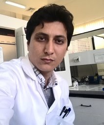 Pagheh Abdol Sattar合作论文数Molecular and Cell Biology Research Center, Mazandaran University of Medical Sciences4
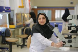 Hanaee-Ahvaz Hana合作论文数Department of Stem Cell Biology, Stem Cell Technology Research Center4
 Shapouri Moghadam Abbas合作论文数Immunology Research Center, Mashhad University of Medical Sciences4
 Vahid Mansouri合作论文数Faculty of Paramedical Sciences, Shahid Beheshti University of Medical Sciences4
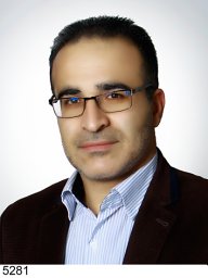 Samad Nadri合作论文数Department of Stem Cells and Tissue Engineering, Stem Cell Technology Institute4
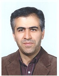 Parviz Kokhaei合作论文数Department of Immunology, Semnan University of Medical Sciences4Elham Sabouri合作论文数3Mohammad mehdi Khani合作论文数3Abdol-Mohammad Kajbafzadeh合作论文数3Mojdeh Amini Faskhodi合作论文数3
 Ayoob Karimizade合作论文数Dept Tissue Engn & Regenerat Med, Mazandaran Univ Med Sci3
 Fatemeh Ahmadi合作论文数Fac Pharm, Mazandaran Univ Med Sci3Shahrzad Nojehdehi合作论文数Stem Cell Technology Research Center3




