BACKGROUND:It is well-known patients with atrial fibrillation (AF) have significant fatigue and impaired quality of life (QoL). Because central sleep apnea (CSA) could be associated with AF, CSA could be a contributory but treatable comorbidity. OBJECTIVES:Retrospective assessment of the impact of CSA treatment with transvenous phrenic nerve stimulation (TPNS) on sleep and QoL in a subgroup of patients with CSA and AF from the remedē® System pivotal trial. METHODS:Patients were implanted with a TPNS device and randomized to treatment or control. Therapy was activated in the treatment arm and remained off in control for 6 months, when TPNS was also activated. Patients were followed through 12 months. RESULTS:Sixty-four of 151 implanted participants had AF, including 32 per arm. The apnea hypopnea index and central apnea index decreased significantly from medians of 49 and 20 events/hour of sleep at baseline to 21 and 1/hour after 6 months of TPNS therapy (p < 0.001 for each), respectively. In parallel, changes in arousal index (48 at baseline vs. 25/hour of sleep at 6 months, p < 0.001) and percent of sleep time with oxygen saturation less than 90% (8% vs. 4%, p = 0.071) improved. Compared to the control group, Patient Global Assessment and Epworth Sleepiness Scale improved significantly with treatment. Improvements were sustained for 12 months and results were similar in the control group after therapy activation. CONCLUSIONS:AF was highly prevalent in patients with CSA. TPNS therapy may significantly improve sleep metrics, QoL, and daytime sleepiness in patients with CSA and AF. REGISTRATION:ClinicalTrials.gov identifier NCT01816776.
STUDY OBJECTIVES:Central (CSA) and obstructive (OSA) sleep apnea are common in heart failure (HF) patients, and treatment options depend on differential classification. To better characterize CSA vs OSA, we evaluated the discriminative value of established and novel polysomnographic traits in patients with HF. METHODS:We analyzed LOFT-HF trial participants with reduced left ventricular ejection fraction (LVEF) and apnea-hypopnea index (AHI) >15 events/hr. Central versus obstructive events were classified with expert investigator input. Multivariable logistic regression tested whether CSA (>50% of central/mixed events; otherwise OSA) was associated with increased loop gain, and other mechanistic traits (arousal threshold, collapsibility, muscle compensation). Mechanistic traits together with novel clinically-informed traits (e.g. event periodicity, flow limitation) were modeled to provide a probability score "Pr(CSA)" for objective CSA-versus-OSA discrimination. RESULTS:Among 122 HF patients (CSA:OSA=84:38), CSA was associated with higher loop gain (OR=4.4[2.2-9.1]), lower arousal threshold (6.4[2.4-16.9]), and greater muscle compensation (2.5[1.0-6.2]). Novel traits-greater event periodicity, larger event-related drive reduction, and reduced flow limitation-further improved discrimination (likelihood ratio test P=0.013 vs. mechanistic model). The final model (AUC=0.88) identified a subgroup enriched for true CSA (OR=8.4[3.5-20.1]). Sensitivity analyses using stricter central apnea-based definitions yielded similar or stronger associations with loop gain and modestly improved discrimination, although fewer individuals met CSA criteria. CONCLUSIONS:Patients with HF and CSA exhibit a distinct set of polysomnographic characteristics compared to OSA, which may facilitate their objective physiology-based characterization along the CSA-OSA continuum.
Abstract Introduction Individuals on opioids are found dead in bed and respiratory depression is a potential cause. In contrast to other opioids, buprenorphine, is a partial μ-opioid receptor agonist and it has been suggested to have reduced respiratory depression with a ceiling threshold. However, death has also been reported in individuals using buprenorphine; yet, it is not known if chronic use of buprenorphine causes significant respiratory depression. The purpose of this study was to determine, if chronic use of buprenorphine is associated with sleep disordered breathing Methods A retrospective study of 11 consecutive patients who were using buprenorphine chronically were referred for evaluation suffering from snoring and daytime sleepiness. All underwent full night polysomnography which was scored page by page by a sleep specialist, according to standard criteria set by the American Academy of Sleep medicine. Results Two patterns of breathing were observed First, four of the 11 patients had significant CSA with an average of 13/h of sleep. This pattern was ataxic, similar to other opioids. Those with CSA, compared to those without CSA, suffered from severe sleep apnea (AHI, 35 vs.9/ hour of sleep), which was associated with a significantly low nadir of saturation (75% vs. 87%). Second, the group without CSA, in spite of a low AHI, had significantly more sustained desaturation and were on a relatively higher dose of buprenorphine (8.7 vs.5.2 mg/day). Conclusion This is the first study to report chronic use of buprenorphine is associated with sleep-disordered breathing. We observed two specific patterns. A subset with CSA and most severe sleep apnea and very low saturation. Second, buprenorphine was associated with sustained nocturnal hypoxemia, potentially related to hypoventilation. These disordered breathing events could be a potential cause of buprenorphine-associated death reported in the literature. Support (if any) none
Despite AASM scoring guidelines encouraging classifying hypopneas as central or obstructive, classification is underutilized in practice. However, without that effort patients may receive an incorrect primary diagnosis and suboptimal treatment. The remedē® System Pivotal Trial studied transvenous phrenic nerve stimulation (TPNS) to treat adult patients with moderate to severe central sleep apnea (CSA). Entry criteria required apnea hypopnea index (AHI)≥20 with the central apnea index (CAI) greater than the obstructive apnea index and obstructive apneas< 20% of AHI but did not consider hypopnea differentiation. This analysis re-examined sleep studies from the trial to assess how hypopnea classification might lead to better patient selection for this therapy. Hypopneas were classified as central versus obstructive by a sleep core laboratory following a modified version of AASM recommended criteria. The AHI composition was assessed at baseline and after 6 months of therapy. At baseline, 91% (138/151) of patients had ≥50% of events classified as central when accounting for hypopnea classification. If all hypopneas were assumed to be obstructive, only 63% (95/151) would have had ≥50% central events. Additionally, 95% (144/151) of patients had central AHI ≥15/hour, compared to 73% (110/151) with a central apnea index (CAI) ≥15/hour. The likelihood of achieving a ≥50% AHI reduction increased with the percentage of baseline events that were central: responder rates were 37.5% for patients with < 50% central events at baseline, incrementally increasing to 76.5% for those with ≥90% central events. At 6 months, the residual AHI predominantly consisted of obstructive events. Central events decreased by 89% with treatment, from a baseline median of 32/hour [1st and 3rd quartiles: 22, 50]. Obstructive apneas rose by 2/hour and obstructive hypopneas increased by 2/hour from a baseline median of 5 obstructive events/hour [2, 12]. Distinguishing central from obstructive hypopneas is required to accurately determine if a patient has central sleep apnea/Cheyne Stokes respiration, and is crucial for epidemiologic studies, appropriate therapy selection, and managing patient expectations about treatment outcomes. This analysis suggests that accurate hypopnea classification may prevent disqualification of patients who could benefit from TPNS therapy, which effectively treats central sleep apnea syndrome. ZOLL Respicardia, Inc.
While not all sleep laboratories distinguish between obstructive and central hypopneas, recent research suggests that patients may receive an incorrect primary diagnosis without this effort. The remedē System Pivotal Trial studied transvenous phrenic nerve stimulation in patients with moderate-to-severe central sleep apnea. Entry criteria required apnea-hypopnea index (AHI) ≥ 20 events/h with central apnea index greater than obstructive apnea index and obstructive apneas < 20 https://www.clinicaltrials.gov/study/NCT01816776 , Identifier: NCT01816776. Dupuy-McCauley K, Schwartz AR, Javaheri S, Germany R, McKane S, Morgenthaler TI. Classifying hypopneas as obstructive or central can enhance transvenous phrenic nerve stimulation therapy patient selection and outcomes. J Clin Sleep Med. 2025;21(12):2113–2120.
Central sleep apnea, a rare polysomnographic finding in the general population, is prevalent in certain cardiovascular conditions including systolic and diastolic left ventricular dysfunction, atrial fibrillation, coronary artery disease, carotid artery stenosis, stroke, and use of certain cardiac-related medications. Polysomnographic findings of central sleep apnea with adverse cardiovascular impacts include nocturnal hypoxemia and arousals, which can lead to increased sympathetic activity both at night and in the daytime. Among cardiovascular diseases, central sleep apnea is most prevalent in patients with left ventricular systolic dysfunction; a large study of more than 900 treated patients has shown a dose-dependent relationship between nocturnal desaturation and mortality. Multiple small randomized controlled trials have shown mitigation of sympathetic activity when central sleep apnea is treated with nocturnal oxygen, continuous positive airway pressure, and adaptive servoventilation. However, two early randomized controlled trials with positive airway pressure devices have shown either a neutral effect on survival or excess premature mortality in the active treatment arm, compared to untreated central sleep apnea. In contrast, the results of the most recent trial using an advanced adaptive servoventilation device showed improved quality of life and no signal for mortality suggesting that treatment of central sleep apnea was at least safe. In addition to positive airway pressure devices, multiple medications have been shown to improve central sleep apnea, but no long-term trials of pharmacologic therapy have been published. Currently, phrenic nerve stimulation is approved for the treatment of central sleep apnea, and the results of a randomized controlled trial showed significant improvement in sleep metrics and quality of life.
Idiopathic central sleep apnea or primary central sleep apnea (ICSA) has been an officially recognized sleep disorder since the 2005 International Classification of Sleep Disorders 2nd edition (ICSD-2) and remains in the International Classification of Sleep Disorders 3rd edition (ICSD-3). The literature supports the etiology as "high loop gain or increased controller gain," along with central sleep apnea (CSA) associated with common diagnoses (e.g. heart failure). Available data place the adult population prevalence at about 0.05% (men) and 0.003% (women), while up to 11% of patients diagnosed with CSA may be classified as ICSA. Symptoms may include nocturnal choking, witnessed apneas, awakenings with the sensation of shortness of breath, restless sleep, insomnia, non-restorative sleep, daytime sleepiness, fatigue, and variable degrees of snoring. Per the ICSD-3, ICSA may only be diagnosed if "The disorder [CSA] is not better explained by another current sleep disorder, medical or neurologic disorder, medication use, or substance use disorder." However, a putative diagnosis of ICSA should prompt a comprehensive search for asymptomatic left ventricular dysfunction without heart failure, atrial fibrillation, carotid artery disease, ischemic central nervous system pathology, acromegaly, and licit or illicit respiratory depressant drug use, which could be potential causes of CSA. Systematic studies are needed to determine the cost effectiveness of this approach. However, if present, ICSA is excluded and intervention can be initiated for the underlying diagnosis, which may resolve CSA and, importantly, improve outcomes specific to the causative disease. Treatment options for ICSA include devices such as adaptive servo-ventilation or transvenous phrenic nerve stimulation, medications such as acetazolamide, and sleep position training. Statement of Significance Idiopathic central sleep apnea (ICSA) has not been comprehensively reviewed in peer-reviewed journals. As many as 11% of patients with central sleep apnea (CSA) may fulfill the criteria for ICSA, although the key requirement that "The disorder is not better explained by another … medical or neurologic disorder…" is often incompletely established in published studies. Although considered a rare entity, early and more recent studies have shown that CSA could be associated with asymptomatic left ventricular dysfunction without heart failure, carotid artery disease, and asymptomatic subtle brain infarcts. Therefore, to label CSA idiopathic, these diseases should be ruled out and prompt management specific to the causative disease may improve disease-specific outcomes. It is of great importance that sleep physicians be aware of this aspect of ICSA and act accordingly.
Rationale: There are insufficient data to inform the management of central sleep apnea (CSA) in patients with heart failure with reduced ejection fraction (HFrEF). Nocturnal oxygen therapy (NOT) has been postulated to benefit CSA patients with HFrEF but has not been rigorously studied. Objectives: To compare NOT with sham NOT (control) in heart failure (HF) patients receiving guideline-based HF therapy on the composite outcome of first occurrence of either mortality due to any cause, a lifesaving cardiovascular intervention, or an unplanned hospitalization for worsening HF, together with other secondary outcomes. Methods: A multisite, double-blind, sham-controlled randomized clinical trial was conducted from September 2019 to December 2021, when the study was terminated prematurely because of slow enrollment. Cox proportional-hazards regression models were used to analyze time-to-event outcomes. Results: Ninety-eight participants (mean left ventricular ejection fraction, 27.8 ± 9.6%; mean central apnea-hypopnea index, 30.6 ± 18.2 events/h) were randomized and followed for an average of 10.8 ± 6.3 months. A total of 22 events met the criteria for the primary composite endpoint. The hazard ratio comparing the NOT group with the control group according to time to first event, adjusted for the stratification factor (hospitalization for HF in the past 12 mo and/or elevated outpatient brain natriuretic peptide or N-terminal pro-B-type natriuretic peptide concentration) was 1.46 (95% confidence interval, 0.65-3.29). No group differences in changes in patient-reported outcomes (HF-specific quality of life [Kansas City Cardiomyopathy Questionnaire], sleep disturbance and sleep-related impairment [Patient-reported Outcomes Measurement Information System], generic health [EQ-5D], or mood [Patient Health Questionnaire-8]) were observed at 6 months. Polysomnography showed improved indices of sleep-disordered breathing (apnea-hypopnea index, central apnea-hypopnea index, and time at oxygen saturation < 90%) with oxygen compared with room air. Conclusions: Although NOT improves CSA and overnight oxygenation, this prematurely terminated study does not provide support for the clinical effectiveness of NOT in patients with CSA and HFrEF. Clinical trial registered with www.clinicaltrials.gov (NCT03745898).
STUDY OBJECTIVES:Central sleep apnea (CSA) is common in heart failure (HF) patients, but its treatment's impact on cardiac function is unclear. Transvenous phrenic nerve stimulation (TPNS) is an emerging CSA therapy that may improve long-term left ventricular systolic function (LVEF) in HF. Given that the cardiovascular risk of sleep apnea appears contingent on respiratory event-related heart rate surges ("high ∆HR"), we hypothesized that TPNS treatment may preferentially improve LVEF in CSA patients with high ∆HR. METHODS:In the remedē System pivotal trial, ∆HR was calculated from baseline polysomnography in patients with HF. Primary analysis quantified whether treatment-related change in left ventricular ejection fraction (∆LVEF; echocardiography, biplane method) versus control was greater in "high ∆HR" (>14.6 beats/min, i.e. fourth quartile) versus "low ∆HR (≤4.2 beats/min, i.e. first quartile)" at 6 months (treatment × "high ∆HR" interaction). Longitudinal analysis quantified whether favorable LVEF changes from baseline were maintained longer term (6-12 months). RESULTS:In primary analysis (N = 79, M:F = 74:5, LVEF = 34 ± 12% [mean ± SD]), TPNS versus control was associated with a markedly greater improvement in LVEF in patients with high ∆HR versus low ∆HR (estimate [95% CI]: +7.8% [0.37, 15.2], pinteraction = 0.04). In longitudinal analysis, LVEF increased in patients with high ∆HR at 6, 9, and 12 months (+2.5% [-0.1, 5.1]; +3.9% [1.2, 6.5]; and +3.7% [1.0, 6.4] from baseline, respectively) but not among low ∆HR (-0.1% [-2.8, 2.6]; -0.3% [-3.1, 2.4]; and -0.8% [-3.7, 2.1]). CONCLUSIONS:Compared to low ∆HR, patients with high ∆HR showed greater LVEF improvement with TPNS for CSA. High ∆HR, a potential reflection of CSA-related sympathetic overactivity, may identify those who benefit most from CSA treatment.
Background Observational and retrospective studies suggest that people with narcolepsy may have an increased prevalence of cardiovascular and cardiometabolic comorbidities and may be at greater risk for future cardiovascular events. An expert consensus panel was formed to establish agreement on the risk of hypertension and cardiovascular/cardiometabolic disease in people with narcolepsy and to develop strategies to mitigate these risks. Methods and Results Experts in sleep medicine and cardiology were selected to participate in the panel. After reviewing the relevant literature, the experts identified key elements, drafted recommendation statements, and developed discussion points to provide supporting evidence for the recommendations. The draft and final recommendations were rated on a scale from 0 (not at all agree) to 4 (very much agree). All experts had an agreement rating of 4.0 for all 14 revised recommendation statements for patients with narcolepsy. These statements comprised 3 themes: (1) recognize the risk of hypertension and cardiovascular/cardiometabolic disease, (2) reduce the risk of hypertension and cardiovascular/cardiometabolic disease, and (3) reduce sodium intake to lower the risk of hypertension and cardiovascular disease. Conclusions These consensus recommendations are intended to increase awareness of potential cardiovascular/cardiometabolic risks in patients with narcolepsy for all clinicians. Early monitoring for, and prevention of, cardiovascular risks in this population are of great importance, especially as narcolepsy usually develops in adolescents and young adults, who will be exposed to adverse effects of the disease for decades. Prospective systematic studies are needed to determine association and causation of narcolepsy with cardiovascular/cardiometabolic disorders.
Many studies have shown an association of obstructive sleep apnea (OSA) with incident cardiovascular diseases, particularly when comorbid with insomnia, excessive sleepiness, obesity hypoventilation syndrome, and chronic obstructive pulmonary disease. Randomized controlled trials (RCTs) have demonstrated that treatment of OSA with positive airway pressure devices (CPAP) improves systemic hypertension, particularly in those with resistant hypertension who are adherent to CPAP. However, large RCTs have not shown long-term benefits of CPAP on hard cardiovascular outcomes, but post hoc analyses of these RCTs have demonstrated improved hard outcomes in those who use CPAP adequately. In theory, low CPAP adherence and patient selection may have contributed to neutral results in intention-to-treat analyses. Only by further research into clinical, translational, and basic underlying mechanisms is major progress likely to continue. This review highlights the various treatment approaches for sleep disorders, particularly OSA comorbid with various other disorders, the potential reasons for null results of RCTs treating OSA with CPAP, and suggested approaches for future trials.
The American Heart Association considers sleep health an essential component of cardiovascular health, and sleep is generally a time of cardiovascular quiescence, such that any deviation from normal sleep may be associated with adverse cardiovascular consequences. Many studies have shown that both impaired quantity and quality of sleep, particularly with obstructive sleep apnea (OSA) and comorbid sleep disorders, are associated with incident cardiometabolic consequences. OSA is associated with repetitive episodes of altered blood gases, arousals, large negative swings in intrathoracic pressures, and increased sympathetic activity. Recent studies show that OSA is also associated with altered gut microbiota, which could contribute to increased risk of cardiovascular disease. OSA has been associated with hypertension, atrial fibrillation, heart failure, coronary artery disease, stroke, and excess cardiovascular mortality. Association of OSA with chronic obstructive lung disease (overlap syndrome) and morbid obesity (obesity hypoventilation syndrome) increases the odds of mortality.
Abstract Introduction It has been well documented that opiate use is a risk factor for developing sleep apnea. We suspect that buprenorphine being a partial-opioid-agonist to also be a risk factor for developing sleep apnea. Study by Farney et al showed an association between Buprenorphine use and sleep apnea. But the literature also has contradictory findings in which a publication by Grunstein et al showed that by switching from methadone to buprenorphine pre-existing sleep apnea was eliminated, although this study only observed the effects of only short-term buprenorphine use. There is a gap in understanding the prevalence and specific risk factors associated with sleep apnea in chronic buprenorphine. Report of case(s) Patient 1: 46-year-old male. Medical History: Obesity, hypertension, coronary artery disease. Former smoker (15 pack year smoking history) Buprenorphine Use: 6 years for opioid dependence. Sleep Study: Central sleep apnea- hypopnea index of 10.3, unresponsive to CPAP titration. Patient 2: 66-year-old male Medical history: Chronic back pain, smoker Buprenorphine Use: 6 years for chronic back pain. Sleep Study: Central sleep apnea- hypopnea index of 4.5, unresponsive to CPAP titration. Patient 3: 47-year-old female. Medical History: Obesity, narcolepsy. Buprenorphine Use: 6 years. Sleep Study: No evidence of central sleep apnea on initial and subsequent studies. Patient 4: 28-year-old male. Medical History: Obesity. Buprenorphine Use: 8 years for chronic pain or opioid dependence. Sleep Study: No central sleep apnea on initial study. Subsequent study revealed central sleep apnea with central apnea- hypopnea index (AHI) of 2.9. Patient 5: 68-year-old male. Medical History: Atrial fibrillation, hyperlipidemia, chronic kidney disease, cigarette smoker, obesity. Buprenorphine Use: 8 years for chronic pain or opioid dependence. Sleep Study: Initial study showed central AHI of 0.5. Subsequent study revealed an increase in central AHI to 13.1. Conclusion The presented cases raise awareness of a possible link between long- term buprenorphine use and sleep apnea. This case series underscores the importance of monitoring individuals on long-term buprenorphine therapy for the potential development of sleep apnea. Further research is needed to elucidate the underlying mechanisms and explore management strategies for individuals facing this complication of buprenorphine therapy. Support (if any)
Medication-induced central sleep apnea (CSA) is one of the eight categories of causes of CSA but in the absence of awareness and careful history may be misclassified as primary CSA. While opioids are a well-known cause of respiratory depression and CSA, non-opioid medications including sodium oxybate, baclofen, valproic acid, gabapentin, and ticagrelor are less well-recognized. Opioids-induced respiratory depression and CSA are mediated primarily by µ-opioid receptors, which are abundant in the pontomedullary centers involved in breathing. The non-opioid medications, sodium oxybate, baclofen, valproic acid, and gabapentin, act upon brainstem gamma-aminobutyric acid (GABA) receptors, which co-colonize with µ-opioid receptors and mediate CSA. The pattern of ataxic breathing associated with these medications is like that induced by opioids on polysomnogram. Finally, ticagrelor also causes periodic breathing and CSA by increasing central chemosensitivity and ventilatory response to carbon dioxide. Given the potential consequences of CSA and the association between some of these medications with mortality, it is critical to recognize these adverse drug reactions, particularly because discontinuation of the offending agents has been shown to eliminate CSA.
Central sleep apnea (CSA) is associated with increased mortality, particularly in heart failure. This review discusses current treatment options with a focus on different positive airway pressure (PAP) modalities, the clinical implication of continuous PAP (CPAP) failure, and key advancements in adaptive servo-ventilation (ASV). CPAP reduces CSA by about 50
 Rami Khayat合作论文数The Ohio State University, Ohio State University32
 William Abraham合作论文数Division of Cardiovascular Medicine, Department of Internal Medicine, College of Medicine, The Ohio State University;Wexner Medical Center, The Ohio State University;V-Wave20Lee K Brown合作论文数19
 Piotr Ponikowski合作论文数Institute of Heart Diseases, Wroclaw Medical University;Institute of Heart Diseases, University Hospital18
 Winfried J. Randerath合作论文数Krankenhaus Bethanien Klinik für Lungen- und Bronchialerkrankungen17
 Sogol Javaheri合作论文数Brigham and Women’s Hospital, Harvard Medical School15
 Robin Germany合作论文数14Olaf Oldenburg合作论文数14Ralph Augostini合作论文数14
 Virend Somers合作论文数Division of Preventive Cardiology, Department of Cardiovascular Medicine, Mayo Clinic;Department of Physiology & Biomedical Engineering, Mayo Clinic13
 Lee R. Goldberg合作论文数Department of Medicine, Perelman School of Medicine, University of Pennsylvania;Penn Cardiovascular Institute, University of Pennsylvania;Penn Medicine Center for Sleep and Circadian Neurobiology, University of Pennsylvania11Maria Rosa Costanzo合作论文数11Scott McKane合作论文数10
 Atul Malhotra合作论文数University of California San Diego10
 Francisco Campos-Rodriguez合作论文数10
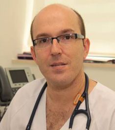 Miguel Angel Martinez Garcia合作论文数Hospital Universitario y Politécnico La Fe de Valencia9Reena Mehra合作论文数8
 Susan Redline合作论文数Division of Sleep Medicine, Harvard Medical School;Division of Sleep and Circadian Disorders, Departments of Medicine and Neurology, Brigham and Women's Hospital;Division of Pulmonary Medicine, Department of Medicine, Beth Israel Deaconess Medical Center7
 Andrew Kao合作论文数7
 Phyllis C Zee合作论文数Department of Neurology, Feinberg School of Medicine, Northwestern University;Division of Sleep Medicine, Northwestern Medicine;Center for Circadian and Sleep Medicine, Feinberg School of Medicine, Northwestern University7
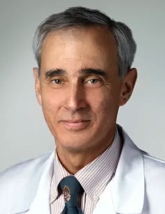 Alan Richard Schwartz合作论文数St. Joseph Medical Center, University of Maryland7
 Babak Mokhlesi合作论文数7
 Scott Sands合作论文数Monash Institute of Medical Research, Monash University6Badr M Safwan合作论文数5
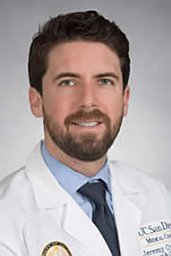 Orr Jeremy E合作论文数UCLA Medical Center5
 Jerome Dempsey合作论文数Department of Population Health Sciences, School of Medicine and Public Health, University of Wisconsin-Madison5Paul E Wylie合作论文数4
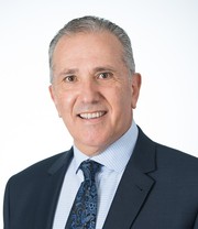 Peter Cistulli合作论文数School of Medicine, Faculty of Medicine and Health, The University of Sydney4Holcomb Richard合作论文数4
 David Gozal合作论文数Joan C. Edwards School of Medicine, Marshall University4
 Bradley A Edwards合作论文数Monash Biomedicine Discovery Institute, School of Psychological Sciences, Monash University4Walter T Mcnicholas合作论文数4
 Michelle Cao合作论文数Neurology & Neurological Sciences Department, Stanford University4Laura Wexler合作论文数4
 Sairam Parthasarathy合作论文数4
 Andrew Wellman合作论文数3andrew merliss合作论文数3Morgenthaler Timothy I合作论文数3
 Klaus-Jürgen Gutleben合作论文数Mühlenkreiskliniken AöR3Kolodziej Adam合作论文数4th Military Hospital3
 Christoph Stellbrink合作论文数3
 Michele Emdin合作论文数Scuola Superiore Sant’Anna3
 Ali Javaheri合作论文数Department of Molecular Genetics and Cell Biology, The University of Chicago3
 Alberto Giannoni合作论文数Scuola Superiore Sant'Anna3
 Yuksel Peker合作论文数3
 Ali Azarbarzin合作论文数3
 Eugene S. Chung合作论文数The Christ Hospital3




