The emergence of groundbreaking large language models capable of performing complex reasoning tasks holds significant promise for addressing various scientific challenges, including those arising in complex clinical scenarios. To enable their safe and effective deployment in real-world healthcare settings, it is urgently necessary to benchmark the diagnostic capabilities of current models systematically. Given the limitations of existing medical benchmarks in evaluating advanced diagnostic reasoning, we present DiagnosisArena, a comprehensive and challenging benchmark designed to rigorously assess professional-level diagnostic competence. DiagnosisArena consists of 1,113 pairs of segmented patient cases and corresponding diagnoses, spanning 28 medical specialties, deriving from clinical case reports published in 10 top-tier medical journals. The benchmark is developed through a meticulous construction pipeline, involving multiple rounds of screening and review by both AI systems and human experts, with thorough checks conducted to prevent data leakage. Our study reveals that even the most advanced reasoning models, o3, o1, and DeepSeek-R1, achieve only 51.12
Dose calculation, as the core of radiotherapy, remains a timeconsuming and experience-dependent process. While deep learning methods have been proposed to improve efficiency, they often face challenges in global modeling and computational complexity, especially with 3D data, limiting their clinical applicability. Moreover, their predicted dose maps are often over-smoothed due to pixel-wise loss functions such as L1 loss that ignore inter-pixel relationship. To address these issues, we propose an efficient framework DoseGLA for 3D dose prediction. First, we propose a Gated Linear Attention-based UNet (GLAUNet) that incorporates an improved gating mechanism inspired by Mamba into linear attention to enhance global modeling capacity while retaining linear computational complexity. Second, a Wavelet-domain Constraint Loss (WDCL) is proposed to guide the model in learning inter-pixel relationship with better prediction contrasts in frequency domain. Experiments on the OpenKBP dataset show that our framework significantly outperforms the state-of-theart model with around 80% fewer parameters, achieving a Dose Score of 2.33 and a DVH Score of 1.46.
Foundation models (FMs) are large-scale deep learning models trained on massive datasets, often using self-supervised learning techniques. These models serve as a versatile base for a wide range of downstream tasks, including those in medicine and healthcare. FMs have demonstrated remarkable success across multiple healthcare domains. However, existing surveys in this field do not comprehensively cover all areas where FMs have made significant strides. In this survey, we present a comprehensive review of FMs in medicine, focusing on their evolution, learning strategies, flagship models, applications, and associated challenges. We examine how prominent FMs, such as the BERT and GPT families, are transforming various aspects of healthcare, including clinical large language models, medical image analysis, and omics research. Additionally, we provide a detailed taxonomy of FM-enabled healthcare applications, spanning clinical natural language processing, medical computer vision, graph learning, and other biology- and omics-related tasks. Despite the transformative potential of FMs, they also pose unique challenges. This survey delves into these challenges and highlights open research questions and lessons learned to guide researchers and practitioners. Our goal is to provide valuable insights into the capabilities of FMs in health, facilitating responsible deployment and mitigating associated risks.
Computed tomography (CT) plays a crucial role in medical diagnosis, but minimizing radiation exposure while maintaining image quality remains a critical challenge. Low-dose CT (LDCT) protocols reduce radiation risks but inevitably suffer from severe noise and artifacts that compromise diagnostic accuracy. While existing deep learning methods have achieved promising results, there remains a continuous quest for generative paradigms that intrinsically capture global-to-local structural dependencies to better preserve fine anatomical details. To this end, we propose DeVAR, a novel generative framework that applies visual autoregressive modeling (VAR) to LDCT denoising for the first time. Conditioned on global context provided by LDCT prefix tokens, DeVAR progressively generates discrete token maps of the target normal-dose CT (NDCT) via next-scale prediction. Because quantization inherently discards high-frequency information, we introduce a residual refiner to capture subtle anatomical structures beyond the capacity of a discrete codebook. Finally, empowered by a dual-representation hybrid training strategy, our hybrid NDCT decoder seamlessly integrates continuous and discrete latents to reconstruct high-fidelity, detail-preserved images. Extensive experiments on two public datasets demonstrate that DeVAR consistently achieves superior qualitative and quantitative performance compared to state-of-the-art LDCT denoising methods.
Multimodal large language models (MLLMs) demonstrate considerable potential in clinical diagnostics, a domain that inherently requires synthesizing complex visual and textual data alongside consulting authoritative medical literature. However, existing benchmarks primarily evaluate MLLMs in end-to-end answering scenarios. This limits the ability to disentangle a model's foundational multimodal reasoning from its proficiency in evidence retrieval and application. We introduce the Clinical Understanding and Retrieval Evaluation (CURE) benchmark. Comprising 500 multimodal clinical cases mapped to physician-cited reference literature, CURE evaluates reasoning and retrieval under controlled evidence settings to disentangle their respective contributions. We evaluate state-of-the-art MLLMs across distinct evidence-gathering paradigms in both closed-ended and open-ended diagnosis tasks. Evaluations reveal a stark dichotomy: while advanced models demonstrate clinical reasoning proficiency when supplied with physician reference evidence (achieving up to 73.4% accuracy on differential diagnosis), their performance substantially declines (as low as 25.4%) when reliant on independent retrieval mechanisms. This disparity highlights the dual challenges of effectively integrating multimodal clinical evidence and retrieving precise supporting literature. CURE is publicly available at https://github.com/yanniangu/CURE.
Due to their high resolution, pathology images incur substantial storage costs. Although most vendor formats have already adopted lossy compression (typically JPEG) to process such images, the storage requirements remain considerable. However, achieving further reductions in storage requirements while ensuring truly loss-less recompression of already lossy-compressed JPEG images remains a critical yet largely unexplored research problem. To address this challenge, we propose a Parallel Hyper-Prior and Dual-Domain Autoregressive Transformer model that processes the three discrete cosine transform (DCT) components in parallel, thereby reducing the complexity of the attention mechanism. The Dual-Domain Autoregressive Transformer module employs a spatial fractal autoregressive transformer, which not only substantially lowers the computational complexity of attention but also captures the inherent hierarchical structure of the spatial domain, and a frequency-domain autoregressive transformer that more effectively models dependencies among coefficients at different frequencies. In addition, we introduce a parameter-sharing and scale-regularized Gaussian–Laplace mixture entropy model to achieve more accurate entropy estimation, thereby enhancing the network’s conditional modeling capability and improving compression performance. We validate the proposed method on the public CAMELYON16 dataset (in-distribution) and multiple TCGA datasets (out-of-distribution). Experimental results show that, for lossless JPEG recompression, our approach achieves state-of-the-art performance compared with both traditional and learned JPEG lossless recompression methods, attaining up to 33.4% additional lossless compression on standard JPEG files. Moreover, the method maintains strong generalization to out-of-distribution data. The source code has been made publicly available at: https://github.com/zhuiguangzhe123/PLR.
Multimodal MR image synthesis aims to generate missing modality images by effectively fusing and mapping from a subset of available MRI modalities. Most existing methods adopt an image-to-image translation paradigm, treating multiple modalities as input channels. However, these approaches often yield sub-optimal results due to the inherent difficulty in achieving precise feature- or semantic-level alignment across modalities. To address these challenges, we propose an Adaptive Group-wise Interaction Network (AGI-Net) that explicitly models both inter-modality and intra-modality relationships for multimodal MR image synthesis. Specifically, feature channels are first partitioned into predefined groups, after which an adaptive rolling mechanism is applied to conventional convolutional kernels to better capture feature and semantic correspondences between different modalities. In parallel, a cross-group attention module is introduced to enable effective feature fusion across groups, thereby enhancing the network's representational capacity. We validate the proposed AGI-Net on the publicly available IXI and BraTS2023 datasets. Experimental results demonstrate that AGI-Net achieves state-of-the-art performance in multimodal MR image synthesis tasks, confirming the effectiveness of its modality-aware interaction design. We release the relevant code at: https://github.com/zunzhumu/Adaptive-Group-wise-Interaction-Network-for-Multimodal-MRI-Synthesis.git.
The non-invasive assessment of renal masses remains a critical challenge in urologic oncology, where diagnostic uncertainty frequently causes overtreatment. Here, we develop RenalCLIP, a vision-language foundation model for precision oncology in kidney cancer. Utilizing 27,866 computed tomography scans from 8809 patients across diverse multi-center cohorts, we employ a two-stage pre-training strategy to align domain-specific visual and textual representations. RenalCLIP achieves enhanced performance and generalizability across ten core clinical tasks, spanning anatomical assessment, diagnostic classification, and survival prediction, significantly outperforming state-of-the-art general-purpose foundation models. Furthermore, RenalCLIP demonstrates strong data efficiency in diagnostic classification, achieving peak baseline performance using only 20% of the training data. The model also exhibits robust zero-shot diagnostic capabilities, effective image-text retrieval, and high-quality medical report generation. Our findings establish RenalCLIP as a powerful, generalizable tool to enhance diagnostic precision, refine prognostic stratification, and personalize the management of renal masses.
Cone-beam CT (CBCT) is routinely acquired in radiotherapy but suffers from severe artifacts and unreliable Hounsfield Unit (HU) values, limiting its direct use for dose calculation. Synthetic CT (sCT) generation from CBCT is therefore an important task, yet paired CBCT--CT data are often unavailable or unreliable due to temporal gaps, anatomical variation, and registration errors. In this work, we introduce rectified flow (RF) into unpaired CBCT-to-CT translation in medical imaging. Although RF is theoretically compatible with unpaired learning through distribution-level coupling and deterministic transport, its practical effectiveness under small medical datasets and limited batch sizes remains underexplored. Direct application with random or batch-local pseudo pairing can produce unstable supervision due to semantically mismatched endpoint samples. To address this challenge, we propose Retrieval-Augmented Flow Matching (RAFM), which adapts RF to the medical setting by constructing retrieval-guided pseudo pairs using a frozen DINOv3 encoder and a global CT memory bank. This strategy improves empirical coupling quality and stabilizes unpaired flow-based training. Experiments on SynthRAD2023 under a strict subject-level true-unpaired protocol show that RAFM outperforms existing methods across FID, MAE, SSIM, PSNR, and SegScore. The code is available at https://github.com/HiLab-git/RAFM.git.
Bidirectional unpaired image translation must preserve source-specific structure while learning coherent transformations in both directions without paired supervision. Existing methods typically employ two direction-specific generators or train separate one-way models. Even when linked by cycle consistency, such models constrain only the round-trip endpoint reconstruction, without requiring the two directions to obey a common local transformation rule. We propose UniCycleFlow, a rectified-flow framework that represents bidirectional translation as forward and reverse integration of a single time-conditioned velocity field. This formulation organizes both directions within the same continuous dynamics, rather than coupling otherwise separate endpoint mappings. A key challenge is that unpaired data provide no meaningful source–target coupling from which rectified-flow trajectories can be constructed. UniCycleFlow addresses this challenge by learning deterministic source-conditioned endpoints whose marginal distributions are adversarially matched to the opposite domains. The resulting paths are regularized by stop-gradient self-flow matching for intermediate velocity supervision, discrete cycle closure for forward–reverse consistency, and representation path-velocity regularization for controlling localized feature changes along the trajectory. Across ten translation directions, UniCycleFlow achieves the lowest FID on 7 of 10 tasks using a single Euler evaluation and obtains the best average FID of 55.1.
Concept segmentation models like Segment Anything Model 3 (SAM3) show strong generalization on natural images, yet their performance degrades in medical imaging due to the domain gap caused by different imaging principles and styles. Test-Time Adaptation (TTA) is essential for improving the testing performance by updating the model on the fly without annotations. However, existing vision-language TTA methods are mainly driven by image-level uncertainty minimization, which does not necessarily reflect region-level semantic correctness in medical segmentation. Moreover, they often lack mechanisms to maintain stability in continual one-pass adaptation, leading to limited performance when reliable dense supervision is missing for segmentation. To address these issues, we propose Concept Alignment Contrast and LongShort Prompt Memory for Test-Time Adaptation (CM-TTA) of SAM3 for medical images. First, for a test sample with multiple augmentations, we introduce a novel Concept Alignment Contrast (CAC) metric, which leverages textual-visual semantic consistency to robustly evaluate prediction quality to select the best augmented view as the supervision. Second, to balance rapid and stable adaptation, we design a Long-Short Prompt Memory (LSPM) module. The short memory dynamically fuses recent prompts based on CAC scores for agile local adaptation, while the long memory maintains a stable global prompt to generate enhanced pseudo-labels. Finally, a Densely Supervised Prompt Update (DSPU) strategy is proposed to optimize the prompt embeddings with enhanced pseudo labels as dense supervision. Extensive experiments on prostate and skin lesion segmentation demonstrate that our CM-TTA framework significantly outperforms existing methods for TTA of SAM3.
Generating synthetic CT images from CBCT or MRI has a potential for efficient radiation dose planning and adaptive radiotherapy. However, existing CNN-based models lack global semantic understanding, while Transformers often overfit small medical datasets due to high model capacity and weak inductive bias. To address these limitations, we propose a DINOv3-Guided Cross Fusion (DGCF) framework that integrates a frozen self-supervised DINOv3 Transformer with a trainable CNN encoder-decoder. It hierarchically fuses global representation of Transformer and local features of CNN via a learnable cross fusion module, achieving balanced local appearance and contextual representation. Furthermore, we introduce a Multi-Level DINOv3 Perceptual (MLDP) loss that encourages semantic similarity between synthetic CT and the ground truth in DINOv3's feature space. Experiments on the SynthRAD2023 pelvic dataset demonstrate that DGCF achieved state-of-the-art performance in terms of MS-SSIM, PSNR and segmentation-based metrics on both MRI→CT and CBCT→CT translation tasks. To the best of our knowledge, this is the first work to employ DINOv3 representations for medical image translation, highlighting the potential of self-supervised Transformer guidance for semantic-aware CT synthesis. The code is available at https://github.com/HiLab-git/DGCF.
We present PULSE, a medical reasoning agent that combines a domain-tuned large language model with scientific literature retrieval to support diagnostic decision-making in complex real-world cases. To evaluate its capabilities, we curated a benchmark of 82 authentic endocrinology case reports encompassing a broad spectrum of disease types and incidence levels. In controlled experiments, we compared PULSE's performance against physicians with varying levels of expertise-from residents to senior specialists-and examined how AI assistance influenced human diagnostic reasoning. PULSE attained expert-competitive accuracy, outperforming residents and junior specialists while matching senior specialist performance at both Top@1 and Top@4 thresholds. Unlike physicians, whose accuracy declined with disease rarity, PULSE maintained stable performance across incidence tiers. The agent also exhibited adaptive reasoning, increasing output length with case difficulty in a manner analogous to the longer deliberation observed among expert clinicians. When used collaboratively, PULSE enabled physicians to correct initial errors and broaden diagnostic hypotheses, but also introduced risks of automation bias. The study explores both serial and concurrent collaboration workflows, revealing that PULSE offers robust support across common and rare presentations. These findings underscore both the promise and the limitations of language model-based agents in clinical diagnosis, and offer a framework for evaluating their role in real-world decision-making.
Medical image segmentation is vital for clinical diagnosis and treatment; however, current solutions face three major limitations: (1) the lack of a universal framework capable of handling diverse modalities and anatomical targets, (2) the limited scalability to adapt to evolving clinical needs and new datasets, and (3) the lack of instructive interfaces that make models usable for non-expert users. To address these challenges, this paper presents MedSegAgent, a universal and scalable multi-agent system for instructive medical image segmentation. Specifically, MedSegAgent comprises five agents: one query parsing agent that processes natural language requests, three coarse-to-fine filtering agents (modality filtering, anatomical filtering, and label selection) for identifying relevant datasets and label values, and one execution agent responsible for model inference and result integration. Based on this framework, MedSegAgent utilizes 23 diverse datasets and pre-trained models to perform 343 types of segmentation across various modalities and anatomical targets. Experimental results demonstrate that MedSegAgent simplifies model selection while maintaining high performance, accurately identifying matching datasets and labels in 94.27% of queries and locating at least one suitable match in 99.03% of queries. MedSegAgent offers a universal and scalable solution for diverse medical image segmentation tasks, bridging the gap between user-friendly queries and the complexities of model selection and deployment. Our code is publicly available at https://github.com/uni-medical/MedSegAgent.
Semi-Supervised Learning (SSL) is important for reducing the annotation cost for medical image segmentation models. State-of-the-art SSL methods such as Mean Teacher, FixMatch and Cross Pseudo Supervision (CPS) are mainly based on consistency regularization or pseudo-label supervision between a reference prediction and a supervised prediction. Despite the effectiveness, they have overlooked the potential noise in the labeled data, and mainly focus on strategies to generate the reference prediction, while ignoring the heterogeneous values of different unlabeled pixels. We argue that effectively mining the rich information contained by the two predictions in the loss function, instead of the specific strategy to obtain a reference prediction, is more essential for SSL, and propose a universal framework MetaSSL based on a spatially heterogeneous loss that assigns different weights to pixels by simultaneously leveraging the uncertainty and consistency information between the reference and supervised predictions. Specifically, we split the predictions on unlabeled data into four regions with decreasing weights in the loss: Unanimous and Confident (UC), Unanimous and Suspicious (US), Discrepant and Confident (DC), and Discrepant and Suspicious (DS), where an adaptive threshold is proposed to distinguish confident predictions from suspicious ones. The heterogeneous loss is also applied to labeled images for robust learning considering the potential annotation noise. Our method is plug-and-play and general to most existing SSL methods. The experimental results showed that it improved the segmentation performance significantly when integrated with existing SSL frameworks on different datasets. Code is available at https://github.com/HiLab-git/MetaSSL.
The emergence of tool-calling-based agent systems introduces a more evidence-driven paradigm for pathology image analysis in contrast to the coarse-grained text-image diagnostic approaches. With the recent large-scale experimental adoption of spatial transcriptomics technologies, molecularly validated pathological diagnosis is becoming increasingly open and accessible. In this work, we propose LAMMI-Pathology (LVLM-Agent System for Molecularly Informed Medical Intelligence in Pathology), a scalable agent framework for domain-specific agent tool-calling. LAMMI-Pathology adopts a tool-centric, bottom-up architecture in which customized domain-adaptive tools serve as the foundation. These tools are clustered by domain style to form component agents, which are then coordinated through a top-level planner hierarchically, avoiding excessively long context lengths that could induce task drift. Based on that, we introduce a novel trajectory construction mechanism based on Atomic Execution Nodes (AENs), which serve as reliable and composable units for building semi-simulated reasoning trajectories that capture credible agent-tool interactions. Building on this foundation, we develop a trajectory-aware fine-tuning strategy that aligns the planner's decision-making process with these multi-step reasoning trajectories, thereby enhancing inference robustness in pathology understanding and its adaptive use of the customized toolset.
Accurate delineation of Gross Tumor Volume (GTV), Lymph Node Clinical Target Volume (LN CTV), and Organ-at-Risk (OAR) from Computed Tomography (CT) scans is essential for precise radiotherapy planning in Nasopharyngeal Carcinoma (NPC). Building upon SegRap2023, which focused on OAR and GTV segmentation using single-center paired non-contrast CT (ncCT) and contrast-enhanced CT (ceCT) scans, the SegRap2025 challenge aims to enhance the generalizability and robustness of segmentation models across imaging centers and modalities. SegRap2025 comprises two tasks: Task01 addresses GTV segmentation using paired CT from the SegRap2023 dataset, with an additional external testing set to evaluate cross-center generalization, and Task02 focuses on LN CTV segmentation using multi-center training data and an unseen external testing set, where each case contains paired CT scans or a single modality, emphasizing both cross-center and cross-modality robustness. This paper presents the challenge setup and provides a comprehensive analysis of the solutions submitted by ten participating teams. For GTV segmentation task, the top-performing models achieved average Dice Similarity Coefficient (DSC) of 74.61% and 56.79% on the internal and external testing cohorts, respectively. For LN CTV segmentation task, the highest average DSC values reached 60.24%, 60.50%, and 57.23% on paired CT, ceCT-only, and ncCT-only subsets, respectively. SegRap2025 establishes a large-scale multi-center, multi-modality benchmark for evaluating the generalization and robustness in radiotherapy target segmentation, providing valuable insights toward clinically applicable automated radiotherapy planning systems. The benchmark is available at: https://hilab-git.github.io/SegRap2025_Challenge.
 Dimitris N. Metaxas合作论文数Department of Computer Science, School of Arts and Sciences, Rutgers University138
 Guotai Wang (王国泰)合作论文数School of Mechanical and Electrical Engieering, University of Electronic Science and Technology of China103
 Xiaofan Zhang (张晓凡)合作论文数Qing Yuan Research Institute, School of Computer Science, Shanghai Jiao Tong University51
 Kang Li (李康)合作论文数Sichuan University-Pittsburgh Institute;West China Hospital, Sichuan University47
 Junzhou Huang合作论文数Scalable Modeling & Imaging & Learning Lab, Department of Computer Science and Engineering, University of Texas at Arlington37
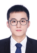 Xiangde Luo合作论文数Department of Radiation Oncology, School of Medicine, Stanford University31
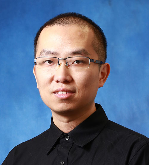 Hongsheng Li (李鴻升)合作论文数Department of Electronic Engineering, The Chinese University of Hong Kong;Multimedia Laboratory, The Chinese University of Hong Kong30
 Shichuan Zhang (张石川)合作论文数School of Medicine, University of Electronic Science and Technology of China27Wenjun Liao合作论文数Department of Radiation Oncology, Nanfang Hospital, Southern Medical University, Guangzhou, China.25
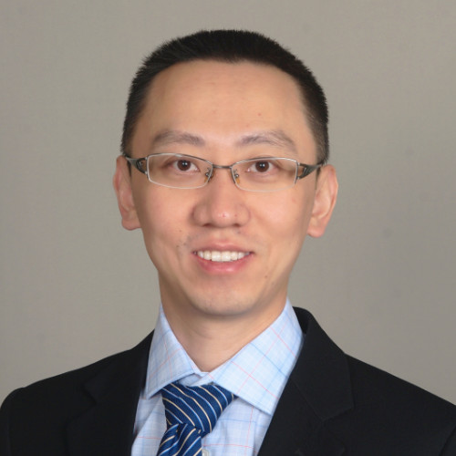 Zhennan Yan合作论文数Helm.ai25
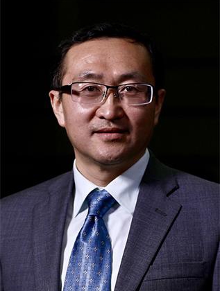 Lixu Gu (顾力栩)合作论文数School of Biomedical Engineering, Shanghai Jiao Tong University;Institute of Medical Robotics, Shanghai Jiao Tong University19Zhiqiang Hu合作论文数SenseTime Research19
 Wenhui Lei合作论文数Univ Elect Sci & Technol China, Chengdu, Peoples R China18
 Leon Axel合作论文数Department of Radiology, Grossman School of Medicine, New York University18
 Ran Gu合作论文数Univ Elect Sci & Technol China, Sch Mech & Elect Engn, Chengdu 611731, Peoples R China16
 Mu Zhou (周牧)合作论文数School of Medicine, Tsinghua University16
 Sharon X. Huang合作论文数Department of Informatics and Intelligent Systems, College of Information Sciences and Technology, Penn State University;Institute for Computational and Data Sciences, Penn State University;Huck Institutes of the Life Sciences, Penn State University16
 Zhongzhen Huang合作论文数Qing Yuan Research Institute, Shanghai Jiao Tong University;Shanghai Artificial Intelligence Laboratory15
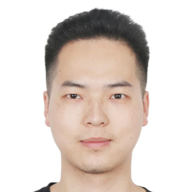 Junjun He (何军军)合作论文数Shanghai AI Laboratory15
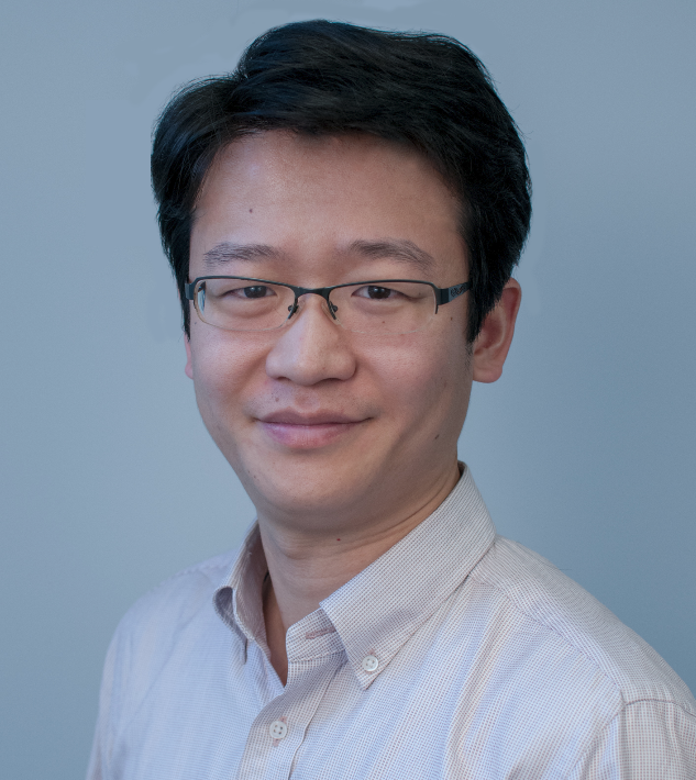 Xiaosong Wang (王潚崧)合作论文数Shanghai AI Laboratory15
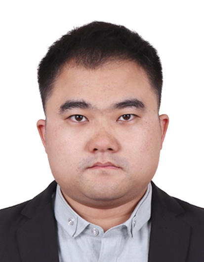 Zhongyu Li (李钟毓)合作论文数School of Computer Science, Shanghai Jiao Tong University;Qing Yuan Research Institute, Shanghai Jiao Tong University13
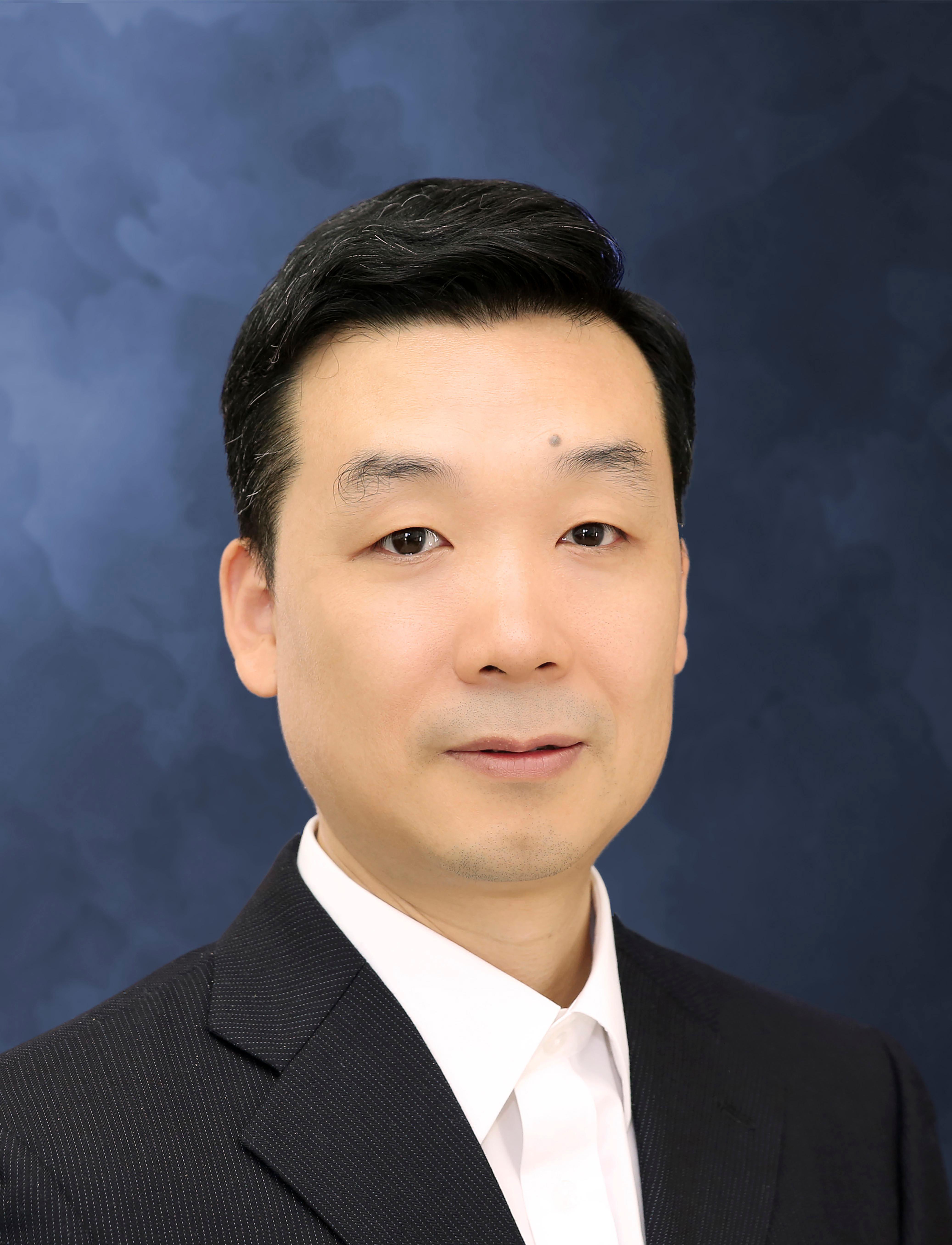 Yu Qiao (乔宇)合作论文数Multimedia Laboratory, Shenzhen Institutes of Advanced Technology, Chinese Academy of Sciences;Shanghai AI Laboratory;Shenzhen University of Advanced Technology13
 Tao Song (宋弢)合作论文数College of Computer Science and Technology, China University of Petroleum (East China)13
 Yanzhou Su合作论文数12
 Yiqiang Zhan合作论文数Shanghai Jiao Tong University12Ziyan Huang合作论文数Institute of Medical Robotics, Shanghai Jiao Tong University11
 Yinan Chen合作论文数[SenseTime Research]11
 Qing Xia合作论文数School of Mathematics and Statistics, Xi’an Jiaotong University11
 Yuanjie Zheng (郑元杰)合作论文数School of Information Science and Engineering, Shandong Normal University10
 Zhongying Deng (邓重英)合作论文数Centre for Vision, Speech and Signal Processing, University of Surrey;IFlyTek-Surrey Joint Research Center on Artificial Intelligence10
 Zhen Qian (钱震)合作论文数Site powered by a free Open Source Project / Non-profit License (more) of Confluence - the Enterprise wiki10
 Ruogu Fang合作论文数Department of Electrical and Computer Engineering, Cornell University10
 Kexin Ding合作论文数Department of Computer Science, The University of North Carolina at Charlotte9
 Mingchen Gao (高明辰)合作论文数Department of Computer Science and Engineering, School of Engineering and Applied Sciences, University at Buffalo9
 Tom Vercauteren合作论文数Department of Surgical & Interventional Engineering, School of Biomedical Engineering & Imaging Sciences, Faculty of Life Sciences & Medicine, King’s College London;University College London9
 Peter Peng (彭曦)合作论文数Deep Robust & Explainable AI Lab, Department of Computer & Information Sciences, University of Delaware;Data Science Institute, University of Delaware9
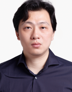 Wei Liu (刘威)合作论文数Video Rebirth9Jianghao Wu合作论文数8
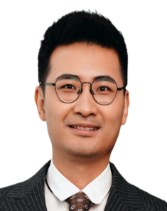 Qicheng Lao (劳琪成)合作论文数Department of Intelligent Science and Technology, School of Artificial Intelligence, Beijing University of Posts and Telecommunications8
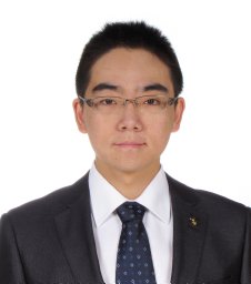 Menglin Jiang合作论文数8
 Chaowei Tan合作论文数8
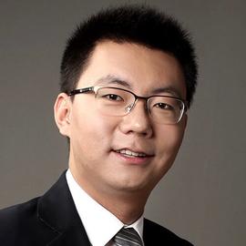 Dequan Wang (王德泉)合作论文数Qing Yuan Research Institute, Shanghai Jiao Tong University;Shanghai Artificial Intelligence Laboratory8
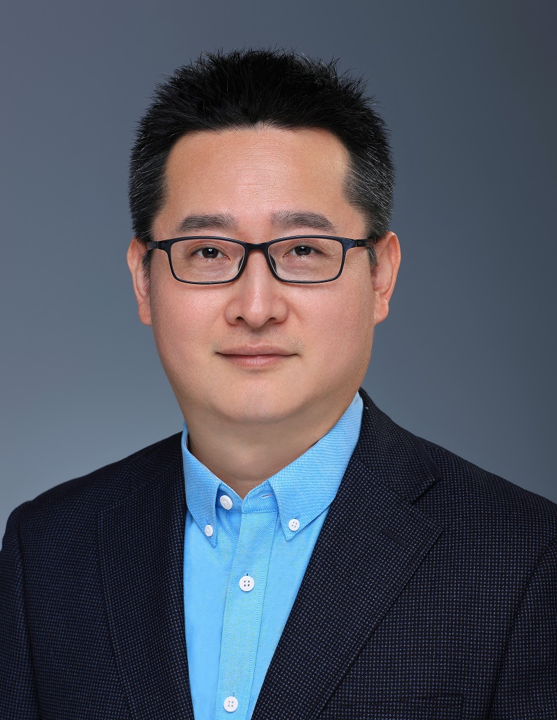 Ye Jin (金野)合作论文数Pattern Recognition and Intelligent System Research Center, School of Computer Science and Technology, Faculty of Computing, Harbin Institute of Technology8
 Sebastien Ourselin合作论文数London Institute for Healthcare Engineering, School of Biomedical Engineering & Imaging Sciences, Faculty of Life Sciences & Medicine, King’s College London;Wellcome EPSRC Centre for Medical Engineering, King’s College London;Artificial Intelligence Centre for Value Based Healthcare;Hypervision Surgical;Elaitra;OxVent8Lijian Xu合作论文数8Haoyu Wang合作论文数7
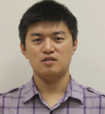 Han Zhang合作论文数Reve AI7Tao Lu合作论文数7
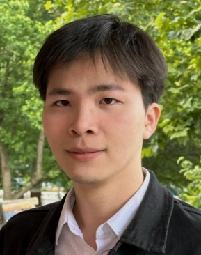 Jieneng Chen合作论文数The Johns Hopkins University7




