Medical vision-language models (MVLMs) offer promise in clinical practice but face limitations in generalizability, data quality, and clinically meaningful evaluation. We propose RadiSim-CL, an MVLM trained via curriculum learning by simulating the three-phase pathway of a radiologist: foundational knowledge understanding, anatomical knowledge, and advanced diagnostic reasoning. To support this, we curate RadiSim, a 12-million image-text pair dataset aligned to these phases. We evaluate the model using a five-stage coarse-to-fine validation framework: (1) modality recognition, (2) anatomical recognition, (3) anatomical localization, (4) abnormality and disease diagnosis, and (5) disease differentiation and grading. This framework spans 24 zero-shot subtasks across MR, CT, and DR imaging. RadiSim-CL achieves comparable performance to state-of-the-art baselines in both foundational and anatomical tasks, and demonstrates superior capabilities in complex reasoning (e.g., an AUC of 0.953 for brain tumor diagnosis and an accuracy of 0.764 for meningioma grading). Ablation studies further confirm the curriculum’s effectiveness. RadiSim-CL thus offers a scalable, clinically aligned solution to enhance diagnostic precision.
Lung cancer is the leading cause of cancerrelated mortality worldwide. In addition to localizing and segmenting lung nodules, a non-invasive risk assessment system can also help clinicians tailor treatment decisions in a timely manner, ultimately improving patient outcomes. Artificial intelligence (AI) technologies are increasingly being used in medical imaging to assess the risk of lung nodules, especially for malignancy classification. However, little research has been conducted on the assessment of other related risks. This work comprehensively reviews AI applications in lung nodule risk assessment, including malignancy diagnosis, pathological subtype assessment, metastasis risk evaluation, specific receptor expression identification, and disease progression tracking. It details common public databases used and state-of-the-art AI techniques, along with their benefits and challenges like data scarcity, generalizability, and interpretability. We anticipate that future research will tackle these issues, thereby increasing the improved interpretability and generalizability of AI methods in clinical workflows.
Automatic anatomical localization is critical for radiology report generation. While many studies focus on lesion detection and segmentation, anatomical localization—accurately describing lesion positions in radiology reports—has received less attention. Conventional segmentation-based methods are limited to organ-level localization and often fail in severe disease cases due to low segmentation accuracy. To address these limitations, we reformulate anatomical localization as an image-to-text retrieval task. Specifically, we propose a CLIP-based framework that aligns lesion image patches with anatomically descriptive text embeddings in a shared multimodal space. By projecting lesion features into the semantic space and retrieving the most relevant anatomical descriptions in a coarse-to-fine manner, our method achieves fine-grained lesion localization with high accuracy across the entire body. Our main contributions are as follows: (1) hierarchical anatomical retrieval, which organizes 387 locations into a two-level hierarchy, by retrieving from the first level of 124 coarse categories to narrow down the search space and reduce localization complexity; (2) augmented location descriptions, which integrate domain-specific anatomical knowledge for enhancing semantic representation and improving visual—text alignment; and (3) semi-hard negative sample mining, which improves training stability and discriminative learning by avoiding selecting the overly similar negative samples that may introduce label noise or semantic ambiguity. We validate our method on two whole-body PET/CT datasets, achieving an 84.13% localization accuracy on the internal test set and 80.42% on the external test set, with a per-lesion inference time of 34 ms. The proposed framework also demonstrated superior robustness in complex clinical cases compared to segmentation-based approaches.
Accurate diagnosis of brain disorders (BDs) is challenging in clinical practice. Most existing deep learning-based methods perform diagnosis only in a one-step manner, ignoring the step-wise, multi-level diagnosis processes as performed by radiologists. This oversight often leads to a high risk of misdiagnosis, especially for long-tail or challenging BDs. In this work, we introduce a Hierarchical Prompt and Prototype Learning (HP2L) framework for BD diagnosis, which emulates multi-level diagnostic procedures. HP2L explicitly captures hierarchical relationships among 23 BDs and groups them into three diagnostic levels: coarse classes (e.g., vascular lesions), intermediate classes (e.g., hemorrhage), and fine-grained classes (e.g., chronic hemorrhage). HP2L integrates three key innovations: (1) Hierarchical Prompting Vision Transformer (ViT) backbone, which performs coarse-to-fine feature extraction for step-wise BD classification; (2) Prompt Learning, which employs optimizable prompt tokens that encode diagnostic knowledge, guiding the classification at each level of the hierarchy; (3) Prototype Learning, which enriches the prompt token with BD-specific prototypes by injecting diagnostic information to enhance diagnosis performance. Extensive evaluations on 54,360 subjects across six multi-center datasets show that HP2L consistently outperforms state-of-the-art methods, achieving a balanced accuracy of 88.43% for both common and long-tail BDs, 8.42 percentage points higher than the best-performing benchmark. Furthermore, HP2L improves interpretability by aligning its predictions and attention visualizations with the clinical hierarchical reasoning process. The code and a portion of data (more data will be released after the decision of the paper) are available under: code, data.
Lengthy acquisition time remains a key bottleneck for the widespread use of MRI in clinics. While accelerated MRI can reduce scan duration, it often introduces increased noise, compromising image quality and diagnostic reliability. In this study, we present a unified deep learning-based denoising model for multi-organ accelerated MRI, designed to operate directly on reconstructed images from commercial MRI systems. Our model was trained on a prospectively collected, large-scale real-world dataset comprising 148,930 noisy-clean image pairs from six clinical centers and four major MRI vendors, spanning six organs and 96 MRI protocols. On a test set of 20,143 real-world image pairs, our model consistently outperforms state-of-the-art denoising methods. Importantly, downstream evaluation using tissue segmentation demonstrates a 7.05% improvement in Dice score across multiple organs compared to noisy images. The model further generalizes effectively to 46,870 external clinical images from four independent cohorts, highlighting its robustness across various scanners and acquisition protocols. To assess clinical utility, two experienced radiologists conducted blinded evaluations across multiple organs, focusing on overall image quality, diagnostic confidence, and disease diagnosis. The denoised images retained high visual fidelity and yielded diagnostic performance equivalent to clean images even with acceleration factor of 3× compared to clinical scanning setup, such that many acquisitions can be completed within one minute. This unified MRI denoising model holds great potential for various clinical applications.
Coronary artery disease poses a significant public health threat, and coronary computed tomography angiography is the preferred imaging modality for diagnosis and risk assessment of coronary artery disease through plaque evaluation. However, understandings of how atherosclerotic characteristics vary by age and sex remains limited due to challenges in manual quantitative plaque assessment. Here, we conducted a retrospective, consecutive, multi-center Chinese cohort study of 16,300 patients undergoing clinically indicated coronary computed tomography angiography that revealed multi-level quantitative patterns of atherosclerosis stratified by age and sex. We found that females experienced a delayed atherosclerosis onset by approximately 20 years compared to males, with plaque burden increasing nonlinearly with age and accelerating more evidently after menopause. The built coronary atlas identified plaque clusters, primarily within proximal segments of major coronary arteries, slightly upstream side branch bifurcations. Our findings provide deeper insights into coronary atherosclerosis in the Chinese population, supporting more tailored prevention strategies.
Manual interpretation of CT images for bone metastasis (BM) detection in primary cancer remains challenging. We present an automated Bone Lesion Detection System (BLDS) developed using CT scans from 2518 patients (9177 BMs; 12,824 non-BM lesions) across five hospitals. The system, developed on 1271 patients and tested on 1247 multicenter cases, demonstrates 89.1% lesion-wise sensitivity (1.40 false-positives/case [FPPC]) in detecting bone lesions on non-contrast CT scans, with 92.3% and 91.1% accuracy in classifying BM/non-BM lesions for internal and external test sets, respectively. Outperforming radiologists in lesion detection (40.5% sensitivity; 0.65 FPPC), BLDS shows lower BM detection sensitivity than junior radiologists, though comparable to trainees. BLDS improves radiologists' lesion-wise sensitivity by 22.2% in BM detection and reduces reading time by 26.4%, while maintaining 90.2% patient-wise sensitivity and 98.2% negative predictive value in real-world validation (n = 54,610). The system demonstrates significant potential to enhance CT-based BM interpretation, particularly benefiting trainees.
Accurate identification of lesions, including anatomical lesion localization, is critical for automated radiology report generation. However, this task is particularly challenging in whole-body PET/CT imaging due to large amount of diverse anatomical regions throughout the whole body. Existing studies mainly rely on anatomical detection or segmentation. These methods are generally limited to only a small subset of anatomical regions due to difficulty of manual segmentation and annotation for large set of anatomical regions in the training stage. To address this issue, we propose a hierarchical CLIP-based 3D model to precisely and efficiently identify 387 anatomical lesion locations within whole-body PET/CT scans. Our model is built on three strategies: (1) Hierarchical localization, based on which anatomical locations are identified from coarse to fine to improve localization accuracy, robustness, and scalability; (2) Semantic location augmentation, which incorporates anatomical knowledge of relative location to adjacent regions to encourage neighborhood preservation of text feature representations; and (3) Location ambiguity mitigation, which excludes penalties on the top K ambiguous localizations in a modified CLIP loss to alleviate the cases with lesions residing at the boundaries of multiple regions. Notably, this work is the first to achieve accurate, robust, and efficient whole-body anatomical lesion localization, with significant performance improvement compared to the SOTA methods on a large whole-body PET/CT dataset comprising 1748 subjects acquired from multiple scanner makers.
Aneurysm segmentation in computed tomography angiography (CTA) images is essential for medical intervention aimed at preventing subarachnoid hemorrhages. However, most existing studies tend to overlook the topological characteristics of arteries related to aneurysms, often resulting in suboptimal performance in aneurysm segmentation. To address this challenge, we propose an arteryaware global-to-local framework for aneurysm segmentation (AASeg) using CTA images of head and neck. This framework consists of two key components: 1) a centerline graph network (CG-Net) for aneurysm global localization, and 2) a point cloud network (PC-Net) for local aneurysm segmentation. The centerline graph is generated by extracting artery centerline structures from vessel masks obtained through a pre-trained model for head and neck vessel segmentation. This representation serves as a high-level representation of the artery structure, allowing for analysis of aneurysms along the entire arteries. It facilitates aneurysm localization via aneurysm-segment graph classification along the arteries. Then, local region of aneurysm segment can be sampled from the vessel mask according to the aneurysm-segment graph. Subsequently, aneurysm segmentation is performed on the point cloud constructed from the aneurysm segment through the PC-Net. Extensive experiments show that the proposed framework achieves state-of-the-art performance in aneurysm localization on a main dataset and an external testing dataset, with Recall of 84.1% and 80.7%, false positives per case of 1.72 and 1.69, and segmentation DSC of 66.1% and 60.2%, respectively.
In clinical work, there are difficulties in distinguishing pulmonary contusion(PC) from bacterial pneumonia(BP) on CT images by the naked eye alone when the history of trauma is unknown. Artificial intelligence is widely used in medical imaging, but its diagnostic performance for pulmonary contusion is unclear. In this study, artificial intelligence was used for the first time to identify lung contusion and bacterial pneumonia, and its diagnostic performance was compared with that of manual. In this retrospective study, 2179 patients between April 2016 and July 2022 from two hospitals were collected and divided into a training set, an internal validation set, an external validation set. PC and BP were automatically recognized, segmented using VB-net and radiomics features were automatically extracted. Four machine learning algorithms including Decision Trees, Logistic Regression, Random Forests and Support Vector Machines(SVM) were using to built the models. De-long test was used to compare the performance among models. The best performing model and four radiologists diagnosed the external validation set, and compare the diagnostic efficacy of human and artificial intelligence. VB-net automatically detected and segmented PC and BP. Among the four machine learning models we’ve built, De-long test showed that SVM model had the best performance, with AUC, accuracy, sensitivity, and specificity of 0.998 (95
Whole-body PET/CT imaging provides detailed metabolic and anatomical information, which is critical for accurate cancer staging, treatment evaluation, and radiotherapy planning. Automated lesion captioning for whole-body PET/CT is essential for reducing radiologists' workload and assisting personalized treatment decisions. Unlike previous works that focus on captioning body-part images, we propose a novel automated lesion captioning framework for whole-body PET/CT images, which usually have large volume and high anatomical variability. Our framework first leverages CLIP for lesion localization, upon which we introduce two location-guided strategies: Confidence-Guided Location Prompts (CGLP), which select top-1 or top-3 anatomical location prompts based on confidence scores to guide captioning, and Dynamic Window Setting (DWS), which applies appropriate intensity windowing to enhance visual representation of the localized regions. To our knowledge, our work is the first to achieve whole-body PET/CT lesion captioning. Experimental results on a large dataset comprising 1867 subjects from Siemens, GE, and United Imaging show that our method not only yields higher BLEU scores compared to state-of-the-art methods, but also produces consistent improvements across multiple scanner makers. This advancement has the potential to streamline radiology reporting and enhance clinical decision-making using whole-body PET/CT images.
A deep learning model enabled automated CT myocardial blood flow quantification and accurate evaluation of ischemic myocardial volume, which was an independent predictor for major adverse cardiovascular events and mildly improved cardiovascular risk stratification.
The widespread implementation of low-dose computed tomography (LDCT) in lung cancer screening has led to the increasing detection of pulmonary nodules. However, precisely evaluating the malignancy risk of pulmonary nodules remains a formidable challenge. Here we propose a triage-driven Chinese Lung Nodules Reporting and Data System (C-Lung-RADS) utilizing a medical checkup cohort of 45,064 cases. The system was operated in a stepwise fashion, initially distinguishing low-, mid-, high- and extremely high-risk nodules based on their size and density. Subsequently, it progressively integrated imaging information, demographic characteristics and follow-up data to pinpoint suspicious malignant nodules and refine the risk scale. The multidimensional system achieved a state-of-the-art performance with an area under the curve (AUC) of 0.918 (95% confidence interval (CI) 0.918-0.919) on the internal testing dataset, outperforming the single-dimensional approach (AUC of 0.881, 95% CI 0.880-0.882). Moreover, C-Lung-RADS exhibited a superior sensitivity compared with Lung-RADS v2022 (87.1% versus 63.3%) in an independent cohort, which was screened using mobile computed tomography scanners to broaden screening accessibility in resource-constrained settings. With its foundation in precise risk stratification and tailored management, this system has minimized unnecessary invasive procedures for low-risk cases and recommended prompt intervention for extremely high-risk nodules to avert diagnostic delays. This approach has the potential to enhance the decision-making paradigm and facilitate a more efficient diagnosis of lung cancer during routine checkups as well as screening scenarios.
Numerous deep-learning models have been developed using task-specific data, but they ignore the inherent connections among different tasks. By jointly learning a wide range of segmentation tasks, we prove that a general medical image segmentation model can improve segmentation performance for computerized tomography (CT) volumes. The proposed general CT image segmentation (gCIS) model utilizes a common transformer-based encoder for all tasks and incorporates automatic pathway modules for task prompt-based decoding. It is trained on one of the largest datasets, comprising 36,419 CT scans and 83 tasks. gCIS can automatically perform various segmentation tasks using automatic pathway modules of decoding networks through text prompt inputs, achieving an average Dice coefficient of 82.84%. Furthermore, the proposed automatic pathway routing mechanism allows for parameter pruning of the network during deployment, and gCIS can also be quickly adapted to unseen tasks with minimal training samples while maintaining great performance.
To examine the associations of apolipoprotein E (APOE) carrier status and lipid profiles with sleep disorders, including excessive daytime sleepiness (EDS) and probable rapid eye movement sleep behavior disorder (pRBD), among patients with early Parkinson's disease (PD) over 5-year follow-up periods. The Parkinson's Progression Markers Initiative is a multicenter cohort study based on an ongoing and open-ended registry. Data from baseline and 5-year follow-up visits from participants of de novo PD were analyzed. Longitudinal associations of APOE carrier status and lipid profiles with sleep disorders were estimated via linear mixed-effects models. A total of 657 participants with complete APOE genotypes were enrolled at baseline. Among them, 153 (25.3%) had available lipid profiles at baseline. In the linear mixed-effects models, baseline APOE ε2/ε3/ε4 carrier status did not exhibit significant associations with EDS and pRBD (all p > 0.05) in all models. However, reduced high-density lipoprotein (HDL) and elevated triglycerides (TG) were associated with developing EDS (β = -0.04, 95% CI: -0.07, -0.00) and pRBD (β = 0.01, 95% CI: 0.00, 0.02) in PD patients, respectively. In the APOE ε4+ subgroup, decreased HDL and increased TG displayed substantial associations with developing EDS and sleep disorders (all p < 0.05) in all models, respectively, whereas no significant differences were noted in the APOE ε4- subgroup (all p > 0.05). Our study did not demonstrate a clear association between APOE ε2/ε3/ε4 and sleep disorders in PD patients. However, the presence of APOE ε4 was associated with changes in lipid profiles, notably affecting TG and HDL levels.
To develop and validate a deep learning (DL)-model for automatic reconstruction for coronary CT angiography (CCTA) in patients with origin anomaly, stent or bypass graft. In this retrospective study, a DL model for automatic CCTA reconstruction was developed with training and validation sets from 6063 and 1962 patients. The algorithm was evaluated on an independent external test set of 812 patients (357 with origin anomaly or revascularization, 455 without). The image quality of DL reconstruction and manual reconstruction (using dedicated cardiac reconstruction software provided by CT vendors) was compared using a 5-point scale. The successful reconstruction rates and post-processing time for two methods were recorded. In the external test set, 812 patients (mean age, 64.0 ± 11.6, 100 with origin anomalies, 152 with stents, 105 with bypass grafts) were evaluated. The successful rates for automatic reconstruction were 100
Vessel Wall Magnetic Resonance Imaging (VW-MRI) is an emerging technique for visualizing lumen and vessel wall structures and facilitating the diagnosis of vascular diseases such as atherosclerosis. However, annotations on VW-MRI are usually sparse due to their labor-intensive nature. On the other hand, computed tomography angiography (CTA) images are widely used in atherosclerosis analysis, where data and annotation are relatively sufficient. To this end, we propose a multi-modality transfer learning network (MT-Net) to transfer anatomical knowledge of vessels from CTA to MR, based on fully-annotated training CTA images and sparsely-annotated training MR images. Furthermore, in the MR branch, we utilize the vessel lumen results to guide the multi-channel network for final vessel wall segmentation. Experimental results on the COSMOS Challenge dataset demonstrate advantage of our method in producing robust lumen and vessel wall segmentations with sparse annotation.
Early identification of malignant pulmonary nodules is of great interest in the lung cancer screening process. However, the surrounding contextual information is usually complex, but could be better preserved in multiple spatial scales. Hence, we propose a multi-scale supervised contrastive learning framework to effectively extract inter-scale and intra-scale contextual information of nodules. First, we employ three hierarchical scales from a 3D CT scan to obtain representations, respectively. Second, a newly designed projection network is used to extract pairwise features and map them to the latent space. Third, a supervised contrastive loss is further applied to pull nodules of same classes closer while push nodules of different classes much more away, which effectively guarantees consistency and also augments performance. Based on 1,226 nodules (benign/malignant: 556/670), our proposed method achieves superior diagnosis performance with an accuracy of 91.8%, and AUC of 96.1%. The proposed method shows its potential for computer-assisted lung cancer diagnosis on CT images.
IntroductionMedical image analysis is of tremendous importance in serving clinical diagnosis, treatment planning, as well as prognosis assessment. However, the image analysis process usually involves multiple modality-specific software and relies on rigorous manual operations, which is time-consuming and potentially low reproducible.MethodsWe present an integrated platform - uAI Research Portal (uRP), to achieve one-stop analyses of multimodal images such as CT, MRI, and PET for clinical research applications. The proposed uRP adopts a modularized architecture to be multifunctional, extensible, and customizable.Results and DiscussionThe uRP shows 3 advantages, as it 1) spans a wealth of algorithms for image processing including semi-automatic delineation, automatic segmentation, registration, classification, quantitative analysis, and image visualization, to realize a one-stop analytic pipeline, 2) integrates a variety of functional modules, which can be directly applied, combined, or customized for specific application domains, such as brain, pneumonia, and knee joint analyses, 3) enables full-stack analysis of one disease, including diagnosis, treatment planning, and prognosis assessment, as well as full-spectrum coverage for multiple disease applications. With the continuous development and inclusion of advanced algorithms, we expect this platform to largely simplify the clinical scientific research process and promote more and better discoveries.
Pulmonary vessel segmentation in computerized tomography (CT) images is essential for pulmonary vascular disease and surgical navigation. However, the existing methods were generally designed for contrast-enhanced images, their performance is limited by the low contrast and the non-uniformity of Hounsfield Unit (HU) in non-contrast CT images, meanwhile, the varying size of the vessel structures are not well considered in current pulmonary vessel segmentation methods. To address this issue, we propose a hierarchical enhancement network (HENet) for better image- and feature-level vascular representation learning in the pulmonary vessel segmentation task. Specifically, we first design an Auto Contrast Enhancement (ACE) module to adjust the vessel contrast dynamically. Then, we propose a Cross-Scale Non-local Block (CSNB) to effectively fuse multi-scale features by utilizing both local and global semantic information. Experimental results show that our approach achieves better pulmonary vessel segmentation outcomes compared to other state-of-the-art methods, demonstrating the efficacy of the proposed ACE and CSNB module. Our code is available at https://github.com/CODESofWenqi/HENet .
 Xiang Zhou (周翔)合作论文数School of Biomedical Engineering, Shanghai Tech University;United Imaging Intelligence, Shanghai59
 Dinggang Shen (沈定刚)合作论文数School of Biomedical Engineering, ShanghaiTech University;Image Display, Enhancement and Analysis Lab, ShanghaiTech University36
 Feng Shi (石峰)合作论文数School of Biomedical Engineering, ShanghaiTech University;Department of Research and Development, Shanghai United Imaging Intelligence Co19
 Maneesh Dewan合作论文数CAD-IKM,Siemens HealthCare Jan19
 Gerardo Hermosillo Valadez合作论文数Siemens Healthcare18
 Dimitris N. Metaxas合作论文数Department of Computer Science, School of Arts and Sciences, Rutgers University15
 Arun Krishnan合作论文数Computer-Aided Diagnosis and Therapy Solutions, Siemens Medical Solutions13
 Xue Zhong (薛忠)合作论文数School of Biomedical Engineering, ShanghaiTech University12
 Shaoting Zhang (张少霆)合作论文数Qing Yuan Research Institute, School of Computer Science, Shanghai Jiao Tong University12
 Qian Wang (王乾)合作论文数School of Biomedical Engineering, ShanghaiTech University11
 Yaozong Gao (高耀宗)合作论文数United Imaging Intelligence, Shanghai;School of Biomedical Engineering, Shanghai Tech University11
 Shu Liao (廖术)合作论文数School of Biomedical Engineering, ShanghaiTech University10
 Zhigang Peng (彭志刚)合作论文数College of Chemistry and Chemical Engineering, Southwest Petroleum University9
 Dijia Wu (吴迪嘉)合作论文数School of Biomedical Engineering, ShanghaiTech University;United Imaging Intelligence9
 Zhong Xue合作论文数Medical Image Lab,The Methodist Hospital Research Institute7Martin Harder合作论文数6
 Xi Ouyang (欧阳熹)合作论文数United Imaging Intelligence6Ying Wei (魏颖)合作论文数Northeastern University6Yoshihisa Shinagawa合作论文数6
 Zhennan Yan合作论文数Helm.ai6Matthias Fenchel合作论文数6
 Bing Jian合作论文数FocusAI6
 Xiaodan Xing合作论文数Shanghai United Imaging Intelligence Co Ltd, Shanghai, Peoples R China5Yanbo Chen合作论文数Shanghai United Imaging Intelligence Co Ltd, Shanghai, Peoples R China5Marcos Salganicoff合作论文数5Kaicong Sun合作论文数4Jiaojiao Wu合作论文数4
 Liang Zhao合作论文数Corresponding authors.4
 Minqing Zhang合作论文数Shanghai United Imaging Intelligence Co Ltd, Shanghai, Peoples R China4
 Dongming Wei合作论文数Shanghai Jiao Tong Univ, Inst Med Imaging Technol, Sch Biomed Engn, Shanghai 200030, Peoples R China4
 Junzhou Huang合作论文数Scalable Modeling & Imaging & Learning Lab, Department of Computer Science and Engineering, University of Texas at Arlington4
 Christos Davatzikos合作论文数Department of Radiology, Perelman School of Medicine, University of Pennsylvania4
 Fitsum Reda合作论文数NVIDIA4
 Xiaohuan Cao (曹晓欢)合作论文数School of Biomedical Engineering, ShanghaiTech University3Miaofei Han合作论文数3Matthias Wolf合作论文数3
 Hao Wei合作论文数Cent S Univ, Sch Informat Sci & Engn, Changsha 410083, Hunan, Peoples R China3Meixin Zhao合作论文数3Dongdong Gu合作论文数Hunan Univ, Changsha, Hunan, Peoples R China3
 Zhou Yan (周燕)合作论文数3
 Kent P. Friedman合作论文数Division of Nuclear Medicine, Department of Radiology, New York University3
 Timothy M. Shepherd合作论文数Department of Radiology, Grossman School of Medicine, New York University3
 Zhengyi Sun (孙正义)合作论文数Institue of Advanced Materials, Nanjing Tech University3Thomas Kosters合作论文数3
 Le Lu (吕乐)合作论文数Ant Group Research Institute, Ant Group3Xinlu Wang (王欣璐)合作论文数3
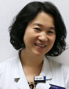 Weifang Zhang (张卫方)合作论文数Department of Nuclear Medicine, Peking University Third Hospital3
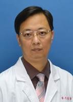 Yuehua Li (李跃华)合作论文数School of Health Science and Engineering, University of Shanghai for Science and Technology;Shanghai Sixth People’s Hospital Affiliated to Shanghai Jiao Tong University3




