Increased sensory nerve density has been described in type 2 inflammatory conditions and is linked to eosinophil and mast cell infiltration and neuropathic pain. To examine these relationships in eosinophilic gastrointestinal diseases, we developed a protocol using Ce3D (clearing-enhanced 3D) tissue clearing to evaluate mucosal gastrointestinal (GI) nerve remodeling and interactions with eosinophils and mast cells in whole-mount GI biopsies. Human gastric and esophageal biopsies were processed for 3-dimensional imaging. Tissues were fixed, permeabilized, and immunolabeled for mucosal nerves (Protein Gene Product 9.5), sensory neurons (substance P), eosinophils (eosinophil peroxidase), mast cells (tryptase), and epithelial cells (pan-cytokeratin). Vacuum-microwave-assisted staining was employed to enhance antibody penetration and protocol efficiency. Samples were cleared with Ce3D and imaged using confocal microscopy. Machine learning-enhanced quantitative analysis and modeling of nerve morphology and cellular interactions, including nerve length, branching, and spatial relationships between eosinophils, mast cells, and sensory neurons were performed. We established a 3-dimensional imaging method for whole-mount GI biopsies to characterize nerve architecture and eosinophil and mast cell interactions in the human esophagus and stomach. This approach enables high resolution and volumetric analyses and can be modified to assess other spatial neuroimmune interactions in human GI mucosal biopsies.
BACKGROUNDS AND AIMS:No guidelines exist for gastric acid assessment (GAA) or endoscopic surveillance for patients with Multiple Endocrine Neoplasia Type 1-Zollinger-Ellison Syndrome (MEN1-ZES). We aimed to analyze how GAA via nasogastric tube (NGT) and esophagogastro-duodenoscopy (EGD) altered acid suppression therapy and identify pre-GAA factors associated with post-GAA medication changes to inform which patients benefit from GAAs and/or EGDs for surveillance. METHODS:We assessed the following data from patients at our institution with MEN1-ZES from 2004-2018: 1) pre-GAA gastrointestinal symptoms; 2) serum gastrin levels; 3) gastric acid output (GAO); 4) EGD findings; 5) post-GAA changes in acid suppressing medication. GAO of <10 milliequivalents (mEq) of hydrochloric acid per hour (hr) indicated adequate acid suppression. RESULTS:Fifty-one patients who underwent 313 EGD/GAAs were identified; 263 EGD/GAAs were included. 51/263 EGD/GAAs (19.4%) led to increased acid suppression medication. Of these, 47.1% had GAO > 10 mEq/hr. Patients who had increases in acid suppression medication had significantly more symptoms, abnormal endoscopic findings, and higher GAOs compared to patients treated with same or decreased dose after endoscopy. All patients without symptoms prior to EGD/GAA were adequately suppressed. 8 NGT/GAAs done in 6 asymptomatic patients demonstrated adequate suppression. CONCLUSION:GAA is inadequately sensitive for detecting which patients with MEN1-ZES will benefit from increased acid suppression medication. Patients with symptoms should undergo EGD. A majority of patients without symptoms will not require increased acid suppressing medication following GAA, but a few asymptomatic patients may have endoscopic findings warranting increased therapy. There is no clear clinical benefit for NGT/GAA in asymptomatic patients.
BACKGROUNDAmong patients with multiple endocrine neoplasia type 1 (MEN1), 80% develop duodenopancreatic neuroendocrine tumors (dpNETs), of whom 15%-25% die of metastasis. There is a need to identify biomarkers to predict aggressive disease. MEN1 genotype affords an attractive possibility as a biomarker, as it remains constant during life. Currently, patients are clinically diagnosed with MEN1 by the presence of ≥2 primary endocrine tumors (pituitary, parathyroid, and pancreas) or ≥1 primary endocrine tumor with a positive family history. From 10% to 30% of patients diagnosed clinically with MEN1 have no pathogenic germline MEN1 variants.METHODSThis was a retrospective study of 162 index patients or probands with genotype-positive and 47 with genotype-negative MEN1 enrolled from 1977 to 2022.RESULTSCompared with patients with genotype-negative disease, patients with genotype-positive disease were younger at diagnosis and had an increased frequency of recurrent parathyroid tumors, dpNETs, and angiofibromas or collagenomas. We propose a weighted scoring system to diagnose genotype-positive MEN1 based on clinical characteristics. No evidence of MEN1 mosaicism was seen in 30 tumors from 17 patients with genotype-negative MEN1. Patients with germline MEN1 variants in exons 2 and 3 had a reduced risk of distant metastases.CONCLUSIONThe clinical course of genotype-negative MEN1 is distinct from genotype-positive disease, raising uncertainty about the benefits of lifetime surveillance in patients with genotype-negative disease. MEN1 mosaicism is rare.TRIAL REGISTRATION ClinicalTrials.gov NCT04969926FUNDINGIntramural Research Program of National Institute of Diabetes and Digestive and Kidney Diseases, NIH (ZIA DK043006-46).
A 50-year-old Central American man presented with 2 months of persistent nausea, weight loss, epigastric abdominal pain, and fluctuating diarrhea-constipation. Medical history was significant for HIV/AIDS and stage IV diffuse large B-cell lymphoma. On physical examination, the patient appeared cachectic with temporal wasting and had epigastric tenderness without rebound. Laboratory evaluation revealed 2400 leukocytes/μL with 33% eosinophils, HIV-RNA viral load 20 copies/mL, CD4 count 50 cells/mm3, and C-reactive protein 44.3 mg/L. Esophagogastroduodenoscopy demonstrated gastric mucosal atrophy with patchy gastric antral nodularity (Figure A). Histologic examination of gastric biopsies revealed gastric antral mucosal invasion with Strongyloides stercoralis colonizing acini (Figures B and C). Stool testing revealed Strongyloides filariform larvae confirming hyperinfection (Supplementary Video 1). Treatment with daily oral ivermectin 200 μg/kg for 2 weeks led to clinical improvement and parasitic elimination on stool examination.
 Harold Frucht合作论文数21
 Aimee L. Lucas合作论文数19Caroline Hwang合作论文数18John A. Chabot合作论文数15Lauren Khanna合作论文数15
 Amy Klion合作论文数Human Eosinophil Section, Laboratory of Parasitic Diseases, National Institute of Allergy and Infectious Diseases, National Institutes of Health15
 Khoury Paneez合作论文数Laboratory of Parasitic Diseases, National Institute of Allergy and Infectious Diseases14
 Christopher Koh合作论文数Division of Intramural Research, National Institute of Diabetes and Digestiveand Kidney Diseases12Hanna Blaney合作论文数10Theo Heller合作论文数Liver Dis Branch, NIH9
 Wendy Chung合作论文数Department of Pediatrics, Columbia University;Division of Genetics and Genomics, Boston Children’s Hospital;Harvard Medical School9Gregory M. Constantine合作论文数9Elana B Mitchel合作论文数7
 Disha Sharma合作论文数Translat Hepatol Sect, NIH7
 Martha Quezado合作论文数Laboratory of Pathology, Center for Cancer Research, National Cancer Institute, National Institutes of Health7Monica Passi合作论文数National Institute of Diabetes and Digestive and Kidney Diseases, National Institutes of Health.6
 Uri Ladabaum合作论文数Medicine Department, Stanford University6
 Chunwei Walter Lai合作论文数5
 Anusha Vittal合作论文数5
 Fei Li Kuang合作论文数Allergy and Immunology, Department of Medicine, Feinberg School of Medicine, Northwestern University5
 Jeremy L. Davis合作论文数National Institutes of Health Surgery Branch, National Cancer Institute5Louis Y. Korman合作论文数5Alexander Yang合作论文数5Stephen A. Wank合作论文数5Mian M. Khalid合作论文数4Bryan F Curtin合作论文数4Elana Levinson合作论文数4Laura Frado合作论文数4JeanAnne Ware合作论文数4
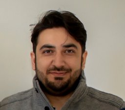 Bilal Asif合作论文数National Institute of Diabetes and Digestive and Kidney Diseases, National Institutes of Health4Farial Rahman合作论文数4Sunil Amin合作论文数4Jenny E Blau合作论文数4Mian Khalid合作论文数Dept Med, Mt Sinai Hosp4Auh Sungyoung合作论文数4Joanna Martinez-Gomez合作论文数4
 Elizabeth C. Verna合作论文数Transplant Clinical Research Center, Columbia University;Center for Liver Disease and Transplantation, Columbia University;Department of Surgery, Columbia University Irving Medical Center4Thomas Brown合作论文数3Amber Famiglietti合作论文数3
 Samuel A Schueler合作论文数3Udit Asija合作论文数3
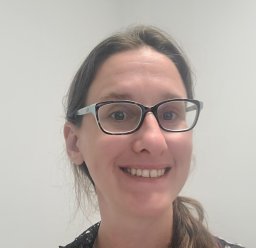 Astin Powers合作论文数Center for Cancer Research, National Cancer Institute3
 Y. Ann Chen合作论文数Department of Internal Medicine, School of Medicine, University of Utah;Department of Population Health Sciences, School of Medicine, University of Utah;Huntsman Cancer Institute3
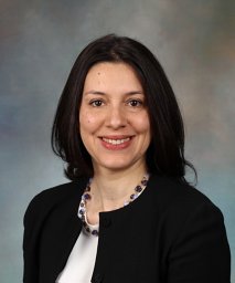 Marcela Salomao合作论文数Div Pathol, Mayo Clin3
 Ellen J Scherl合作论文数Joan and Sanford I. Weill Department of Medicine, Weill Cornell Medicine, Cornell University3
 David E. Kleiner合作论文数Laboratory of Pathology, Center for Cancer Research, National Cancer Institute3Holland-Thomas Nicole合作论文数3
 Hawwa Alao合作论文数3
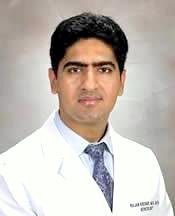 Kochar Rajan合作论文数3




